. 2025 Jun 16;80(7):433-444.
doi: 10.1136/thorax-2024-221634.

Inflammation in preschool cystic fibrosis is of mixed phenotype, extends beyond the lung and is differentially modified by CFTR modulators

Affiliations

Inflammation in preschool cystic fibrosis is of mixed phenotype, extends beyond the lung and is differentially modified by CFTR modulators

Shivanthan Shanthikumar et al. Thorax. .

Abstract

Background: Early-life inflammation has long been recognised as a key pathophysiological process in the evolution of cystic fibrosis (CF) lung disease. Despite this, no CF-specific anti-inflammatory treatments have been developed. This is crucial even in the era of highly effective modulator therapy as recent evidence suggests that modulators alter, but may not fully resolve, pulmonary inflammation.

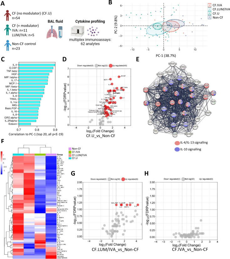
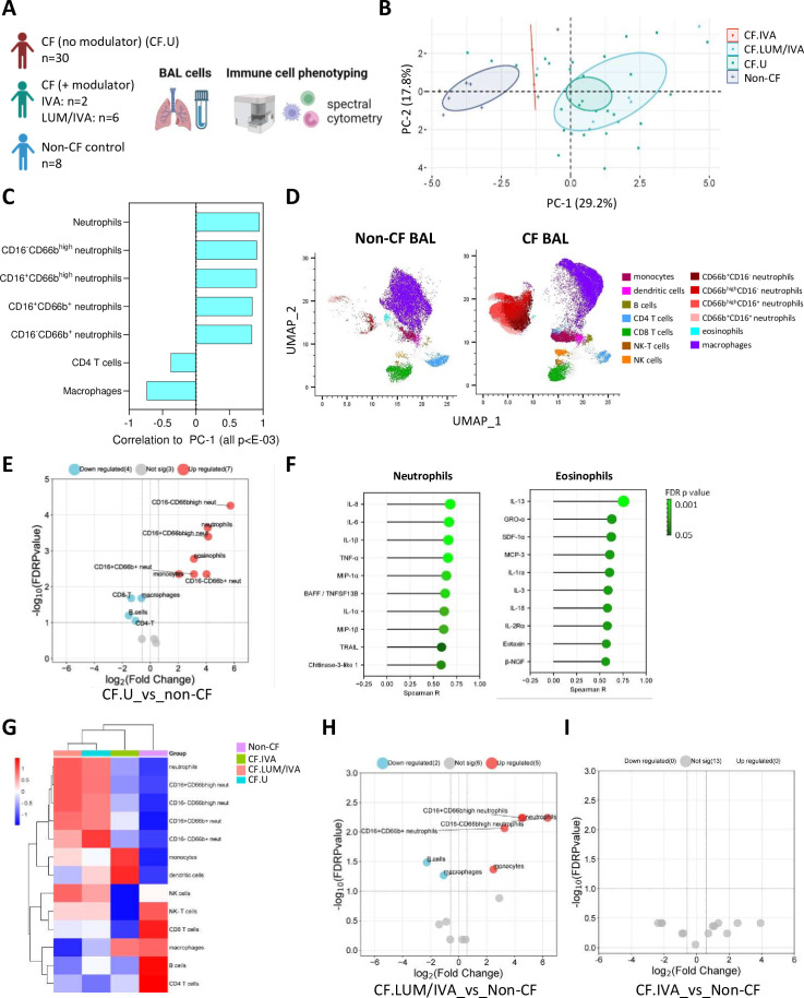
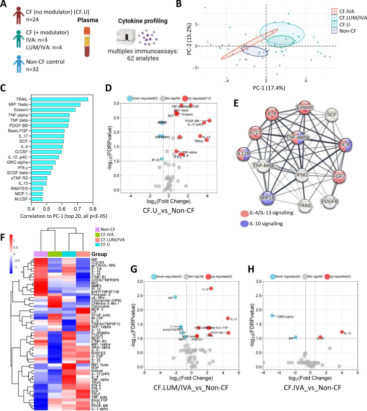
Methods: In this study, we used clinical microbiology data, high-dimensional flow cytometry and multiplex immunoassays to compare pulmonary (bronchoalveolar lavage (BAL)) and systemic immunity in 70 preschool children with CF and a total of 32 age-matched preschool controls.

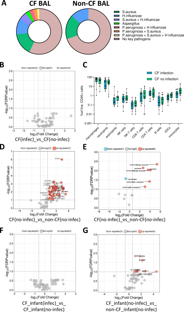
Results: We show that inflammation in the early-life CF lung is characterised by innate cell infiltration (neutrophils: 31.31 vs 1.8% of BAL in CF compared with controls, FDRp=0.0001; eosinophils: 0.55 vs 0.06%, FDRp=0.001, and monocytes: 1.91 vs 0.45%, FDRp=0.004) and widespread upregulation of both traditional and type 2 inflammatory soluble signatures (40 analytes significantly elevated in BAL of CF compared with controls, all FDRp<0.1). Key targetable features of this response included pulmonary interleukin (IL)-8 and IL-13 which were most significantly associated with neutrophilic and eosinophilic infiltration, respectively (IL-8 and neutrophils; Spearman rho=0.68, FDRp=0.002: IL-13 and eosinophils; Spearman rho=0.75, FDRp=0.01). Signatures of type 2 inflammation, as identified by REACTOME pathway analysis, including IL-4, IL-13 and FGF-2, were highly elevated in both the lungs and circulation in early CF. When exploring the efficacy of Cystic Fibrosis Transmembrane Conductance Regulator modulators to resolve pulmonary and systemic inflammation in early life, we showed that different classes of modulators have varying effects on inflammation, with ivacaftor showing a more significant effect in the lungs and circulation than lumacaftor/ivacaftor. Finally, we showed that CF children with pathogen colonisation had similar levels of pulmonary inflammation as CF children without pathogen colonisation (no significant differences), and that inflammation was evident during infancy even without evidence of colonisation (as observed by significant increases in levels of SDF-1alpha, M-CSF, IL-2, IL-9, IL-12p40, IL-17, MCP-1 and LIGHT/TNFSF14, all FDRp<0.1), highlighting a role for intrinsic dysregulation of inflammation that begins in early life.

Conclusions: We provide a rationale for targeted anti-inflammatory intervention in early-life CF.

Keywords: Allergic lung disease; Cystic Fibrosis; Cytokine Biology; Innate Immunity; Paediatric Lung Disaese.


Conflict of interest statement

Competing interests: None declared.

Figures

Figure 1
Figure 1. Soluble pulmonary immune signatures of early-life cystic fibrosis (CF). (A) Experimental workflow: bronchoalveolar lavage (BAL) cell-free fluid from children with CF (with (n=16) or without (n=54) Cystic Fibrosis Transmembrane Conductance Regulator (CFTR) modulator therapy) and non-CF controls (n=23) was analysed for immune-related analytes using commercially available multiplex immunoassays and the Bio-Plex 200 system. (B) Principal components analysis of BAL analyte data reveals clear separation between CF and non-CF samples, as well as between CF lumacaftor/ivacaftor (LUM/IVA) and CF IVA samples in principal component 1 (PC-1). (C) Correlation coefficients of the top 20 signatures that significantly correlated with PC-1 (all p<E-19). (D) Volcano plot depicting analytes that were significantly different in BAL cell-free fluid from children with CF (not on a modulator therapy) compared with controls. (E) Protein-protein interaction (PPI) analysis using STRING reveals significant interactions between analytes elevated in CF BAL (the strength of the line indicates the confidence of the interaction) and shows enrichment of interleukin (IL)-4/IL-13 and IL-10 signalling pathways in the data set. (F) Heatmap depicting unsupervised clustering analysis of median BAL cell-free fluid analytes in children with CF not on modulator therapy (blue), children with CF on LUM/IVA therapy (peach), children with CF on IVA therapy (green) and non-CF controls (purple). (G) Volcano plot depicting BAL cell-free fluid analytes that were significantly different in children with CF on LUM/IVA (n=5) therapy compared with non-CF controls (n=23). (H) Volcano plot depicting BAL cell-free fluid analytes that were significantly different in children with CF on IVA therapy (n=11) compared with non-CF controls (n=23) (none were significantly different). The p-values were calculated by Mann-Whitney U test and corrected for false discovery rate using the Benjamini-Hochberg approach. FDR-corrected p<0.1 and a fold change >1.5 were required to reach significance.
Figure 2
Figure 2. Cellular pulmonary signatures of early-life cystic fibrosis (CF). (A) Experimental workflow: bronchoalveolar lavage (BAL) cells from children with CF (with (n=8) or without (n=30) Cystic Fibrosis Transmembrane Conductance Regulator (CFTR) modulator therapy) and non-CF controls (n=8) were analysed by flow cytometry. (B) Principal components analysis of BAL cell data reveals clear separation between CF and non-CF samples, as well as between CF lumacaftor/ivacaftor (LUM/IVA) and CF IVA samples in principal component 1 (PC-1). (C) Correlation coefficients of the cellular signatures that significantly correlated with PC-1 (all p<E-13). (D) Uniform Manifold Approximation and Projection (UMAP) depicting the immune cell profile of BAL from children with CF (not on a CFTR modulator therapy) and non-CF controls. (E) Volcano plot depicting BAL immune cell populations that were significantly different in children with CF (not on a modulator therapy) compared with non-CF controls. (F) Two-tailed Spearman correlations of infiltrating granulocytes (neutrophils and eosinophils) and BAL cell-free fluid analytes. (G) Heatmap depicting unsupervised clustering analysis of median BAL immune cell populations in children with CF not on modulator therapy (blue), children with CF on LUM/IVA therapy (peach), children with CF on IVA therapy (green) and non-CF controls (purple). (H) Volcano plot depicting BAL immune cell populations that were significantly different in children with CF on LUM/IVA (n=6) therapy compared with non-CF controls (n=8). (I) Volcano plot depicting BAL immune cell populations that were significantly different in children with CF on IVA therapy (n=2) compared with non-CF controls (n=8) (none were significantly different). The p-values were calculated by Mann-Whitney U test and corrected for false discovery rate using the Benjamini-Hochberg approach. FDR-corrected p<0.1 and a fold change>1.5 were required to reach significance. Correlation p-values were determined by two-tailed Spearman test and corrected for false discovery rate using the Benjamini-Hochberg approach.
Figure 3
Figure 3. Systemic signatures of early-life cystic fibrosis (CF). (A) Experimental workflow: plasma from children with CF (with (n=7) or without (n=24) Cystic Fibrosis Transmembrane Conductance Regulator (CFTR) modulator therapy) and non-CF controls (n=32) was analysed for immune-related analytes using commercially available multiplex immunoassays and the Bio-Plex 200 system. (B) Principal components analysis of plasma analyte data reveals clear separation between CF and non-CF samples, as well as between CF lumacaftor/ivacaftor (LUM/IVA) and CF IVA samples in principal component 1 (PC-1). (C) Correlation coefficients of the top 20 signatures that significantly correlated with PC-1 (all p<E-05). (D) Volcano plot depicting analytes that were significantly different in plasma from children with CF (not on a modulator therapy) compared with controls. (E) Protein-protein interaction (PPI) analysis using STRING reveals significant interactions between analytes elevated in CF BAL (the strength of the line indicates the confidence of the interaction) and highlights interleukin (IL)-4/IL-13 and IL-10 signalling pathways as enriched in the data set. (F) Heatmap depicting unsupervised clustering analysis of plasma analytes in children with CF not on modulator therapy (blue), children with CF on LUM/IVA therapy (peach), children with CF on IVA therapy (green) and controls (purple). (G) Volcano plot depicting plasma analytes that were significantly different in children with CF on LUM/IVA therapy (n=4) compared with controls (n=32). (H) Volcano plot depicting plasma analytes that were significantly different in children with CF on IVA therapy (n=3) compared with controls (n=32). The p-values were calculated by Mann-Whitney U test and corrected for false discovery rate using the Benjamini-Hochberg approach. FDR-corrected p<0.1 and a fold change>1.5 were required to reach significance.
Figure 4
Figure 4. Relationship between clinically important pathogen colonisation and pulmonary inflammation in early-life cystic fibrosis (CF). (A) Proportion of colonisation with four key pathogens (Pseudomonas aeruginosa, Staphylococcus aureus, Haemophilus influenzae and Aspergillus) in CF and non-CF bronchoalveolar lavage (BAL) samples. (B) Volcano plot depicting BAL analytes that were significant between CF children who had evidence of one or more of these pathogens in BAL (n=22) and children with CF who had no evidence of these pathogens (n=32) (none were significant). (C) Box plots depicting immune cell populations in BAL of CF children who had evidence of one or more of these pathogens in BAL (n=15) and CF children who had no evidence of these pathogens in BAL (n=15). There were no significant differences. (D) Volcano plot depicting BAL analytes that were significant between CF children with no evidence of these pathogens (n=32) and non-CF children with no evidence of these pathogens (n=16). (E) Volcano plot depicting BAL immune cell populations that were significantly different between CF children with no evidence of these pathogens (n=15) and non-CF children with no evidence of these pathogens (n=7). (F) Volcano plot depicting BAL analytes that were significant between CF infants with evidence of these pathogens (n=12) and CF infants with no evidence of these pathogens (n=19) (none were significant). (G) Volcano plot depicting BAL analytes that were significant between CF infants with no evidence of these pathogens (n=19) and non-CF infants with no evidence of these pathogens (n=6). The p-values were calculated by Mann-Whitney U test and corrected for false discovery rate using the Benjamini-Hochberg approach. FDR-corrected p<0.1 and a fold change>1.5 were required to reach significance.

Similar articles

Cited by

References

    1. Shteinberg M, Haq IJ, Polineni D, et al. Cystic fibrosis. Lancet. 2021;397:2195–211. doi: 10.1016/S0140-6736(20)32542-3. - DOI - PubMed
    1. Sly PD, Gangell CL, Chen L, et al. Risk factors for bronchiectasis in children with cystic fibrosis. N Engl J Med. 2013;368:1963–70. doi: 10.1056/NEJMoa1301725. - DOI - PubMed
    1. Ranganathan SC, Hall GL, Sly PD, et al. Early Lung Disease in Infants and Preschool Children with Cystic Fibrosis. What Have We Learned and What Should We Do about It? Am J Respir Crit Care Med. 2017;195:1567–75. doi: 10.1164/rccm.201606-1107CI. - DOI - PMC - PubMed
    1. Cystic Fibrosis Foundation Drug development pipeline. 2023. [20-Sep-2023]. https://apps.cff.org/trials/pipeline Available. Accessed.
    1. Taylor-Cousar JL, Robinson PD, Shteinberg M, et al. CFTR modulator therapy: transforming the landscape of clinical care in cystic fibrosis. Lancet. 2023;402:1171–84. doi: 10.1016/S0140-6736(23)01609-4. - DOI - PubMed

MeSH terms