. 2025 Feb 10;16(1):1484.
doi: 10.1038/s41467-025-56317-8.

Macrocycle-based PROTACs selectively degrade cyclophilin A and inhibit HIV-1 and HCV

Affiliations

Macrocycle-based PROTACs selectively degrade cyclophilin A and inhibit HIV-1 and HCV

Lydia S Newton et al. Nat Commun. .

Abstract

Targeting host proteins that are crucial for viral replication offers a promising antiviral strategy. We have designed and characterised antiviral PROteolysis TArgeting Chimeras (PROTACs) targeting the human protein cyclophilin A (CypA), a host cofactor for unrelated viruses including human immunodeficiency virus (HIV) and hepatitis C virus (HCV). The PROTAC warheads are based on fully synthetic macrocycles derived from sanglifehrin A, which are structurally different from the classical Cyp inhibitor, cyclosporine A. Our Cyp-PROTACs decrease CypA levels in cell lines and primary human cells and have high specificity for CypA confirmed by proteomics experiments. Critically, CypA degradation facilitates improved antiviral activity against HIV-1 in primary human CD4+ T cells compared to the non-PROTAC parental inhibitor, at limiting inhibitor concentrations. Similarly, we observe antiviral activity against HCV replicon in a hepatoma cell line. We propose that CypA-targeting PROTACs inhibit viral replication potently and anticipate reduced evolution of viral resistance and broad efficacy against unrelated viruses. Furthermore, they provide powerful tools for probing cyclophilin biology.


Conflict of interest statement

Competing interests: The Ciulli laboratory receives or has received sponsored research support from Almirall, Amgen, Amphista Therapeutics, Boehringer Ingelheim, Eisai, Merck KGaA, Nurix Therapeutics, Ono Pharmaceutical and Tocris-BioTechne. A.C. is a scientific founder, advisor, and shareholder of Amphista Therapeutics, a company that is developing targeted protein degradation therapeutic platforms. The other authors declare no competing interests.

Figures

Fig. 1
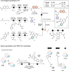
Fig. 1. Design of macrocycle-based PROTACs with high affinity for cyclophilins.
a X-ray crystal structure of sanglifehrin A-CypA complex (PDB 1YND) (left) and TWH106 docked into CypA (right). The TWH106 nitrophenyl moiety fills a subpocket near residues K82 and E81 and the nitro group H-bonds with K82 (arginine in CypB). Two solvent-exposed methyl groups ✱ and # of TWH106 were used to synthetically grow PROTACs. b Chemical structure of our two TWH106-based PROTACs CG167 and RJS308.
Fig. 2
Fig. 2. Synthesis of enantiopure macrocycle-based PROTACs.
a HATU, i-Pr2NEt, DMF, 96% b THF, 97% or 28% (CG167) c dichloro(p-cy)Ru(II) dimer, (1 R, 2 R)-(− )-N-p-tosyl−1,2-diphenylethylenediamine, THF, H2O, HCOONa, 90% or 86% (CG167) d n-BuLi, THF, 90% e LDA, THF, DMPU f THF, H2O, 64% (2 steps) g1 HATU, OxymaPure, i-Pr2NEt, MeCN, 52% g2 i. TFA, CH2Cl2 ii. Like G1, 69% (RJS308) h i. TFA, CH2Cl2 ii. PyAOP, i-Pr2Net, CH3CN, 41%, 57% (RJS308) i 2 M HCl, dioxane, H2O, 66% j EDC, DMAP, i-Pr2NEt, CH2Cl2, 78% or 95% (CG167) k i. TFA, CH2Cl2 ii. HATU, i-Pr2NEt, OxymaPure, 55% (TWH106), 38% (CG167), 54% (RJS308) l CuSO4, L-ascorbic acid, THF, H2O, 91% (RJS308), 82% (CG167) m Pd(OAc)2, piperazine, DMF, μ-wave, 8% (TWH106), 27% (RJS308), 7% (CG167), n HATU, i-Pr2NEt, DMF, 30% (RJS308), 80% (CG167). Characterisation data is provided in the Supplementary Information file.
Fig. 3
Fig. 3. PROTACs CG167 and RJS308 degrade CypA in a dose-dependent manner.
af Immunoblots detecting CypA, or β–actin loading control, in Jurkat cells treated with 5 μM TWH106, CG167 or RJS308 for up to 6 days (ac) or 20 h (df). g CypA densities from (af) adjusted for loading by reference to β–actin densities, mean (n = 2 independent experiments). hm Immunoblots detecting CypA in Jurkat cells treated with 0.01 – 10 μM TWH106, CG167 or RJS308 for 24 h (hj) or 48 h (km). n CypA densities relative to β-actin (hj) top and (km) bottom, mean (n = 2 independent experiments). Additional immunoblots in Supplementary Fig. 5. Source data are provided in the Source Data file.
Fig. 4
Fig. 4. Degradation of CypA by CG167 and RJS308 is via a PROTAC mechanism.
ae Immunoblots detecting CypA, or β-actin as loading control, in Jurkat cells. Cells were pretreated with (a) 1 μM NEDD8-activating enzyme inhibitor MLN4924 for 6 h or (b) 50 μM VHL inhibitor VH298 (VHLi) for 2 h, followed by 5 μM PROTAC CG167 or RJS308 for 48 h, or cells were treated with (c) 10 μM TWH106 and 1 μM PROTAC CG167 or RJS308 for 48 h, or cells were treated with (d) MLN4924 as in (a) or with (e) VHLi as in (b). CypA densities (top) adjusted for loading by reference to β–actin densities, mean (n = 2 independent experiments). Additional immunoblots in Supplementary Fig. 6. f, g Size-exclusion chromatography traces for complex formation between VCB (pVHL/elongin C/elongin B, 41 kDa) and CypA (20 kDa) with (f) RJS308 (1.2 kDa, blue) vs DMSO (red) or (g) CG167 (1.2 kDa, green) vs RJS308 (1.2 kDa, blue) addition. h, i Dose-dependent binding between (h) RJS308 or (i) RJS308/CypA complex (flow) and VCB (immobilised on chip) in representative SPR experiments. Binding affinities were calculated from kinetic fits (black curves, 1:1 binding model), mean ± SD (n = 5 (h) or n = 4 (i) independent experiments). Source data are provided in the Source Data file.
Fig. 5
Fig. 5. Cyp-PROTACs are selective for CypA.
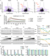
ac Volcano plots of THP-1 cell proteomics analysis after 72 h treatment with 5 μM (a) TWH106 (parental Cyp ligand), (b) CG167 (Cyp-PROTAC), or (c) RJS308 (Cyp-PROTAC), compared to DMSO control, significant change (red), other proteins of interest (purple). Two-sided two-sample student t-test, s0 = 0.5; FDR = 0.05 (n = 3 independent experiments). d Dose-dependent binding (SPR) of RJS308/N-truncated CypB complex (flow) and VCB (immobilised on chip) with kinetic fits (black curves, 1:1 binding model) (n = 1). Slow complex dissociation indicates sub-nanomolar affinity but prevents accurate binding affinity determination. eg, ik Immunoblots detecting CypB, or β-actin as loading control, in Jurkat cells treated with TWH106, CG167 or RJS308 at (eg) 5 μM for 6 days or (ik) 0.01–10 μM for 48 h. h, l CypB densities (h) from (eg), (l) from (ik) adjusted for loading by reference to β–actin densities, mean (n = 2 independent experiments). m, n CypA densities (m) or CypB densities (n) relative to β-actin from immunoblots after 48 h treatment with 5 μM CG167 or RJS308 on cells, mean (n = 2 independent experiments). Additional immunoblots for (el) in Supplementary Fig. 9, and immunoblots for (m, n) in Supplementary Fig. 11. Source data are provided in the Source Data file.
Fig. 6
Fig. 6. Cyp-PROTACs exhibit improved anti-HIV-1 activity compared to parental Cyp ligand TWH106.
a Experimental design for (bx): activated primary CD4+ T cells were pretreated for 48 h with TWH106 (orange), CG167 (blue), RJS308 (green) or DMSO (black), washed and infected with HIV-1 NL4.3 (2000 mU RT/106 cells). Cells were treated with 1 μM or 5 μM Cyp inhibitor throughout the experiment (solid circles) or only pretreated with 5 μM Cyp inhibitor (dashed circle). Alternatively, cells were pretreated with 5 μM Cyp inhibitor and 50 μM VHL inhibitor VH298 (5 μM + VHLi, dashed circle). bm Representative data from one donor at indicated days post infection (dpi), mean (n = 1 independent experiment performed in duplicate), (be) % Gag+ cells (fi) virus levels in supernatant measured by SG-PERT (jm) CypA mean fluorescence intensity (MFI), normalised to DMSO. nx Data from 4 donors (1 μM, 5 μM and 5 μM pretreatment only) or 3 donors (5 μM pretreatment only + VHL inhibitor), all data normalised to DMSO, mean ± SD, (nq) % Gag+ cells at 4 dpi, (ru) virus levels in supernatant at 4 dpi measured by SG-PERT, (vx) CypA MFI at time of infection (0 dpi). Statistical comparison using RM one-way ANOVA with Dunnett’s multiple comparisons test comparing to TWH106, * (P ≤ 0.05), ** (P ≤ 0.01), *** (P ≤ 0.001), **** (P ≤ 0.0001), P-values shown. Data from additional donors in Supplementary Fig. 14. w (right) histogram showing the spread of CypA-FITC fluorescence at 0 dpi, representative data from one donor. Gating strategies are shown in Supplementary Fig. 15a–c. Source data are provided in the Source Data file.

References

    1. Kumar, N. et al. Host-directed antiviral therapy. Clin. Microbiol. Rev.33, e00168 (2020). - PMC - PubMed
    1. Lou, Z., Sun, Y. & Rao, Z. Current progress in antiviral strategies. Trends Pharmacol. Sci.35, 86 (2014). - PMC - PubMed
    1. Dwek, R. A., Bell, J. I., Feldmann, M. & Zitzmann, N. Host-targeting oral antiviral drugs to prevent pandemics. Lancet399, 1381 (2022). - PMC - PubMed
    1. Chitalia, V. C. & Munawar, A. H. A painful lesson from the COVID-19 pandemic: the need for broad-spectrum, host-directed antivirals. J. Transl. Med.18, 390 (2020). - PMC - PubMed
    1. Bekes, M., Langley, D. R. & Crews, C. M. PROTAC targeted protein degraders: the past is prologue. Nat. Rev. Drug Discov.21, 181 (2022). - PMC - PubMed

MeSH terms

LinkOut - more resources