. 2025 Feb 10;16(1):1511.
doi: 10.1038/s41467-025-56782-1.

Loss-of-function mutations in the dystonia gene THAP1 impair proteasome function by inhibiting PSMB5 expression

Affiliations

Loss-of-function mutations in the dystonia gene THAP1 impair proteasome function by inhibiting PSMB5 expression

Dylan E Ramage et al. Nat Commun. .

Abstract

The 26S proteasome is a multi-catalytic protease that serves as the endpoint for protein degradation via the ubiquitin-proteasome system. Proteasome function requires the concerted activity of 33 distinct gene products, but how the expression of proteasome subunits is regulated in mammalian cells remains poorly understood. Leveraging coessentiality data from the DepMap project, here we characterize an essential role for the dystonia gene THAP1 in maintaining the basal expression of PSMB5. PSMB5 insufficiency resulting from loss of THAP1 leads to defects in proteasome assembly, impaired proteostasis and cell death. Exploiting the fact that the toxicity associated with loss of THAP1 can be rescued upon exogenous expression of PSMB5, we define the transcriptional targets of THAP1 through RNA-seq analysis and perform a deep mutational scan to systematically assess the function of thousands of single amino acid THAP1 variants. Altogether, these data identify THAP1 as a critical regulator of proteasome function and suggest that aberrant proteostasis may contribute to the pathogenesis of THAP1 dystonia.


Conflict of interest statement

Competing interests: The authors declare no competing interests.

Figures
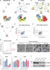
Fig. 1
Fig. 1. Transcriptional regulation of PSMB5 by THAP1 explains their co-essential relationship.
AC Co-essential relationships involving UPS genes predict biological relationships, as exemplified by three E3 ligase complexes: the BAG6 complex (A), Cul2VHL (B) and the CTLH complex (C). Network diagrams were produced using NetworkX; numbers annotating the edges indicate pairwise correlation coefficients as calculated in ref. . DF THAP1 exhibits a strong positive co-essential relationship with both PSMB5 and PSMB6 across DepMap data. G, H THAP1 disruption is toxic in HEK-293T cells. Cells were transduced with a lentiviral vector expressing Cas9 and the indicated sgRNAs, followed by puromycin selection to eliminate untransduced cells commencing 48 h later. A further 48 hours later, cells were counted, plated in equal numbers, and their viability assessed by counting (G) and brightfield microscopy (H). Data in (G) represent mean values of n = 3 biological replicates ± s.d. (***P < 0.001, two-tailed t-test) (Scale bar = 100 µm). I, J Ablation of THAP1 decreases PSMB5 expression. HEK-293T expressing Cas9 and the indicated sgRNAs were analyzed by qRT-PCR (I) and immunoblot (J). Data in (I) are presented as mean values of n = 3 technical replicates ± s.d. Source data are provided as a Source Data file.
Fig. 2
Fig. 2. Lethality resulting from THAP1 loss can be rescued by exogenous expression of PSMB5.
A Exogenous expression of PSMB5 rescues cell viability upon THAP1 ablation. HEK-293T cells were first transduced with lentiviral vectors expressing either PSMB5 or PSMB6; then, following transduction with Cas9 and the indicated sgRNAs, cell numbers were monitored over time. B Loss of THAP1 is broadly toxic across cell types. CRISPR/Cas9-mediated ablation of THAP1 adversely affects the viability of 946/1100 cancer cell lines (blue dots, representing effect scores < -0.25) examined by DepMap. C, D The toxicity associated with THAP1 ablation is rescued by exogenous PSMB5 expression in HeLa cells (C) and A549 cells (D). E–H Like PSMB5, expression of PSMB8 also protects against the toxic effects of THAP1 loss. THP-1 cells do not exhibit any substantial growth defect following THAP1 ablation (E). High levels of PSMB8 expression are observed in the cell lines whose growth is not significantly affected by THAP1 loss (orange dots, representing effect scores >-0.25) in DepMap data (F), and THP-1 cells strongly express PSMB8 as assessed by qRT-PCR (G). Exogenous expression of PSMB8 can rescue the viability of HEK-293T cells following THAP1 disruption (H). Data in (G) represent mean values of n = 3 technical replicates ± s.d.; data in (A, CE, and H) represent mean values of n = 3 biological replicates ± s.d. (***P < 0.001, two-tailed t-test; ns, not significant). Source data are provided as a Source Data file.
Fig. 3
Fig. 3. A fluorescent reporter measures endogenous PSMB5 expression in live cells.
AC CRISPR/Cas9-mediated knock-in of GFP into the endogenous PSMB5 locus. A schematic representation of the procedure is shown in (A). Transfection of HEK-293T cells with Cas9, an sgRNA targeting PSMB5 and a donor template resulted in ~10% GFP-positive cells (B), which were purified by FACS and single cell cloned (C). DG THAP1 ablation reduces PSMB5 expression. CRISPR/Cas9-mediated disruption of THAP1 in a PPSMB5-GFP reporter clone (expressing exogenous PSMB5 to ensure viability) reduced PPSMB5-GFP expression (D), permitting the derivation of GFPdim THAP1 KO clones (E). These THAP1 KO clones exhibited greatly reduced expression of PSMB5 by qRT-PCR (using primers annealing to the 3’UTR to ensure selective amplification of the endogenous transcripts) (F), and an absence of THAP1 protein by immunoblot (a non-specific band is indicated by an asterisk) (G). Data in (F) represent mean values of n = 3 technical replicates ± s.d. Source data are provided as a Source Data file.
Fig. 4
Fig. 4. THAP1 binds cognate motifs within the PSMB5 promoter to regulate its expression.
A THAP1 binds the PSMB5 promoter. THAP1 ChIP-seq data in K562 cells reveals an intense peak representing THAP1 occupancy at the PSMB5 transcription start site (TSS) (top). PSMD8 is the only other proteasome subunit at which concordant binding of THAP1 is observed (bottom). B Consensus THAP1 binding site (THABS) motif (adapted from). C Schematic representation of the three consensus THABS motifs located near the PSMB5 TSS. DF THAP1 targets cognate sites in the PSMB5 promoter to activate gene expression. D Schematic representation of a lentiviral reporter system in which ~1 kb of the PSMB5 promoter drives the expression of GFP. Removal of the three THABS motifs (“ΔTHABS”) reduced GFP expression (E); this effect was mediated through THAP1, as THAP1 ablation decreased GFP expression driven by the wild-type promoter but not the ΔTHABS promoter (F). Source data are provided as a Source Data file.
Fig. 5
Fig. 5. PSMB5 insufficiency resulting from THAP1 loss impairs proteasome function.
A The catalytic activity of PSMB5 is required to rescue viability upon THAP1 loss. Exogenous expression of wild-type PSMB5, but not a catalytically-inactive mutant, restored the viability of HEK-293T cells following THAP1 ablation. Data are presented as mean values of n=3 biological replicates +/− s.d. (***P < 0.001, two-tailed t-test; ns, not significant). B, C Loss of THAP1 impairs proteasome assembly. B THAP1 ablation decreases the abundance of mature, processed PSMB6 and PSMB7 as assessed by immunoblot, but leads to the accumulation of the uncleaved proproteins (indicated with asterisks). C Native PAGE analysis reveals the accumulation of proteasome assembly intermediates (indicted with asterisks) following THAP1 disruption, mimicking the defects observed upon PSMB5 knockdown. DF THAP1 impairs the proteasomal degradation of short-lived proteins. D Schematic representation of the lentiviral Global Protein Stability (GPS) two-color fluorescent reporter system to monitor protein stability. E Stabilization of two model GFP-degron fusion proteins upon ablation of THAP1, as assessed by flow cytometry; the N-terminal peptide derived from PTGS1 harbors an N-terminal degron targeted by UBR-family E3 ligases, while the C-terminal peptide derived from TNNC2 harbors a C-terminal degron targeted by Cul4DCAF12. F Increased abundance of endogenous HIF-1α upon THAP1 disruption, as assayed by immunoblot. All immunoblot data is representative of at least two independent experiments. Source data are provided as a Source Data file.
Fig. 6
Fig. 6. Defining the transcriptional targets of THAP1.
A Schematic representation of the RNA-seq experiment. Created in BioRender. Timms, R. (2025) https://BioRender.com/ x85w665. B, C Identifying the transcriptional targets of THAP1. A summary of the RNA-seq dataset is shown in (B): genes exhibiting differential expression between sgControl and sgTHAP1 cells (n = 277) are highlighted in yellow, with the subset of those genes that display THAP1 occupancy as assessed by ChIP-seq (n = 42) colored red. Expression changes amongst all differentially expressed genes are summarized in (C), with circles representing genes bound by THAP1. D Consensus binding sites for YY1 and THAP1 are enriched amongst the promoters of the 42 direct THAP1 target genes. Each circle represents an individual YY1 (purple) or THAP1 (red) binding site (see also Supplementary Fig. 4C); bubble size is proportional to the number of the gene promoters containing the motif. EI Validation of THAP1 target genes. Five target genes directly activated by THAP1 binding are shown: ChIP-seq data indicating THAP1 occupancy is shown on the left; the RNA-seq expression data is summarized in the center, and qRT-PCR validation is shown on the right. Data are presented as mean values of n = 3 technical replicates +/− s.d. Source data are provided as a Source Data file.
Fig. 7
Fig. 7. A deep mutational scan defines the functional landscape of THAP1 mutations.
A Schematic representation of the domain architecture of the THAP1 protein. B Schematic representation of the deep mutational scan, designed to interrogate the ability of all possible single amino acid variants of THAP1 to activate expression of the endogenous PPSMB5-GFP knock-in allele. Created in BioRender. Timms, R. (2025) https://BioRender.com/ x85w665. C Site-saturation mutagenesis reveals critical residues for THAP1 function. Each cell represents the performance of a single THAP1 mutant: the mean performance of all the wild-type proteins is centered at 1 (light gray), with red colors indicating mutants which abrogate activation of the PPSMB5-GFP reporter and blue colors indicating mutants which may enhance the activation of the PPSMB5-GFP reporter. Dark gray cells indicate mutants for which insufficient data was available for analysis.
Fig. 8
Fig. 8. Determining the functional effects of THAP1 mutations found in Dystonia patients.
A, B The deep mutational scan successfully identifies THAP1 residues critical for DNA binding. A Performance of all THAP1 variants targeting zinc-chelating residues of the zinc finger domain. Bars indicate the mean of two replicate experiments. Constructs which encode the wild-type protein are indicated in bold; the mean activity exhibited across all the wild-type THAP1 constructs is set at 1 (dotted line). B Distribution of activity scores across all the THAP1 variants targeting residues previously determined to be important for DNA binding by THAP1. CF Defining structure-function relationships for THAP1. Mean activity scores from the genetic screen were mapped onto the predicted structure of THAP1 (C) or the experimental structure of the THAP1 zinc finger domain (D). Overall, residues predicted to lie in ordered regions of the protein (AlphaFold pLDDT > 60) were much less tolerant of mutations than residues predicted to lie in disordered regions (E). Individual validation of the screen results was performed using flow cytometry: deletion of the coiled-coil and HCFC1-binding motif abrogated THAP1 function, whereas deletion of the disordered C-terminus did not (F). G, H Profiling the activity of THAP1 mutations found in Dystonia patients. G Performance of all missense variants identified in Dystonia patients, displayed as in (A). H Individual validation experiments measuring the activity of THAP1 mutants predicted to be inactive (top row) and THAP1 mutants predicted to be active (bottom row) by flow cytometry. See also Supplementary Fig. 5C–E.

References

    1. Rape, M. Ubiquitylation at the crossroads of development and disease. Nature Reviews Molecular Cell Biology19, 59–70 (2017). - PubMed
    1. Kwon, Y. T. & Ciechanover, A. The Ubiquitin Code in the Ubiquitin-Proteasome System and Autophagy. Trends Biochem Sci42, 873–886 (2017). - PubMed
    1. Popovic, D., Vucic, D. & Dikic, I. Ubiquitination in disease pathogenesis and treatment. Nat Med20, 1242–1253 (2014). - PubMed
    1. Murata, S., Yashiroda, H. & Tanaka, K. Molecular mechanisms of proteasome assembly. Nature Reviews Molecular Cell Biology10, 104–115 (2009). - PubMed
    1. Collins, G. A. & Goldberg, A. L. The Logic of the 26S Proteasome. Cell169, 792–806 (2017). - PMC - PubMed

MeSH terms

Substances

LinkOut - more resources