. 2025 Feb;34(2):e70057.
doi: 10.1111/exd.70057.

The Inflammatory Landscape of a Whole-Tissue Explant Model of Hidradenitis Suppurativa

Affiliations

The Inflammatory Landscape of a Whole-Tissue Explant Model of Hidradenitis Suppurativa

Phoebe E Leboit et al. Exp Dermatol. 2025 Feb.

Abstract

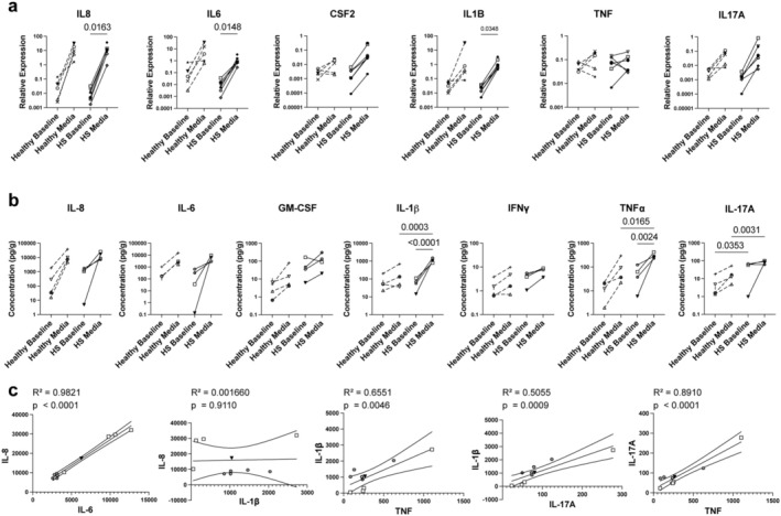
Hidradenitis suppurativa (HS) is a relatively common and highly morbid inflammatory skin disease. Due to the relatively limited understanding of HS's pathogenesis, there are currently insufficient treatment options available, and many patients' medical needs are not being met. This is partly due to the historical scarcity of ex vivo assays and animal models that accurately recapitulate the disease. Thus, we have developed a standardised whole-tissue explant model of HS to examine its pathogenic mechanisms and the efficacy of potential treatments within intact human tissue. We measured cytokine protein and RNA within whole tissue maintained in an agar-media solution, finding that IL-6 and IL-8 concentrations trended upwards in both HS explants and healthy controls, while IL-17A, IL-1β, and TNF-α exhibited increases in HS tissue alone. We also show that the explants were responsive to treatment with both dexamethasone and IL-2. Not only do our results show that this model effectively delivers treatments throughout the explants, but they also elucidate which cytokines are related to the explant process regardless of tissue state and which are related to HS tissue specifically, laying the groundwork for future implementations of this model.

Keywords: cytokine; explant model; hidradenitis suppurativa; immunology; inflammation.


Conflict of interest statement

Michael D. Rosenblum is a consultant and cofounder of TRex Bio Inc., Sitryx Bio Inc., and Radera Bio Inc. He is also a consultant for Mozart Bio Inc. Jarish N. Cohen is a consultant for TRex Bio Inc. and Radera Bio Inc. Haley B. Naik has received consulting fees from Abbvie, Medscape, Sonoma Biotherapeutics, Union Chimique Belge's (UCB) and Novartis; and holds shares in Radera Inc. She is also an Associate Editor for JAMA Dermatology and Vice President of the Hidradenitis Suppurativa Foundation. Ryan L. Kyle and Matthew Kelly are employees of Sitryx Therapeutics.

Figures

FIGURE 1
FIGURE 1
Explants cultured for 24 h maintain live immune cell populations and exhibit complete drug penetration. (a) HS explants cultured for 24 h. (b) H&E‐stained sections of HS (250 μm scale bar) and healthy control (400 μm scale bar) explants (representative of 3 independent healthy samples and 2 independent HS samples). (c) Cell yields following 24‐h culture for donors (a–d) (each bar represents one explant). (d) Quantitative reverse‐transcriptase polymerase chain reaction (RT‐qPCR) analysis of CD3E and PTPRC (CD45) in healthy (n = 5) and HS explants (n = 6) at baseline and after culture relative to housekeeping gene expression (ns, mixed‐effects analysis with Šídák's multiple comparisons test). Each shape represents one donor (averaged for 1–4 explant tissues, solid line = HS donor, dashed line = healthy control). (e) Frozen sections of healthy explants cultured with Cpd A for the specified time (representative of 3 independent samples).
FIGURE 2
FIGURE 2
Concentrations of IL‐6 and IL‐8 trend higher across disease state, while upward trends in the concentrations of IL‐17A, IL‐1β, and TNF‐α are specific to HS. (a) RT‐qPCR analysis of cytokines relative to housekeeping gene expression in healthy (n = 5) and HS (n = 6) explants at baseline and following 24‐h culture (mixed‐effects analysis with Šídák's multiple comparisons test). Each shape represents one donor (averaged for 1–4 explants, solid line = HS donor, dashed line = healthy control). (b) Cytokine protein concentration of healthy (n = 5) and HS (n = 4) explant tissue quantified via Luminex Assay (ordinary one‐way ANOVA with Šídák's multiple comparisons test). Each shape represents one donor (averaged for 1–4 explants, solid line = HS donor, dashed line = healthy control). (c) Simple linear regression of cytokine protein concentrations (pg/g) in HS explants (n = 4) following 24‐h culture in media. Each shape represents one donor; each symbol represents one explant tissue.
FIGURE 3
FIGURE 3
Explant cytokine concentrations are consistent with those from single‐cell suspension cultures. Cytokine protein concentrations from the supernatants of healthy (n = 7) and HS (n = 8) single‐cell ex vivo cultures after 48 h quantified via Luminex assay (Welch's t test). Each dot represents one healthy or HS donor (averaged for 2–3 single‐cell suspension wells).
FIGURE 4
FIGURE 4
Explants cultured in media with added treatment for 24 h showed significant differences on both the RNA and protein levels. (a) RT‐qPCR analysis of cytokines relative to baseline in HS explants (n = 5) after 24‐h culture in the specified condition (mixed‐effects analysis with Šídák's multiple comparisons test). Each shape represents one donor (averaged for 1–4 explant tissues). (b) Cytokine protein concentration of HS explant tissue (n = 5) at baseline or after 24‐h culture in the specified condition quantified via Luminex Assay (linear mixed‐effects analysis). Each shape represents one donor (averaged for 1–3 explant tissues). (c) Healthy explants (n = 5) were cultured in media with or without 3000 IU/mL IL‐2 for 24 h and analysed by flow cytometry (Welch's t test). Each symbol represents one donor (averaged for 1–2 explant tissues).

Update of

References

    1. Garg A., Kirby J. S., Lavian J., Lin G., and Strunk A., “Sex‐ and Age‐Adjusted Population Analysis of Prevalence Estimates for Hidradenitis Suppurativa in the United States,” JAMA Dermatology 153, no. 8 (2017): 760–764, 10.1001/jamadermatol.2017.0201. - DOI - PMC - PubMed
    1. van Straalen K. R., Prens E. P., and Gudjonsson J. E., “Insights Into Hidradenitis Suppurativa,” Journal of Allergy and Clinical Immunology 149, no. 4 (2022): 1150–1161, 10.1016/j.jaci.2022.02.003. - DOI - PubMed
    1. Jiang S. W., Whitley M. J., Mariottoni P., Jaleel T., and MacLeod A. S., “Hidradenitis Suppurativa: Host‐Microbe and Immune Pathogenesis Underlie Important Future Directions,” JID Innovations 1, no. 1 (2021): 100001, 10.1016/j.xjidi.2021.100001. - DOI - PMC - PubMed
    1. Seyed Jafari S. M., Hunger R. E., and Schlapbach C., “Hidradenitis Suppurativa: Current Understanding of Pathogenic Mechanisms and Suggestion for Treatment Algorithm,” Frontiers in Medicine 7 (2020): 68, 10.3389/fmed.2020.00068. - DOI - PMC - PubMed
    1. Naik H. B. and Lowes M. A., “A Call to Accelerate Hidradenitis Suppurativa Research and Improve Care‐Moving Beyond Burden,” JAMA Dermatology 155, no. 9 (2019): 1005–1006, 10.1001/jamadermatol.2019.1105. - DOI - PMC - PubMed