. 2025 Mar;55(3):51.
doi: 10.3892/ijmm.2025.5492. Epub 2025 Jan 24.

Curcumin attenuates myocardial ischemia‑reperfusion‑induced autophagy‑dependent ferroptosis via Sirt1/AKT/FoxO3a signaling

Affiliations

Curcumin attenuates myocardial ischemia‑reperfusion‑induced autophagy‑dependent ferroptosis via Sirt1/AKT/FoxO3a signaling

Shi-Tao Zhao et al. Int J Mol Med. 2025 Mar.

Abstract

Curcumin (Cur) effectively attenuates myocardial ischemia/reperfusion injury (MIRI). MIRI has a complex mechanism and is associated with autophagy‑dependent ferroptosis. Therefore, the present study aimed to determine whether autophagy‑dependent ferroptosis occurs in MIRI and assess the mechanism of Cur in attenuating MIRI. The study was conducted on a Sprague‑Dawley rat MIRI model and H9c2 cell anoxia/reoxygenation (A/R) injury model. The effect of Cur pretreatment on A/R or MIRI induced autophagy‑dependent ferroptosis and its molecular mechanism were investigated. Protein expression, lysosomal, reactive oxygen species, Fe2+, oxidative systems, mitochondrial function, subcellular localization of molecules, and cardiac function assays will be employed. Cur decreased MIRI; improved myocardial histopathology; increased cardiomyocyte viability; inhibited ferroptosis, apoptosis and autophagy; reduced infarct size and maintained cardiac function. MIRI decreased silent information regulator 1 (Sirt1), decreased AKT and forkhead box O3A (FoxO3a) phosphorylation, leading to FoxO3a entry into the nucleus to activate translation of autophagy‑related genes and inducing ferroptosis, apoptosis and autophagy. However, Cur pretreatment activated AKT and FoxO3a phosphorylation via Sirt1, thereby transporting FoxO3a out of the nucleus, reducing autophagy‑related gene translation and attenuating MIRI‑induced ferroptosis, apoptosis and autophagy. Of note, the silencing of Sirt1 and administration of triciribine (an AKT inhibitor) both eliminated the protective effect of Cur. Thus, Cur maintained cardiomyocyte function by inhibiting autophagy‑dependent ferroptosis via Sirt1/AKT/FoxO3a signaling.

Keywords: Silent information regulator 1; autophagy; curcumin; ferroptosis; myocardial ischemia/reperfusion.


Conflict of interest statement

The authors declare that they have no competing interests.

Figures

Figure 1
Figure 1
Cur attenuates A/R injury in H9c2 cells. (A) Chemical structure of Cur. (B) Cell Counting Kit-8 assay for cell viability. (C) LDH activity in H9c2 cells. (D) Viability of H9c2 cells pre-treated with 10 μM Cur. (E) LDH activity assay of H9c2 cells pre-treated with 10 μM Cur. *P<0.05, **P<0.01, ***P<0.001. Cur, Curcumin; A/R, anoxia/reoxygenation; LDH, Lactate dehydrogenase; CON, control.
Figure 2
Figure 2
Cur attenuates A/R injury in H9c2 cardiomyocytes via Sirt1 activation of Akt/FoxO3a. Detection of (A) apoptosis-associated proteins (Bcl2, Bax, caspase 3) and (B) Sirt1 expression in A/R-induced cells after Cur treatment. (C) Apoptosis rate of Cur-pretreated H9c2 cells was measured by flow cytometry with Annexin V-FITC/PI. (D) Proportion of apoptotic cells as determined by flow cytometry. Phosphorylation of AKT (E) and FoxO3a (F). **P<0.05, ***P<0.01. A/R, anoxia/reoxygenation; Cur, curcumin; p-, phosphorylation; Sirt, silent information regulator 1; CON, control.
Figure 3
Figure 3
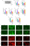
Cur inhibits excessive autophagy due to A/R injury (A) Western blot analysis of (B) expression of autophagy-related proteins. (C) Cur and 3-MA effectively attenuate A/R-induced fluorescence intensity and lysosome production in H9c2 cells (magnification, ×200; scale bar, 400 μm). *P<0.05, **P<0.01, ***P<0.001. Cur, curcumin; A/R, anoxia/reoxygenation; 3-MA, 3-Methyladenine; LCB, microtubule-associated protein 1 light chain 3 beta; CON, control.
Figure 4
Figure 4
Cur attenuates autophagy-dependent ferroptosis induced by A/R injury in H9c2 cells. (A) Expression of autophagy-dependent ferroptosis-associated proteins (FTH1 and NCOA4) was detected in A/R-injured H9c2 cells after Cur or 3-MA treatment. (B) Relative protein expression of FTH1 and NCOA4. Detection of (C) total iron ions, (D) MDA, (E) GSSG, (F) GSH, (G) GSH/GSSG and (H) SOD in H9c2 cells with A/R injury by Cur or 3-MA intervention. (I) ROS and (J) Fe2+ levels in H9c2 cells following AR injury after Cur or 3-MA treatment (magnification, ×200; scale bar, 400 μm). *P<0.05, **P<0.01, ***P<0.001. Cur, curcumin; A/R, anoxia/reoxygenation; FTH1, ferritin heavy chain 1; NCOA4, nuclear receptor coactivator 4; 3-MA, 3-Methyladenine; MDA, malondialdehyde; GSSG, glutathione disulfide; GSH, glutathione; SOD, superoxide dismutase; ROS, reactive oxygen species; CON, control; prot, protein.
Figure 5
Figure 5
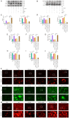
Cur decreases A/R injury in H9c2 cells via Sirt1. Expression of (A) apoptosis-associated proteins in A/R-injured H9c2 cells following Cur pretreatment, Sirt1 silencing and targeted inhibition of AKT activity. (B) Relative protein expression of Bcl2. (C) represents the relative protein expression of Bax. Expression of Sirt1 (D) in A/R-injured H9c2 cells following Cur pretreatment, Sirt1 silencing and targeted inhibition of AKT activity. (E) Relative protein expression of Sirt1. Apoptosis rate of H9c2 cells was measured by flow cytometry (F and G) illustrates the proportion of apoptotic cells as determined by flow cytometry. (H) caspase 3, (I) LDH activity and (J) viability of A/R injured H9c2 cells after Cur pretreatment, silencing of Sirt1 expression and targeted inhibition of AKT activity. *P<0.05, **P<0.01, ***P<0.001. Cur, curcumin; A/R, anoxia/reoxygenation; Sirt, silent information regulator 1; LDH, Lactate dehydrogenase; si, small interfering; NC, non-targeting control; API-2, triciribine; CON, control.
Figure 6
Figure 6
Cur reduces autophagy-dependent ferroptosis in H9c2 cells associated with A/R injury via Sirt1. Expression of autophagy-(A) and ferroptosis-related proteins (B) in A/R-injured H9c2 cells following Cur pretreatment, Sirt1 silencing and targeted inhibition of AKT activity. (C) Relative protein expression of P62 and the ratio of LC3II/I. (D) Relative protein expression of NCOA4 and FTH1. Detection of (E) total iron ions, (F) MDA, (G) GSSG, (H) GSH, (I) GSH/GSSG and (J) SOD in A/R injured H9c2 cells following Cur pretreatment, Sirt1 silencing and targeted inhibition of AKT activity. Fluorescence intensity of (K) lysosomes, (L) ROS and (M) Fe2+ in A/R-injured H9c2 cells following Cur pretreatment, Sirt1 silencing and targeted inhibition of AKT activity (magnification, ×200; scale bar, 400 μm). **P<0.05, ***P<0.01. Cur, curcumin; A/R, anoxia/reoxygenation; Sirt, silent information regulator 1; MDA, malondialdehyde; GSSG, glutathione disulfide; GSH, glutathione; SOD, superoxide dismutase; ROS, reactive oxygen species; NCOA4, nuclear receptor coactivator 4; FTH1, ferritin heavy chain 1; CON, control; si, small interfering; prot, protein; API, triciribine; LC3II, microtubule-associated protein 1 light chain 3 β; NC, non-targeting control.
Figure 7
Figure 7
Cur mediates nuclear localization of FoxO3a via Sirt1/AKT. (A) Western blot analysis of (B) phosphorylation of AKT and FoxO3a in A/R-injured H9c2 cells after Cur pretreatment, Sirt1 silencing and targeted inhibition of AKT activity. (C) Western blot analysis of (D) distribution of FoxO3a in the cytoplasm and nucleus of A/R-injured H9c2 cells following Cur pretreatment, Sirt1 silencing and targeted inhibition of AKT activity. ***P<0.01. Cur, curcumin; PCNA, proliferating cell nuclear antigen; Sirt, silent information regulator 1; A/R, anoxia/reoxygenation; p-, phosphorylated; si, small interfering; NC, non-targeting control; API, Triciribine; CON, control.
Figure 8
Figure 8
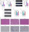
Cur attenuates MIRI. Cur effectively attenuated activities of (A) CK-MB and (B) LDH in rat serum. (C) Evans blue and TTC staining showed (D) Cur effectively decreased infarct size after MIRI. (E) Cur effectively improved (F) LVEF and (G) LVFS after MIRI. Representative images of cardiac injury evaluated using (H) hematoxylin and eosin and (I) TUNEL staining. Arrows indicated TUNEL-positive cells (apoptotic cells). ***P<0.01. Cur, curcumin; MIRI, myocardial ischemia/reperfusion injury; CK-MB, creatine kinase isoenzyme; LDH, Lactate dehydrogenase; TTC, Triphenyl tetrazolium chloride; LVEF, left ventricular ejection fraction; LVFS, left ventricular fractional shortening; I/R, ischemia/reperfusion.
Figure 9
Figure 9
Potential mechanism of action of Cur in MIRI. (A) Western blot was performed to detect protein expression of Sirt1 in MIRI heart tissues after Cur treatment, (B) Relative protein expression of Sirt1. Western blot was performed to detect protein expression of LC3II and P62 (C) in MIRI heart tissues after Cur treatment, (D represents the relative protein expression of P62 and the ratio of LC3II/I. Western blot was performed to detect protein expression of NCOA4 and FTH1 (E) in MIRI heart tissues after Cur treatment, (F) represents the relative protein expression of NCOA4 and FTH1. Western blot was performed to detect the ratios of p-AKT/AKT and p-FoxO3a/FoxO3a (G) in MIRI heart tissues after Cur treatment, (H) showed statistical figures. (I) Cur inhibits MIRI-induced autophagy-dependent ferroptosis and apoptosis in cardiomyocytes via Sirt1/AKT/FoxO3a, which increases cardiomyocyte survival and maintains cardiac function. Part of the figure was created using Figdraw (https://www.figdraw.com). *P<0.05, **P<0.01, ***P<0.001. Cur, curcumin; MIRI, myocardial ischemia/reperfusion injury; Sirt, silent information regulator 1; LC3II, microtubule-associated protein 1 light chain 3 beta; NCOA4, nuclear receptor coactivator 4; FTH1, ferritin heavy chain 1; p-, phosphorylation; I/R, ischemia/reperfusion; ROS, reactive oxygen species; SOD, superoxide dismutase; MDA, malondialdehyde; GSH, glutathione; GSSG, glutathione disulfide; A/R, anoxia/reoxygenation; ATG, autophagy related gene.

References

    1. Abdollahi E, Momtazi AA, Johnston TP, Sahebkar A. Therapeutic effects of curcumin in inflammatory and immune-mediated diseases: A nature-made jack-of-all-trades? J Cell Physiol. 2018;233:830–848. doi: 10.1002/jcp.25778. - DOI - PubMed
    1. Xie W, Gan J, Zhou X, Tian H, Pan X, Liu W, Li X, Du J, Xu A, Zheng M, et al. Myocardial infarction accelerates the progression of MASH by triggering immunoinflammatory response and induction of periosti. Cell Metab. 2024;36:1269–1286.e9. doi: 10.1016/j.cmet.2024.04.020. - DOI - PubMed
    1. Shen H, Yao Z, Zhao W, Zhang Y, Yao C, Tong C. miR-21 enhances the protective effect of loperamide on rat cardiomyocytes against hypoxia/reoxygenation, reactive oxygen species production and apoptosis via regulating Akap8 and Bard1 expression. Exp Ther Med. 2019;17:1312–1320. - PMC - PubMed
    1. Wang Z, Yao M, Jiang L, Wang L, Yang Y, Wang Q, Qian X, Zhao Y, Qian J. Dexmedetomidine attenuates myocardial ischemia/reperfusion-induced ferroptosis via AMPK/GSK-3β/Nrf2 axis. Biomed Pharmacother. 2022;154:113572. doi: 10.1016/j.biopha.2022.113572. - DOI - PubMed
    1. Hausenloy DJ, Garcia-Dorado D, Bøtker HE, Davidson SM, Downey J, Engel FB, Jennings R, Lecour S, Leor J, Madonna R, et al. Novel targets and future strategies for acute cardioprotection: Position paper of the european society of cardiology working group on cellular biology of the heart. Cardiovasc Res. 2017;113:564–585. doi: 10.1093/cvr/cvx049. - DOI - PubMed

MeSH terms

LinkOut - more resources