. 2025 Feb 4:20:1561-1578.
doi: 10.2147/IJN.S497940. eCollection 2025.

Umbilical Cord Mesenchymal Stromal Cell-Derived Small Extracellular Vesicles Modulate Skin Matrix Synthesis and Pigmentation

Affiliations

Umbilical Cord Mesenchymal Stromal Cell-Derived Small Extracellular Vesicles Modulate Skin Matrix Synthesis and Pigmentation

Li Ting Kee et al. Int J Nanomedicine. .

Abstract

Introduction: Research has unveiled the remarkable properties of extracellular vesicles derived from mesenchymal stromal cells (MSCs), particularly in promoting wound healing, aiding re-epithelialization, revitalizing aging skin, and inhibiting hyperpigmentation. However, investigations into the potential of small extracellular vesicles from umbilical cord-derived MSCs (UC-MSC-sEVs) in reducing scarring and preventing hyperpigmentation remain limited. Therefore, this study aims to evaluate the impact of UC-MSC-sEVs on the synthesis of the skin's extracellular matrix (ECM) and pigmentation using in vitro models.

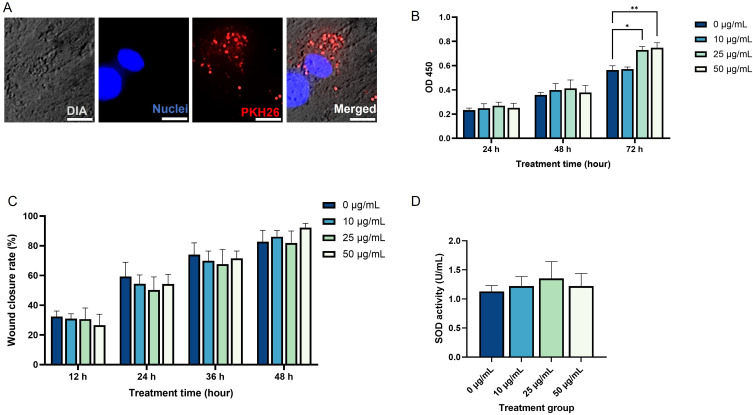
Methods: The study investigated the impact of characterized UC-MSC-sEVs on various aspects including the proliferation, migration, antioxidant activity, and ECM gene expression of human dermal fibroblasts (HDF). Additionally, the effects of UC-MSC-sEVs on the proliferation, melanin content, and tyrosinase (TYR) activity of human melanoma cells (MNT-1) were examined. Furthermore, ex vivo models were employed to evaluate the skin permeation of PKH26-labelled UC-MSC-sEVs.

Results: The findings indicated that a high concentration of UC-MSC-sEVs positively influenced the proliferation of HDF. However, no changes in cell migration rate were observed. While the expressions of collagen type 1 and type 3 remained unaffected by UC-MSC-sEVs treatment, there were dose-dependent increases in the gene expressions of fibronectin, matrix metallopeptidase (MMP) 1, and MMP 3. Furthermore, UC-MSC-sEVs treatment did not impact the antioxidative superoxide dismutase (SOD) expression in HDF. Although UC-MSC-sEVs did not alter the proliferation of MNT-1 cells, it did result in a dose-dependent reduction in melanin synthesis without affecting TYR activity. However, when it was applied topically, UC-MSC-sEVs failed to penetrate the skin barrier and remained localized within the stratum corneum layer even after 18 hours.

Conclusion: These results highlight the potential of UC-MSC-sEVs in stimulating HDF proliferation, regulating ECM synthesis, and reducing melanin production. This demonstrates the promising application of UC-MSC-sEVs in medical aesthetics for benefits such as scar reduction, skin rejuvenation, and skin lightening.

Keywords: anti-scarring; extracellular vesicles; medical aesthetic; mesenchymal stromal cell; pigmentation.


Conflict of interest statement

Ms Li Ting Kee reports a patent PI2024006022 pending to. Dr Min Hwei Ng reports a patent WJMSC exosomes for skin aesthetic use pending to None; and A company, Ming Medical Sdn Bhd, sponsors the study. However, academic researchers have developed the protocol for the derivation of the exosome entirely. Dr Jia Xian Law reports a patent An anti-scarring and anti-pigmentation composition pending to Universiti Kebangaan Malaysia and Ming Medical Sdn Bhd. The authors report no other conflicts of interest in this work.

Figures

Figure 1
Figure 1
Characterization of UC-MSCs and UC-MSC-sEVs. (A) Morphology of pooled UC-MSCs at passage five, observed under 40× magnification. (B) Adipogenic-induced UC-MSCs stained with Oil Red O, viewed under 200× magnification. (C) Osteogenic-induced UC-MSCs stained with Alizarin Red, observed under 40× magnification. (D) Immunophenotyping analysis of pooled UC-MSC at passage five. Characterization of UC-MSCs data represents a single technical replicate from pooled UC-MSCs derived from three independent donors. sEVs harvested from conditioned medium of pooled UC-MSCs using (E) Centricon and (F) TFF. Nanoparticle tracking analysis of (G) sEVs-C and (H) sEVs-TFF. Immunoblotting analysis of (I) sEVs-C and (J) sEVs-TFF using the positive markers CD63, HSP70, and TSG101 as well as the negative marker GP96. (Scale bar represents 100 μm). Characterization of sEVs-C represents four technical replicates, while sEVs-TFF represent two technical replicates.
Figure 2
Figure 2
(A) Cellular uptake of sEVs by HDF, viewed under 400× magnification. Five random fields of view were analyzed from a single biological replicate. Scale bar represents 10 μm. (B) Proliferation rate of HDF treated with sEVs. (C) Wound closure rate of HDF treated with sEVs. (D) SOD activity of HDF treated with sEVs. Four different biological samples were used, each with three technical replicates. (*p<0.05; **p<0.01).
Figure 3
Figure 3
(A) Expression levels of ECM genes in HDF treated with different concentrations of sEVs. The gene expression levels of the treatment groups are shown as fold changes compared to the control group (0 μg/mL). Three biological samples, each with two technical replicates, were used. (B) Protein expression levels of COLI and α-SMA in HDF treated with different concentrations of sEVs. (C) Immunofluorescent image of HDF treated with different concentrations of sEVs. The cells were viewed under 200× magnification and each scale bar represent 100 μm. Representative image from five random fields of view per sample across three biological replicates.
Figure 4
Figure 4
(A) Cellular uptake of sEVs by MNT-1. The cells were viewed under 400× magnification and the scale bars represent 10 μm. Five random fields of view were analyzed from a single biological replicate. (B) Proliferation rate of MNT-1 treated with sEVs. (C) Melanin synthesis of MNT-1 treated with sEVs. (D) Tyrosinase activity of MNT-1 treated with sEVs. Three biological samples, each with three technical replicates, were used (E) Expression levels of genes related to melanin synthesis in MNT-1. The gene expression levels of the treatment groups are presented as fold changes relative to the control group (0 μg/mL). Three biological samples, each with two technical replicates, were used. (*, p<0.05).
Figure 5
Figure 5
Skin permeation analysis of sEVs-C labelled with PKH26 after 18h. The tissue sections were viewed under 200× magnification and each scale bar represented 100 μm. Five random fields of view were analyzed from a single biological replicate.

References

    1. Al-Masawa ME, Alshawsh MA, Ng CY, et al. Efficacy and safety of small extracellular vesicle interventions in wound healing and skin regeneration: a systematic review and meta-analysis of animal studies. Theranostics. 2022;12(15):6455–6508. doi:10.7150/thno.73436 - DOI - PMC - PubMed
    1. Gurung S, Perocheau D, Touramanidou L, Baruteau J. The exosome journey: from biogenesis to uptake and intracellular signalling. Cell Commun Signal. 2021;19(1):1–19. doi:10.1186/s12964-021-00730-1 - DOI - PMC - PubMed
    1. Witwer KW, Van Balkom BWM, Bruno S, et al. Defining mesenchymal stromal cell (MSC)-derived small extracellular vesicles for therapeutic applications. J Extracell Vesicles. 2019;8(1). doi:10.1080/20013078.2019.1609206 - DOI - PMC - PubMed
    1. Welsh JA, Goberdhan DCI, O’Driscoll L, et al. Minimal information for studies of extracellular vesicles (MISEV2023): from basic to advanced approaches. J Extracell Vesicles. 2024;13(2). doi:10.1002/jev2.12404 - DOI - PMC - PubMed
    1. Verweij FJ, Balaj L, Boulanger CM, et al. The power of imaging to understand extracellular vesicle biology in vivo. Nat Methods. 2021;18(9):1013–1026. doi:10.1038/s41592-021-01206-3 - DOI - PMC - PubMed

MeSH terms

LinkOut - more resources