

Standardization of a Model of Vertebral Metastasis of Breast Cancer in CD1/Nu/Nu Mice
- PMID: 39931605
- PMCID: PMC11809943
- DOI: 10.7759/cureus.77291
Standardization of a Model of Vertebral Metastasis of Breast Cancer in CD1/Nu/Nu Mice
Abstract
Introduction: Breast cancer is the leading cause of cancer-related death in Mexico, with high mortality associated with spinal bone metastasis. We propose to standardize a murine model of bone metastasis to study and understand the tumor microenvironment.
Materials and methods: An experimental, prospective, longitudinal study was conducted using 18 CD1/Nu/Nu 30g nude mice. Two cell lines, MCF-7 and 4T1, were inoculated, clinical follow-up was performed, and biopsy samples were obtained for histopathological evaluation.
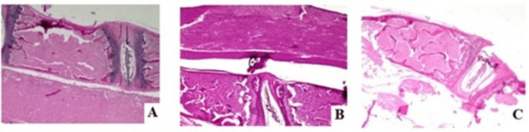
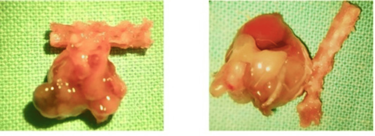
Results: Histopathological evaluation of models inoculated with the MCF-7 cell line showed no tumor development, while inoculation with the 4T1 cell line resulted in tumor development, as evidenced by PET-CT and histopathology, using 5,000 and 1,000 cells, respectively.
Conclusions: The use of this model is proposed for studying the clinical, molecular, and prognostic aspects of breast cancer progression by inoculating 1,000 cells of the 4T1 cell line.
Keywords: 4t1; breast cancer; hematogenous metastasis; pain; pet-ct; spinal cord compression.
Copyright © 2025, Reyes Soto et al.
Conflict of interest statement
Human subjects: All authors have confirmed that this study did not involve human participants or tissue. Animal subjects: Bioethics Committee of National Cancer Institute, Mexico Issued protocol number NOM 062-ZOO-1999. Conflicts of interest: In compliance with the ICMJE uniform disclosure form, all authors declare the following: Payment/services info: All authors have declared that no financial support was received from any organization for the submitted work. Financial relationships: All authors have declared that they have no financial relationships at present or within the previous three years with any organizations that might have an interest in the submitted work. Other relationships: All authors have declared that there are no other relationships or activities that could appear to have influenced the submitted work.
Figures


References
-
- Programa de Acción Específico Prevención y Control del Cáncer de la Mujer (2013-2018). Secretaria de Salud. [ Oct; 2022 ];Programa de Acción Específico Prevención y Control del Cáncer de la Mujer (2013-2018. https://www.gob.mx/salud/acciones-y-programas/informacion-estadistica Updated 8 de septiembre de. 2015 5:2022.
-
- Metástasis óseas múltiples de cáncer de mama: Papel del CA 15.3 y respuesta a la hormonoterapia. López CN, Ramón GN, Sánchez MJI, de Santiago GJ. https://www.scielo.cl/scielo.php?script=sci_arttext&pid=S0717-7526201200... Rev Chil Obstet Ginecol. 2012;77:291–295.
-
- Multidisciplinary approach for bone metastasis: a review. Kimura T. https://www.mdpi.com/2072-6694/10/6/156. Cancers (Basel) 2018;10:156. - PMC - PubMed
LinkOut - more resources
Full Text Sources
Research Materials