Forearm Giant Osteochondromas in a Young Patient With Multiple Hereditary Exostoses: A Case Report
- PMID: 39931628
- PMCID: PMC11810139
- DOI: 10.7759/cureus.77295
Forearm Giant Osteochondromas in a Young Patient With Multiple Hereditary Exostoses: A Case Report
Abstract
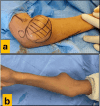
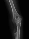
Multiple hereditary exostoses (MHE) is a rare skeletal disorder inherited as an autosomal dominant disorder. It is characterized by widespread multiple osteochondromas that grow near bone growth plates, leading to pain and deformities that significantly impact physical and emotional well-being and disrupt daily activities, social interactions, and psychological health, leading to considerable disability. This case report describes a 15-year-old boy with a family history of MHE who developed a large osteochondroma at his right elbow. We aim to present the surgical management of extraordinarily large-size proximal radius osteochondroma, fortunately, caused by a benign underlying condition despite typically carrying more chances of transformation into malignancy. To the best of our knowledge, it would be the largest proximal radius osteochondroma documented in the literature.
Keywords: elbow; hereditary exostoses; large osteochondroma; proximal radial resection; radial head dislocation.
Copyright © 2025, Sulaiman et al.
Conflict of interest statement
Human subjects: Consent for treatment and open access publication was obtained or waived by all participants in this study. Conflicts of interest: In compliance with the ICMJE uniform disclosure form, all authors declare the following: Payment/services info: All authors have declared that no financial support was received from any organization for the submitted work. Financial relationships: All authors have declared that they have no financial relationships at present or within the previous three years with any organizations that might have an interest in the submitted work. Other relationships: All authors have declared that there are no other relationships or activities that could appear to have influenced the submitted work.
Figures


References
-
- Multiple hereditary exostoses and enchondromatosis. Jurik AG. Best Pract Res Clin Rheumatol. 2020;34:101505. - PubMed
-
- Prediction of radial head subluxation and dislocation in patients with multiple hereditary exostoses. Feldman DS, Rand TJ, Deszczynski J, et al. https://journals.lww.com/jbjsjournal/fulltext/2021/12010/Prediction_of_R... J Bone Joint Surg Am. 2021;103:2207–2214. - PubMed
-
- An evaluation of forearm deformities in hereditary multiple exostoses: factors associated with radial head dislocation and comprehensive classification. Jo AR, Jung ST, Kim MS, Oh CS, Min BJ. J Hand Surg Am. 2017;42:292–298. - PubMed
Publication types
LinkOut - more resources
Full Text Sources
