

Diffusion-Weighted Magnetic Resonance Imaging: A Diagnostic Tool for Auditory (Axonal) Neuropathy
- PMID: 39932015
- PMCID: PMC11811761
- DOI: 10.1111/ene.70083
Diffusion-Weighted Magnetic Resonance Imaging: A Diagnostic Tool for Auditory (Axonal) Neuropathy
Abstract
Background: Axonal neuropathies are disorders that impair neural transmission, leading to substantial sensory deficits. In the auditory system, axonal degeneration can disrupt auditory processing, causing significant hearing difficulties. Understanding the extent of axonal degeneration and its impact on auditory function is crucial for improving diagnosis and management. This study aims to quantify axonal degeneration in the VIIIth nerve using diffusion-weighted MRI and to correlate these findings with auditory function.
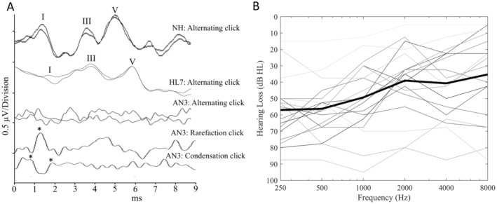
Methods: Fifty-two children and adults participated. A total of, 27 with normal hearing, 7 with cochlear hearing loss and 18 with auditory neuropathy (AN). Hearing thresholds and dMRI data was collected for all participants and the VIIIth nerve was evaluated using the fixel-based analysis metric of Apparent Fibre Density (AFD).
Results: AFD was significantly lower in participants with AN compared to participants with normal hearing and cochlear hearing loss (p < 0.05). 9/18 participants with AN exhibited AFD values ≥ 2 standard deviations below the normal range. Additionally, AFD was strongly correlated with hearing thresholds in participants with no evidence of cochlear dysfunction (r = -0.776, p < 0.001), suggesting reduced auditory nerve fibre density is associated with impaired sound detection.
Conclusions: dMRI-derived AFD is a sensitive marker for axonal degeneration in the VIIIth nerve. This study provides the first in vivo evidence linking VIIIth nerve microstructure with hearing thresholds, highlighting the potential of dMRI in diagnosing and monitoring AN. The findings suggest that dMRI could be a valuable tool in clinical settings for assessing auditory nerve health and guiding treatment strategies for individuals with AN.
Keywords: auditory neuropathy; axonopathy; diffusion‐weighted MRI; hearing loss; neuroimaging.
© 2025 The Author(s). European Journal of Neurology published by John Wiley & Sons Ltd on behalf of European Academy of Neurology.
Conflict of interest statement
The authors declare no conflicts of interest.
Figures


Similar articles
-
Objective Determination of Site-of-Lesion in Auditory Neuropathy.Ear Hear. 2025 Mar-Apr 01;46(2):371-381. doi: 10.1097/AUD.0000000000001589. Epub 2024 Sep 19. Ear Hear. 2025. PMID: 39294863
-
Fiber-Specific Changes in White Matter Microstructure in Individuals With X-Linked Auditory Neuropathy.Ear Hear. 2020 Nov/Dec;41(6):1703-1714. doi: 10.1097/AUD.0000000000000890. Ear Hear. 2020. PMID: 33136644
-
Diffusion-Weighted Magnetic Resonance Imaging (dMRI) and Cochlear Implant Outcomes in Axonal Auditory Neuropathy: A Case Report.J Clin Med. 2024 May 24;13(11):3072. doi: 10.3390/jcm13113072. J Clin Med. 2024. PMID: 38892782 Free PMC article.
-
Vestibular test findings in individuals with auditory neuropathy: review.J Laryngol Otol. 2013 May;127(5):448-51. doi: 10.1017/S0022215113000406. Epub 2013 Mar 22. J Laryngol Otol. 2013. PMID: 23521835 Review.
-
Auditory neuropathy/dys-synchrony and its perceptual consequences.Trends Amplif. 2005;9(1):1-43. doi: 10.1177/108471380500900102. Trends Amplif. 2005. PMID: 15920648 Free PMC article. Review.
References
-
- Marner L., Nyengaard J. R., Tang Y., and Pakkenberg B., “Marked Loss of Myelinated Nerve Fibers in the Human Brain With Age,” Journal of Comparative Neurology 462 (2003): 144–152. - PubMed
-
- Engström B., Hillerdal M., Laurell G., and Bagger‐Sjöbäck D., “Selected Pathological Findings in the Human Cochlea,” Acta Oto‐Laryngologica 104 (1987): 110–116. - PubMed
-
- Rapin I. and Gravel J., “‘Auditory Neuropathy’: Physiologic and Pathologic Evidence Calls for More Diagnostic Specificity,” International Journal of Pediatric Otorhinolaryngology 67 (2003): 707–728. - PubMed
-
- Spoendlin H. and Schrott A., “Analysis of the Human Auditory Nerve,” Hearing Research 43 (1989): 25–38. - PubMed
MeSH terms
Supplementary concepts
LinkOut - more resources
Full Text Sources
