. 2025 Feb 11;272(3):188.
doi: 10.1007/s00415-025-12913-8.

Vestibular paroxysmia: a systematic review

Affiliations

Vestibular paroxysmia: a systematic review

Marianne Dieterich et al. J Neurol. .

Abstract

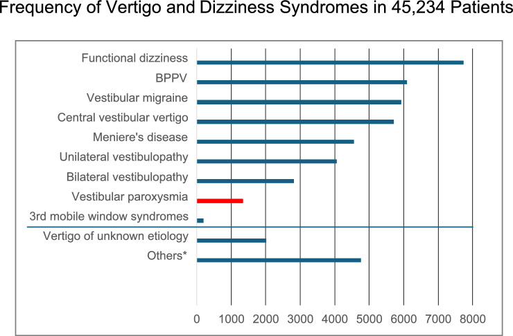
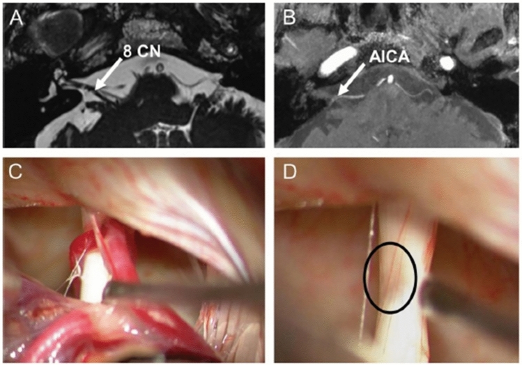
The key symptoms of vestibular paroxysmia (VP) due to neurovascular cross-compression (classical VP) or compression of the eighth nerve by space-occupying cerebellar-pontine angle processes (secondary VP) are frequent short attacks of vertigo and dizziness with unsteadiness which last seconds to minutes. They can be accompanied by unilateral auditory symptoms such as tinnitus or hyperacusis. Head movements and hyperventilation can induce nystagmus and VP attacks that most often occur spontaneously. VP is diagnosed in 3% of patients in a tertiary vertigo care center and very rarely affects children. The mean age of first appearance is 47 to 51 years with equal sex distribution. A combination of high-resolution MRI sequences (with constructive interference in steady-state/fast imaging employing steady-state, 3D-CISS/ FIESTA) of the cerebello-pontine may support the diagnosis although the beneficial treatment with sodium channel blockers is the most reliable clinical sign for classical VP, secondary VP and idiopathic VP (without verification of a causative pathology). Because of the frequency, shortness, and audiovestibular symptomatology of the attacks, the differential diagnosis to other conditions such as paroxysmal brainstem attacks, vestibular epilepsy, rotational vertebral artery compression syndrome or "near"-narrowed internal auditory canal syndrome is only relevant in exceptional cases. However, imaging of the posterior fossa including the inner ear is mandatory to distinguish between classical, secondary and idiopathic VP forms. Randomized controlled trials for medical treatment are still needed. Practical therapy of choice is medical treatment with sodium channel blockers (carbamazepine, oxcarbazepine, lacosamide). A microsurgical decompression is effective in secondary VP but is the ultimate therapy in cases with classical or idiopathic VP when medication is not tolerated.

Keywords: Audiovestibular deficits; Neurovascular cross compression; Sodium channel blockers; Vertigo attacks; Vestibular excitation.


Conflict of interest statement

Declarations. Conflict of interest: The authors report no conflicts of interest.

Figures

Fig. 1
Fig. 1
Frequency of different vertigo and dizziness syndromes in 45,234 patients from a tertiary interdisciplinary vertigo outpatient unit of the German Center for Vertigo and Balance Disorders and the Department of Neurology, LMU University Hospital, Munich, Germany, over 26 years from 1998 to 2023. VP is a rare vestibular disease accounting for 3% (N = 1339) of vertigo/dizziness patients. *Others means dizziness or imbalance due to non-vestibular origin, e.g., polyneuropathy, orthostatic dizziness, ocular motor disorders
Fig. 2
Fig. 2
Example of the cranial MRI of a VP patient. Constructive interference in steady-state sequence (CISS) with 0.5 × 0.5 × 0.5 mm for high-resolution structural imaging (left column) depicting the semicircular canals and the vestibular nerve with the root entry zone into the pontomedullary brainstem. Reconstruction of fractional anisotropy based on 2 × 2 × 2 mm diffusion-weighted imaging (DWI)(right column).While structural MRI (left column) can reveal fine details of vestibulocochlear nerve anatomy, such as the presence and extent of neurovascular contact and associated nerve angulation, it cannot provide information on nerve integrity. For this, DWI data are used (right column) since the reconstruction of specific diffusion tensor maps from raw DWI data can relay further information on voxel-wise physical properties such as fractional anisotropy. Fiber-tracking algorithms allow to determine the three-dimensional directional course of nerve fibers. Paired with high-resolution structural data, the vestibulocochlear nerve can be identified and disease-specific changes in nerve integrity can be measured (bottom). Lower nerve integrity is shown in yellow–red colors
Fig. 3
Fig. 3
Cranial MRI of a patient with VP who had microsurgery because of intolerable medical side effects. A Constructive interference in steady-state sequence (CISS) and B time-of-flight (TOF) shows contact (arrows) between the right eighth cranial nerve (8 CN) and the anterior inferior cerebellar artery (AICA). C Intraoperative micrographs demonstrate vascular contact and D considerable compression of the eighth nerve after removal of the arteries (circle). (Modified from [23])
Fig. 4
Fig. 4
Secondary VP due to an arachnoid cyst in the right cerebellopontine angle that distorted the right vestibulocochlear nerve (A arrow at the nerve above the cyst in MRI scan) causing recurrent episodes of oscillopsia, rotational vertigo, and postural imbalance. These attacks were elicited and modulated by changing horizontal head positions. B Electronystagmography (middle) and posturography (bottom) during different head positions from straight ahead with eyes open (left) and eyes closed to head turn to the right and head turn to the left (right). Note the variation of nystagmus direction and intensity and the postural sway which was largest during head turn to the right. Ocular motor analysis revealed two different types of attacks, first, episodes of vestibular hypofunction (conduction block) for minutes to several hours, and second, B paroxysmal vestibular excitation (ectopic discharges) for seconds with head rotation to the right. One week after resection of the cyst and decompression of the eighth nerve the patient was symptom free and the ocular motor analysis normal. (Modified from [27])

References

    1. Jannetta PJ (1975) Neurovascular cross-compression in patients with hyperactive dysfunction symptoms of the eighth cranial nerve. Surg Forum 26:467–468 - PubMed
    1. Møller MB, Møller AR, Jannetta PJ, Sekhar L (1986) Diagnosis and surgical treatment of disabling positional vertigo. J Neurosurg 64:21–28 - PubMed
    1. Møller MB, Møller AR (1990) Vascular compression syndrome of the eighth nerve. Clinical correlations and surgical findings. Neurol Clin 8(2):421–439 - PubMed
    1. Møller MB, Møller AR, Jannetta PJ, Jho HD, Sekhar LN (1993) Microvascular decompression of the eighth nerve in patients with disabling positional vertigo: selection criteria and operative results in 207 patients. Acta Neurochir 125(1–4):75–82 - PubMed
    1. Brandt T, Dieterich M (1994) Vestibular paroxysmia: vascular compression of the eighth nerve? Lancet 343(8900):798–799 - PubMed

Publication types

LinkOut - more resources