. 2025 Feb 11;135(7):e163730.
doi: 10.1172/JCI163730.

Phosphorylation of CRYAB induces a condensatopathy to worsen post-myocardial infarction left ventricular remodeling

Affiliations

Phosphorylation of CRYAB induces a condensatopathy to worsen post-myocardial infarction left ventricular remodeling

Moydul Islam et al. J Clin Invest. .

Abstract

Protein aggregates are emerging therapeutic targets in rare monogenic causes of cardiomyopathy and amyloid heart disease, but their role in more prevalent heart-failure syndromes remains mechanistically unexamined. We observed mislocalization of desmin and sarcomeric proteins to aggregates in human myocardium with ischemic cardiomyopathy and in mouse hearts with post-myocardial infarction ventricular remodeling, mimicking findings of autosomal-dominant cardiomyopathy induced by the R120G mutation in the cognate chaperone protein CRYAB. In both syndromes, we demonstrate increased partitioning of CRYAB phosphorylated on serine 59 to NP40-insoluble aggregate-rich biochemical fraction. While CRYAB undergoes phase separation to form condensates, the phosphomimetic mutation of serine 59 to aspartate (S59D) in CRYAB mimics R120G-CRYAB mutants with reduced condensate fluidity, formation of protein aggregates, and increased cell death. Conversely, changing serine to alanine (phosphorylation-deficient mutation) at position 59 (S59A) restored condensate fluidity and reduced both R120G-CRYAB aggregates and cell death. In mice, S59D CRYAB knockin was sufficient to induce desmin mislocalization and myocardial protein aggregates, while S59A CRYAB knockin rescued left ventricular systolic dysfunction after myocardial infarction and preserved desmin localization with reduced myocardial protein aggregates. 25-Hydroxycholesterol attenuated CRYAB serine 59 phosphorylation and rescued post-myocardial infarction adverse remodeling. Thus, targeting CRYAB phosphorylation-induced condensatopathy is an attractive strategy to counter ischemic cardiomyopathy.

Keywords: Cardiology; Cardiovascular disease; Cell biology; Chaperones.


Figures
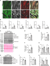
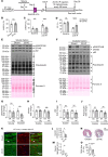
Figure 1
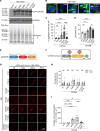
Figure 1. Desmin, α-actinin, and actin and the serine 59 phosphorylated form of their chaperone protein, CRYAB, localize to protein aggregates in human ICM.
(A) Representative immunohistochemical images from left ventricular myocardium of individuals evaluated as controls (donor) or patients with end-stage ICM stained for desmin, α-actinin, and actin. Arrows point to mislocalization of these proteins from their physiologic location on Z-discs and intercalated discs (desmin), Z-disc (α-actinin), and sarcomere (actin) in donor myocardium to protein aggregates in ICM myocardium. (B) Quantitation of striation score and aggregate score for desmin, α-actinin, and actin in ICM and donor hearts. n = 3-4 hearts/group. For striation scoring, normal localization of proteins got scored as 0, and abnormal striation or mislocalization of proteins was scored as 1. For scoring aggregates, absence of aggregates was scored as 0 and presence of aggregates was scored as 2. (CG) Immunoblot (C) and quantitation (fold change as compared with donor mean) depicting total p62 (D), polyUb proteins (E), CRYAB (F), and pS59-CRYAB and pS45-CRYAB (G) in NP40-detergent-insoluble fractions from human hearts from patients with ICM and donors. Ponceau S staining is shown as loading control. (HK) Immunoblot (H) and quantitation for p62 (I), CRYAB (J), and pS59-CRYAB and pS45-CRYAB (K) abundance in NP-40 detergent soluble biochemical fractions from human hearts as in CG. GAPDH was used as loading control. n = 6 samples/group for CK. *P < 0.05; **P < 0.01; ***P < 0.001 versus donor as control by t test.
Figure 2
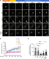
Figure 2. Phosphorylation of CRYAB at S59 makes it aggregate prone and toxic.
(A) Immunoblot (A) demonstrating expression of GFP-fusion proteins in HEK293A cells transfected with GFP-tagged WT CRYAB, its phospho-mimetic mutant (S59D), phosphorylation-deficient mutant (S59A), R120G mutant, or the R120G and S59A double-mutant proteins. (B and C) Representative immunofluorescence images (B) for detection of protein aggregates with quantitation (C) of aggregate area per cell. **P < 0.01; ****P < 0.0001 by Tukey’s post hoc test after 1-way ANOVA. Nuclei are blue (DAPI). (D) Cell death in cells treated in A. **P < 0.01; ****P < 0.0001 by Tukey’s post hoc test after 1-way ANOVA. (E) Schematic depicting generation of optoIDR constructs. mCh indicates mCherry fluorophone and Cry2 encodes for Arabidopsis thaliana protein with light-activated phase separation characteristics. (F) Various domains of CRYAB with localization of serine 59 and arginine 120 residues depicted. (G) Representative time-lapse images at t = 0 seconds, 100 seconds, 200 seconds, and 300 seconds after light activation in HEK293A cells transfected with constructs generated with CRYAB WT, its phosphorylation-deficient mutant (S59A), phospho-mimetic mutant (S59D), R120G mutant, or the R120G and S59A double-mutant proteins as the IDR in the optoIDR constructs. Cry2 fused with mCherry without an IDR was used as the negative control, and FUS-N fused with mCherry-Cry2 was studied as positive control. (H) Average number of condensates/cell at t = 0 versus t = 300 seconds in cells treated as in E. **P < 0.01 by Mann-Whitney test. (I) Average area of condensates/cell at t = 300 seconds in cells treated in E. ***P < 0.001; ****P < 0.0001 by Tukey’s post hoc test after 1-way ANOVA.
Figure 3
Figure 3. Phosphorylation of CRYAB at serine 59 reduces dynamicity of condensates.
(A) Representative images demonstrating recovery of fluorescence after photobleaching in HEK 293A cells transfected with mCherry-Cry2 fused optoIDR constructs generated with CRYAB WT, its phosphorylation-deficient mutant (S59A), phospho-mimetic mutant (S59D), R120G mutant, or the R120G and S59A double-mutant proteins. Representative images demonstrate area of photobleaching (marked with a dotted circle) prior to (prebleach), immediately after, and at 100, 200, 300, 400 and 500 seconds after photobleaching was terminated. Intensity at various time points is depicted as a fraction of intensity prior to bleaching (set at 100%). (B and C) Fluorescence intensity normalized to baseline (B) and quantitation of fluorescence recovery (C, maximum minus immediately after bleach) in condensates of various CRYAB variants indicated in A. *P < 0.05; ****P < 0.0001 by Tukey’s post hoc test after 1-way ANOVA.
Figure 4
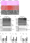
Figure 4. Mice with phosphomimetic aspartic acid residue instead of serine at position 59 in CRYAB demonstrate myocardial protein aggregates.
(A) Representative H&E- (top row) and Masson’s trichrome–stained (middle row) myocardial sections from young adult mice homozygous for alleles bearing serine–to–aspartic acid (phosphomimetic residue; S59D) or serine-to-alanine (phosphorylation deficient; S59A) mutation at position 59 in CRYAB and mice bearing WT CRYAB alleles as controls. Transmission electron micrographs (bottom row) from WT, S59A, and S59D mice. Representative of n = 2 mice per group. Black arrowheads point to abnormal-appearing mitochondria and white arrowheads point to protein aggregates. (B and C) Representative immunoblots depicting expression of ubiquitinated proteins and p62, CRYAB, and GAPDH proteins in NP40-soluble (B) and NP40-insoluble (C) myocardial extracts from WT, S59A, and S59D mice. Ponceau S staining is shown as loading control. (DF) Quantitation of ubiquitinated proteins (D), p62 (E), and CRYAB (F) in soluble and insoluble myocardial extracts from WT, S59A, and S59D mice. *P < 0.05; **P < 0.01; ***P < 0.001; ****P < 0.0001 by Tukey’s post hoc test after 1-way ANOVA, except for the data in the insoluble fraction in D and soluble fraction in E, which were analyzed by Dunn’s multiple-comparison test after Kruskal-Wallis test.
Figure 5
Figure 5. Knockin of phosphorylation-deficient alanine instead of serine at position 59 in CRYAB rescues post-IR left ventricular remodeling in mice.
(A) Schematic depicting experimental strategy for closed-chest IR modeling (90 minutes of ischemia followed by reperfusion) in S59A, S59D, and WT. (B) Quantitative assessment of area-at-risk (AAR) during LAD occlusion in mice treated as in A. (C and D) Quantitative analyses of left ventricular EDV (LVEDV, C) and LV EF (EF (%), D) at baseline (i.e. prior to) and at 4 weeks after IR injury. *P < 0.05; ***P < 0.001 by Tukey’s post hoc testing after 1-way ANOVA. Echocardiographic parameters at baseline and 4 weeks after IR injury were analyzed separately as they were performed under different anesthetic regimens. (E and F) Masson’s trichrome–stained left ventricular sections demonstrating presence of scar at 4 weeks after IR injury (E) with quantitation of scar size (F). *P < 0.05; **P < 0.01 by Tukey’s post hoc testing after 1-way ANOVA.
Figure 6
Figure 6. Knockin of phosphorylation-deficient alanine or phosphomimetic aspartic acid instead of serine at position 59 in CRYAB has opposing effects on desmin localization.
(A) Representative immunohistochemical images demonstrating localization of desmin (top row) and ubiquitinated proteins (middle row) and merged images (bottom row) from S59A, S59D, and WT mice 4 weeks after being subjected to closed-chest IR injury or sham procedure. Arrows point to desmin aggregates. (B and C) Quantitative evaluation of desmin localization with striation score (B) and aggregated desmin (C) in S59A, S59D, and WT mice 4 weeks after being subjected to closed-chest IR injury or sham procedure. For striation scoring, normal localization of proteins got scored as 0, and abnormal striation or mislocalization of proteins was scored as of 1. For scoring aggregates, absence of aggregates was scored as 0 and presence of aggregates was scored as 2. *P < 0.05; **P < 0.01 by Tukey’s post hoc testing after 1-way ANOVA. (D and E) Representative immunoblot (D) and quantitative assessment (E) of CRYAB and desmin expression after IP with desmin from myocardial extracts from young adult S59A, S59D, and WT mice. Expression of CRYAB is assessed as fold of WT control. (F and G) Representative immunoblot (F) and quantitative assessment (G) of CRYAB and desmin expression after IP with CRYAB from myocardial extracts from young adult S59A, S59D, and WT mice. Expression of desmin is assessed as fold of WT control. *P < 0.05; **P < 0.01; ****P < 0.0001 by Tukey’s post hoc testing after 1-way ANOVA.
Figure 7
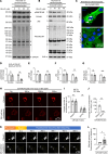
Figure 7. Treatment with 25-HC reduces phosphorylation of CRYAB at S59 and alters the phase-separation characteristics of CRYAB-R120G.
(A and B) Representative immunoblots depicting total pS59-CRYAB, CRYAB, p62, and polyUb proteins in NP40-detergent (A) soluble and (B) insoluble fractions from neonatal rat cardiomyocytes (NRCMs) transduced with adenoviral CRYAB-R120G (MOI=10) for 90 hours and treated with 0, 10, 20, and 40 mM 25-HC for the final 72 hours. GAPDH and actin are shown as loading controls. (C) Representative immunofluorescence images of NRCMs treated in A. Arrows point to GFP-positive aggregates. (DG) Quantitative assessment of pS59-CRYAB (D), total CRYAB (E), p62 (F), and polyUb protein (G) expression in NP-40 soluble and insoluble fractions from NRCMs treated in A. **P < 0.01 by t test. (H) Representative time-lapse images at t = 0 seconds, 100 seconds, 200 seconds, and 300 seconds after light activation in HEK293A cells transfected with OptoIDR constructs generated with CRYAB-R120G as the IDR protein and treated with diluent or 25-HC (40 mM). (I and J) Average number (I) and area (J) of condensates/cell at t = 0 versus t = 300 seconds in cells treated as in H. **P < 0.01 by t test. (K) Representative images demonstrating recovery of fluorescence after photobleaching in HEK 293A cells treated as in H. The area of photobleaching is marked with a dotted circle prior to (prebleach), and at 0, 100, 200, 300, 400, and 500 seconds (s) after photobleaching. (L) Quantitation of fluorescence recovery in condensates of CRYAB variants (as maximum minus immediately after photobleaching) as shown in K. *P < 0.05 by t test.
Figure 8
Figure 8. Treatment with 25-HC rescues adverse left ventricular remodeling after IR injury.
(A) Schematic depicting experimental strategy for closed-chest IR modeling (90 minutes of ischemia followed by reperfusion) in male WT mice followed by intraperitoneal administration of 25-HC (10 mg/kg/mouse, every 48 hours) or diluent, initiated on day 4 after IR injury. (BD) Quantitative analyses of area-at-risk (AAR, B), left ventricular EDV (LVEDV, C) and LV EF (EF) (%), D) prior to and at 4 weeks after IR injury in mice treated as in A. ***P < 0.001; ****P < 0.0001 by t test. (E and F) Immunoblots depicting total CRYAB, pS59-CRYAB, p62, and polyUb protein expression in NP-40 soluble (E) and insoluble (F) fraction in myocardium of mice 4 weeks after IR injury and treated with diluent or 25-HC. (GJ). Quantitative analyses of pS59-CRYAB (G), total CRYAB (H), p62 (I), and polyUb proteins (J) in NP-40 soluble and insoluble fraction in myocardium of mice 4 weeks after IR injury and treated with diluent or 25-HC. **P < 0.01 by t test. (K) Representative images with immunofluorescence staining for desmin and ubiquitinated proteins in the remote myocardium from mice modeled as in A. (L and M) Quantitative evaluation of desmin localization with striation score (L) and aggregated desmin (M) in mice treated as in A. For striation scoring, normal localization of proteins got scored as 0, and abnormal striation or mislocalization of proteins was scored as 1. For scoring aggregates, absence of aggregates was scored as 0, and presence of aggregates was scored as 2. *P < 0.05 by t test. (N and O) Masson’s trichrome–stained left ventricular sections (N) demonstrating presence of scar at 4 weeks after IR injury in mice treated as in A with quantitation of scar size (O). P value shown is by t test.
Figure 9
Figure 9. Schematic depicting consequences of serine 59 phosphorylation on phase separation of CRYAB in ICM.
CRYAB undergoes dynamic phase separation into condensates in physiology with maintenance of cardiac myocyte health in homeostasis. Persistent stress, as observed with myocardial IR injury (or with human disease-causing R120G mutation in CRYAB protein, not shown), results in phosphorylation at serine 59. Phosphorylated CRYAB at serine 59 (pS59-CRYAB) undergoes phase separation into condensates with reduced dynamicity and increased toxicity. pS59-CRYAB partitions into the aggregate-rich NP-40 insoluble fraction with segregation of its client proteins, such as desmin, α-actinin, and actin, in aggregates within cardiac myocytes in ICM. Inhibiting phosphorylation of CRYAB at serine 59 prevents it from becoming aggregate-prone and attenuates development of cardiomyopathy in the setting of IR injury. Image prepared with BioRender software.

Update of

References

    1. Day SM, et al. Myosin modulators: emerging approaches for the treatment of cardiomyopathies and heart failure. J Clin Invest. 2022;132(5):e148557. doi: 10.1172/JCI148557. - DOI - PMC - PubMed
    1. Ali HR, et al. Defining decreased protein succinylation of failing human cardiac myofibrils in ischemic cardiomyopathy. J Mol Cell Cardiol. 2020;138:304–317. doi: 10.1016/j.yjmcc.2019.11.159. - DOI - PMC - PubMed
    1. Kong SW, et al. Heart failure-associated changes in RNA splicing of sarcomere genes. Circ Cardiovasc Genet. 2010;3(2):138–146. doi: 10.1161/CIRCGENETICS.109.904698. - DOI - PMC - PubMed
    1. Ali MA, et al. Titin is a target of matrix metalloproteinase-2: implications in myocardial ischemia/reperfusion injury. Circulation. 2010;122(20):2039–2047. doi: 10.1161/CIRCULATIONAHA.109.930222. - DOI - PMC - PubMed
    1. Neagoe C, et al. Titin isoform switch in ischemic human heart disease. Circulation. 2002;106(11):1333–1341. doi: 10.1161/01.CIR.0000029803.93022.93. - DOI - PubMed

MeSH terms