. 2025 Feb 11;21(2):e1012571.
doi: 10.1371/journal.ppat.1012571. eCollection 2025 Feb.

A guanidine-based coronavirus replication inhibitor which targets the nsp15 endoribonuclease and selects for interferon-susceptible mutant viruses

Affiliations

A guanidine-based coronavirus replication inhibitor which targets the nsp15 endoribonuclease and selects for interferon-susceptible mutant viruses

Benjamin Van Loy et al. PLoS Pathog. .

Abstract

The approval of COVID-19 vaccines and antiviral drugs has been crucial to end the global health crisis caused by SARS-CoV-2. However, to prepare for future outbreaks from drug-resistant variants and novel zoonotic coronaviruses (CoVs), additional therapeutics with a distinct antiviral mechanism are needed. Here, we report a novel guanidine-substituted diphenylurea compound that suppresses CoV replication by interfering with the uridine-specific endoribonuclease (EndoU) activity of the viral non-structural protein-15 (nsp15). This compound, designated EPB-113, exhibits strong and selective cell culture activity against human coronavirus 229E (HCoV-229E) and also suppresses the replication of SARS-CoV-2. Viruses, selected under EPB-113 pressure, carried resistance sites at or near the catalytic His250 residue of the nsp15-EndoU domain. Although the best-known function of EndoU is to avoid induction of type I interferon (IFN-I) by lowering the levels of viral dsRNA, EPB-113 was found to mainly act via an IFN-independent mechanism, situated during viral RNA synthesis. Using a combination of biophysical and enzymatic assays with the recombinant nsp15 proteins from HCoV-229E and SARS-CoV-2, we discovered that EPB-113 enhances the EndoU cleavage activity of hexameric nsp15, while reducing its thermal stability. This mechanism explains why the virus escapes EPB-113 by acquiring catalytic site mutations which impair compound binding to nsp15 and abolish the EndoU activity. Since the EPB-113-resistant mutant viruses induce high levels of IFN-I and its effectors, they proved unable to replicate in human macrophages and were readily outcompeted by the wild-type virus upon co-infection of human fibroblast cells. Our findings suggest that antiviral targeting of nsp15 can be achieved with a molecule that induces a conformational change in this protein, resulting in higher EndoU activity and impairment of viral RNA synthesis. Based on the appealing mechanism and resistance profile of EPB-113, we conclude that nsp15 is a challenging but highly relevant drug target.


Conflict of interest statement

The authors have declared that no competing interests exist.

Figures

Fig 1
Fig 1. Inhibitory activity of EPB-113 on the replication of HCoV-229E and SARS-CoV-2.
(A) Chemical structures of EPB-113 and its structural analogues EPB-102 and JMLv-061. (B) Inhibition of HCoV-229E replication in HEL299 cells was determined by MTS-based CPE reduction assay; infectious virus titration on the supernatant; and RT-qPCR-based viral load assay. Inhibition of SARS-CoV-2 replication in A549ACE2/TMPRSS2 cells was quantified by viral load assay. Cytotoxicity was determined by MTS cell viability assay in mock-infected cells and is shown in dashed lines (open circles). Data points are the mean ± SEM (N=3-12). (C) Anti-HCoV-229E activity and cytotoxicity in HEL299 cells for EPB-102 and JMLv-061, two structural analogues of EPB-113. Data points are the mean ± SEM (N=3). (D) Table summarizing the values for antiviral activity and cytotoxicity of EPB-113, JMLv-061, EPB-102 and reference compound GS-441524.
Fig 2
Fig 2. Resistance to EPB-113 maps to the catalytic core of nsp15 EndoU.
(A) Setup of the resistance selection experiment. (B) Alignment of partial nsp15 sequences from 11 coronaviruses, with EPB-113 and 5h resistance sites marked with triangles. Both EPB-113 resistance sites, G248 and H250, are located at the catalytic core of nsp15 EndoU and strictly conserved in all CoVs, with H250 being part of the catalytic triad. Sequence similarities from Clustal Omega-aligned sequences were rendered using ESPript 3.0. (C) Structure of the catalytic core of SARS-CoV-2 EndoU bound to dsRNA (PDB 7TJ2) [42]. Resistance of HCoV-229E to EPB-113 maps to residues G248 and H250, while that for 5h maps to K60 and T66 (corresponding to residues K60, V66, G247 and H249 in SARS-CoV-2). (D) Evaluation of EPB-113 and reference compound GS-441524 against nsp15-mutant CoVs, based on RT-qPCR-based viral load assay. Left panels: HEL299 cells infected with HCoV-229E WT; the G248Vnsp15 or H250Ynsp15 mutant (obtained in this study); or the K60Rnsp15 or T66Insp15 mutant (previously selected under compound 5h) [42]. Middle panels: 16HBE cells infected with H250Ansp15 mutant HCoV-229E, created by reverse genetics [11]. Right panels: A549ACE2/TMPRSS2 cells infected with K60Rnsp15 or V66Insp15 mutant SARS-CoV-2, created by reverse genetics. Data points are the mean ± SEM (N=2-3). The panel legends show the EC90 values and statistical significance of the difference between mutant and WT, based on (left panels) ordinary one-way ANOVA with Dunnett’s corrections for multiple comparisons and (middle panels) two-tailed unpaired t-test.
Fig 3
Fig 3. EPB-113 is active in both IFN responsive and non-responsive cell types.
EC90 values of EPB-113 and GS-441524 for inhibition of HCoV-229E WT virus in human macrophages, HEL299 cells, 16HBE cells and Huh7-STAT1-KO cells, based on viral load assay. The graph shows the individual values and mean from three independent experiments.
Fig 4
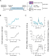
Fig 4. Action point of EPB-113 and reference compounds in a one-cycle time-of-addition assay.
(A) Experimental setup. (B) Timewise, EPB-113 has a similar action point as K22, located at an intermediate stage in HCoV-229E replication. The fold increase in intracellular viral RNA is expressed relative to the amount of input RNA at 2 h p.i. Compound concentrations: 15 µM E64d; 15 µM GS-441524; 15 µM K22 [57]; 3 µM nirmatrelvir; and 15 µM EPB-113. Data points are the mean ± SEM (N=3).
Fig 5
Fig 5. Delaying EPB-113 exposure results in subtle induction of IFN-I.
(A) Compound addition to HEL299 cells infected with HCoV-229E WT was delayed until 24 h p.i. and 48 h later, samples were collected for assessment of (B) viral load and (C) levels of IFN-I (left Y-axis: IFN-β mRNA; right Y-axis: bioactive IFN-I). GS-441524 and nirmatrelvir were included as reference compounds. Data points are the mean ± SEM (N=4). Statistical analysis was performed using a one-way ANOVA with Dunnett’s correction for multiple comparisons, to compare each compound concentration to untreated virus control (0 µM).
Fig 6
Fig 6. EPB-113 binds to hexameric nsp15 protein.
(A) Thermal shift assay showing the decrease in Tm when 6 µM of nsp15 protein was incubated with 100 µM EPB-113. The controls contained 100 µM EPB-102 or the equivalent amount (= 5%) of DMSO. The top panel shows the curves generated from the mean data of three independent experiments. The graph (bottom panel) shows the melting temperatures (individual and mean values) in three independent experiments. Statistical significance was determined with an ordinary one-way ANOVA with Dunnett’s correction. (B and C) Assessment of binding by surface-plasmon resonance. RU: resonance units. Panel B: dose-dependent binding between EPB-113 (top panels) or EPB-102 (bottom panels) and recombinant nsp15 from HCoV-229E (left panels) or SARS-CoV-2 (right panels). Panel C: binding of EPB-113 to HCoV-229E nsp15 was severely affected by the G248V-, H250Y- and H250A mutations.
Fig 7
Fig 7. EPB-113 enhances the EndoU cleavage reaction.
(A) Scheme of the two-step EndoU cleavage reaction. (B) FRET assay in the presence of 100 µM EPB-113, 100 µM EPB-102 or the equivalent amount (= 5%) of DMSO. RFU: relative fluorescence units. An example of the real-time fluorescence curves is shown in the top panel, while the graph in the bottom panel shows the initial reaction velocity (V0) derived from the 5-10 min interval. Individual and mean data from N=3. (C) HPLC-MS analysis on samples collected after 1 h reaction time. In the chromatograms, the colors indicate the FRET substrate; 2′,3′-cyclic intermediate; 5′-hydroxylated product; and 3′-phosphorylated product. The Y-axis shows the % normalized intensity.
Fig 8
Fig 8. HCoV-229E viruses bearing EPB-113 resistance mutations have impaired viral fitness in IFN responsive cells.
(A) Replication kinetics in human macrophages and HEL299, 16HBE and Huh7-STAT1-KO cells, based on RT-qPCR for viral load. Data points are the mean ± SEM (N=3). At each time point, statistical significance is shown for the difference between mutant and WT (multiple unpaired t-tests, with Holm-Šídák’s correction for multiple comparisons). (B) Competitive growth assay. At the start of the experiment, each mutant was mixed with WT virus at a ratio of 9:1, 1:1 or 1:9, then applied to HEL299 cells. After the virus was passaged three more times, Nanopore sequencing was performed on each passage, to determine the percentage of viral genomes that contained the mutant or WT form of nsp15.
Fig 9
Fig 9. The nsp15-mutant HCoV-229E viruses boost the host cell’s innate immune response.
(A) mRNA levels (determined by RT-qPCR) for a range of innate immune factors, in HEL299 cells infected with WT or nsp15-mutant virus. (B) Levels of bioactive IFN-I in the supernatant of human macrophages, HEL299 cells or 16HBE cells infected with WT or mutant virus. For both heatmaps, detailed graphs containing the numeric values and statistical analysis are provided in S6 Fig.
Fig 10
Fig 10. The nsp15 mutations increase the sensitivity to temperature and exogenous IFN-β.
(A) Temperature-dependent replication of WT and nsp15-mutant HCoV-229E viruses in IFN responsive (HEL299) or non-responsive (Huh7-STAT1-KO) cells. The graphs show the mean CCID50 values of two independent virus titration experiments, conducted at the indicated temperatures. (B) The nsp15 mutations render HCoV-229E more sensitive to the antiviral effect of exogenous IFN-β, as determined by viral load assay in HEL299 cells. The graphs show the individual and mean values from four independent experiments. An ordinary one-way ANOVA with Dunnett’s correction was used to analyze the differences in EC90, comparing mutants to WT virus.

References

    1. World Health Organization. WHO COVID-19 dashboard. [cited 2025 Jan 08]. Available from: https://data.who.int/dashboards/covid19/cases.
    1. Infectious Diseases Society of America. IDSA guidelines on the treatment and management of patients with COVID-19. [cited 2024 Aug 23]. Available from: https://www.idsociety.org/COVID19guidelines#null.
    1. V’Kovski P, Kratzel A, Steiner S, Stalder H, Thiel V. Coronavirus biology and replication: implications for SARS-CoV-2. Nat Rev Microbiol. 2021;19(3):155–70. Epub 2020/10/30. doi: 10.1038/s41579-020-00468-6 PMCID: PMC7592455 - DOI - PMC - PubMed
    1. Lou Z, Rao Z. The life of SARS-CoV-2 inside cells: replication-transcription complex assembly and function. Annu Rev Biochem. 2022;91:381–401. doi: 10.1146/annurev-biochem-052521-115653 - DOI - PubMed
    1. Li G, Hilgenfeld R, Whitley R, De Clercq E. Therapeutic strategies for COVID-19: progress and lessons learned. Nat Rev Drug Discov. 2023;22(6):449–75. Epub 20230419. doi: 10.1038/s41573-023-00672-y PMCID: PMC10113999 - DOI - PMC - PubMed

MeSH terms

LinkOut - more resources