. 2025 Feb 11;13(2):e009852.
doi: 10.1136/jitc-2024-009852.

4-1BB agonist targeted to fibroblast activation protein α synergizes with radiotherapy to treat murine breast tumor models

Affiliations

4-1BB agonist targeted to fibroblast activation protein α synergizes with radiotherapy to treat murine breast tumor models

Eneko Garate-Soraluze et al. J Immunother Cancer. .

Abstract

Background: Ionizing radiation (IR) is a double-edged sword for immunotherapy as it may have both immunosuppressive and immunostimulatory effects. The biological effects of IR on the tumor microenvironment (TME) are a key factor for this balance. Fibroblast activation protein (FAP) is expressed on the surface of cancer-associated fibroblasts (CAF) in many cancer types and its abundance is associated with the poor immune response to immune-checkpoint-blockade in patients. We hypothesized that IR increases FAP expression in CAFs, therefore the combination of IR with targeted immunomodulators such as an agonistic anti-FAP-4-1BBL fusion protein could enhance the immune-mediated antitumoral effects of these treatments.

Methods: The murine transplantable TS/A tumor-cell-line co-engrafted with CAFs was used to investigate increases in FAP expression in tumors following irradiation using immunohistochemistry, real-time polymerase chain reaction (RT-PCR) and multiplex tissue immunofluorescence. One lesion of bilateral tumor-bearing mice was only locally irradiated or combined with weekly injections of the bispecific muFAP-4-1BBL fusion protein (a mouse surrogate for RG7826). Tumor sizes were followed over time and TME was assessed by flow cytometry. Selective monoclonal antibody (mAb)-mediated depletions of immune cell populations, neutralizing interferon alpha/beta receptor 1 (IFNAR-I) IFNAR and interferon (IFN)-γ mAbs and gene-modified mice (4-1BB-/-) were used to delineate the immune cell subsets and mechanisms required for efficacy. 67Ga labeled muFAP-4-1BBL tracked by SPECT-CT was used to study biodistribution. In human colorectal carcinoma samples, the inducibility of FAP expression following radiotherapy was explored by multiplex immunofluorescence.

Results: Irradiation of TS/A+CAF tumors in mice showed an increase in FAP levels after local irradiation. A suboptimal radiotherapy regimen in combination with muFAP-4-1BBL attained primary tumor control and measurable abscopal effects. Immune TME landscape analyses showed post-treatment increased infiltration of activated immune cells associated with the combined radioimmunotherapy treatment. Efficacy depended on CD8+ T cells, type I IFN, IFN-γ and ability to express 4-1BB. Biodistribution studies of muFAP-4-1BBL indicated enriched tumor targeting to irradiated tumors. Human colorectal cancer samples pre and post irradiation showed enhanced FAP expression after radiotherapy.

Conclusion: Increased FAP expression in the TME as a result of radiotherapy can be exploited to target agonist 4-1BB immunotherapy to malignant tumor lesions using an FAP-4-1BBL antibody fusion protein.

Keywords: Abscopal; Monoclonal antibody; Radiotherapy/radioimmunotherapy; Tumor microenvironment - TME.


Conflict of interest statement

Competing interests: EG-S, IS-M, LF-R, AR, CEDA, CB-A, CdPH, CLR, AT, JAS, MC, and PS-M declare no competing interests. MER-R reports receiving research funding from Roche and Highlight Therapeutics. She also has received speaker’s bureau honoraria from BMS and ROCHE. IM reports receiving commercial research grants from BMS, Highlight Therapeutics, Alligator, Pfizer Genmab and Roche; has received speaker’s bureau honoraria from MSD; and is consultant or advisory board member for BMS, Roche, AstraZeneca, Genmab, Pharmamar, F-Star, Bioncotech, Bayer, Numab, Pieris, Gossamer, Alligator and Merck Serono. CK, PU, CC, and TT declare employment, stock ownership and patents with Roche.

Figures
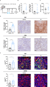
Figure 1
Figure 1. Irradiation increases the presence of FAP+ fibroblasts in engrafted mouse tumors. (A) Scheme of TS/A+CAF co-injection followed by hypofractionated radiotherapy (RT, blue arrows) of the primary tumor (PT) but not a contralateral tumor. Subsequent immunohistochemistry (IHC), flow cytometry and multiplex studies were performed at the indicated days (n=7–9 mice/group). (B) Non-treated (vehicle) and irradiated (RT) tumors were compared in TS/A+CAF model. Shown are the frequency in % of CAF (CD90.2+ in the total population of CD45/CD31/Epcam) and the frequency in % of CAF αSMA+ quantification by flow cytometry of the digested tumor. (C) Median fluorescence intensity (MFI) of FAP on CAFs (n=9–11 mice/group). (D) IHC studies of FAP expression and CD8+ T-cell immune infiltration. Representative images (50 µm). (E) Multiplex immunofluorescence in non-treated (vehicle) and irradiated (RT) tumors showing markers: FAP, collagen type VI, αSMA. The nuclei were counterstained with DAPI. Plots, representative images and their single staining (50 µm) (n=7–11 mice/group). Results show means±SEM, each symbol indicates one mouse, significance is analyzed by one-tailed t-test, *p<0.05, ***p<0.001. Pooled data from two independent experiments. CAF, cancer-associated fibroblast; FAP, fibroblast activation protein.
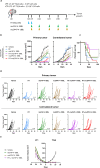
Figure 2
Figure 2. muFAP-4-1BBL synergizes with local radiotherapy exerting partial abscopal control of non-irradiated distant tumors. (A) Scheme of 8-day TS/A+CAF co-injection and subsequent treatments with hypofractionated radiotherapy (RT, blue arrows) of the primary tumor (PT) but not contralateral tumor (CT). Tumor-bearing mice were treated by intraperitoneal administration of muFAP-4-1BBL (green arrows) or DP47-4-1BBL (brown arrows) and tumor size was monitored (n=12 mice/group). Pooled data from two independent experiments. (B) Tumor volumes (mm3) over time are shown per group as means±SEM and statistical comparisons among experimental groups *p<0.05, **p<0.01, (two-way analysis of variance). (C) The percentage of survival over time is shown for the experiment (Mantel-Cox test). (D) Individual tumor sizes in the treated and distant non-irradiated tumors as indicated. (E) Rechallenge of cured mice is shown by 4T1 and TS/A cell line inoculation. CAF, cancer-associated fibroblast; FAP, fibroblast activation protein.
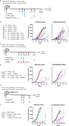
Figure 3
Figure 3. CD8+ T cells, 4-1BB gene-integrity and the functions of type I and II IFNs are necessary for the synergistic therapeutic effects of RT+muFAP-4-1BBL. (A) Scheme of selective depletion of CD8+, CD4+ T cells and IFN-γ (pink arrows) in mice bearing bilateral TS/A+CAFs-derived tumors in which primary tumors (PT) were treated with radiotherapy (RT, blue arrows) but not contralateral tumors (CT). Intraperitoneal administration of muFAP-4-1BBL (green arrows) was given as a combination therapy as indicated. (B) Tumor volumes (mm3) over time are shown per color-coded group as means±SEM and compared (Two-way ANOVA) (n=9–12 mice/group). Pooled data from two independent experiments. (C) Scheme of IFNAR blockade (red arrows) by intratumoral administration in mice bearing bilateral TS/A+CAFs-derived tumors in which PT was treated with RT (blue arrows). Some mice received additional intraperitoneal administration of muFAP-4-1BBL (green arrows) as combination therapy. (D) Tumor volumes (mm3) over time are shown per color-coded group as means±SEM (two-way ANOVA) (n=9–12 mice/group). Pooled data from two independent experiments. (E) Scheme of 8-day tumor co-engraftment and subsequent treatments with hypofractionated radiotherapy (blue arrows) and intraperitoneal injections of muFAP-4-1BBL (green arrows) in bilateral subcutaneous TS/A+CAF-derived tumors in 4-1BBKO mice. Tumor size was monitored over time. BALBc wildtype mice were treated similar as control. (F) Tumor volumes (mm3) over time are shown per color-coded group as means±SEM (two-way ANOVA) (n=6–10 mice/group). Significance is indicated as *p<0.05, ***p<0.001. ANOVA, analysis of variance; CAF, cancer-associated fibroblast; FAP, fibroblast activation protein; IFN, interferon; IFNAR-I, interferon alpha/beta receptor 1.
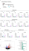
Figure 4
Figure 4. Irradiation and muFAP-4-1BBL reshape the tumor microenvironment into a more immune-stimulating landscape. (A) Scheme of TS/A+CAFs tumor co-engraftment and subsequent treatments with hypofractionated radiotherapy of the primary tumor (RT, blue arrows) but not the contralateral tumor (CT). Intraperitoneal administration of muFAP-4-1BBL (green arrow) was given as combination therapy and tumors were excised at day 11 (RNA sequencing) and day 15 (flow cytometry analysis). (B) Plots showing the absolute numbers of the indicated T cells and their surface markers in primary (top panels) and contralateral (bottom panels) tumors analyzed by flow cytometry and normalized to gram of tumor. Each symbol indicates one mouse, mean±SEM is indicated (two-way ANOVA). (C) Plots showing the absolute numbers of dendritic cells and their surface marker in primary and contralateral tumors analyzed by flow cytometry and normalized to gram of tumor (n=4–6 mice/group). Each symbol indicates one mouse, mean±SEM is indicated (two-way ANOVA) *p<0.05, **p<0.01, ***p<0.001. (D) Volcano plot comparing the most upregulated (logFC 0<4) and downregulated genes (logFC −4<0) at day 11 analyzed by RNA sequencing, whereby comparison was done between RT+muFAP-4-1BBL and muFAP-4-1BBL. Some of the most upregulated genes are highlighted by gene name. (E) Bar plots of p values presenting enrichment of immune-related pathways between RT+muFAP-4-1BBL and muFAP-4-1BBL. The vertical line shows the threshold value at p=0.05. Three mice of each treatment group were analyzed as indicated. ANOVA, analysis of variance; CAF, cancer-associated fibroblast; DC, dendritic cell; FAP, fibroblast activation protein; PT, primary tumor; RT, radiotherapy; Treg, regulatory T cell.
Figure 5
Figure 5. Radiotherapy enhances muFAP-4-1BBL targeting irradiated tumors. (A) Schematic representation of radiolabeling assay in which muFAP-4-1BBL was labeled with (67Ga) and administered intraperitoneally at day 9 (green arrow) into TS/A+CAF tumor-bearing mice treated or non-treated with RT therapy (RT, blue arrows) of the primary tumor (PT) but not contralateral tumor (CT). Biodistribution analysis measuring radioactivity was performed by SPECT/CT images obtained in vivo on day 16 (orange arrows). (B) Representative SPECT/CT images of mice at day 16 of muFAP-4-1BBL monotherapy and RT+muFAP-4-1BBL where the PT was irradiated. The coronal section of mice is shown on SPECT/CT images. The red dotted circles are indicating the location of the CT and PT. (C) Ex vivo biodistribution analysis performed by gamma counter and displayed as per cent of injected dose per gram of tumor (%ID/g) of both primary and contralateral tumors of indicated mice (n=3 mice/group, mean±SEM) (two-way analysis of variance); ***p<0.001. CAF, cancer-associated fibroblast; FAP, fibroblast activation protein; RT, radiotherapy.
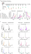
Figure 6
Figure 6. Combinations of systemic PD-1 and CTLA-4 blockade with muFAP-4-1BBL further synergize with radiotherapy to attain curative abscopal effects. (A) Scheme of 10-day tumor co-engraftment and subsequent treatments of TS/A+CAF tumor-bearing mice with hypofractionated radiotherapy (RT, blue arrows) of the primary tumor (PT) but not contralateral tumor (CT). For combination treatment mice received intraperitoneal administration of muFAP-4-1BBL (green arrows) alone or combined with anti-CTLA-4 (orange arrows) or anti-PD-1 (brown arrows). (B) Tumor growth was monitored and means±SEM are shown over time of primary (left) and contralateral tumor (right) (two-way analysis of variance); **p<0.01, ***p<0.001. (C) The percentage of survival over time is shown for experiments in B (Mantel-Cox test); **p<0.01. (D) Individual tumor sizes of the RT-treated primary tumor and distant non-irradiated contralateral tumors receiving additional treatment as indicated (n=6–10 mice/group). Pooled data from two independent experiments. CAF, cancer-associated fibroblast; CTLA-4, cytotoxic T-lymphocyte associated protein 4; FAP, fibroblast activation protein; PD-1, programmed cell death protein-1.
Figure 7
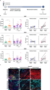
Figure 7. Post-chemoradiotherapy upregulation of FAP expression in patient-derived colorectal carcinoma tissue samples. (A) Schematic representation of patients with colorectal carcinoma’s treatment course from diagnosis, baseline biopsy collection, chemoradiotherapy (CRT) treatment to surgery and post-CRT specimen collection for multiplex immunofluorescence studies (n=12 patients). (B) Plots showing FAP expression and immune infiltration of CD8 and CD3 expressing cells between baseline biopsy and post-CRT specimens in patients with residual disease or complete response evaluated by immunofluorescence imaging of tissue. Each symbol represents one patient, means±SEM are indicated (paired t-test and repeated measured analysis of variance) *p<0.05, **p<0.01, ***p<0.001. (C) Immunofluorescence representative images of tumor samples (scale bar 20 µm). FAP, fibroblast activation protein; RT, radiotherapy.

References

    1. Kalluri R. The biology and function of fibroblasts in cancer. Nat Rev Cancer. 2016;16:582–98. doi: 10.1038/nrc.2016.73. - DOI - PubMed
    1. Orimo A, Weinberg RA. Stromal fibroblasts in cancer: a novel tumor-promoting cell type. Cell Cycle. 2006;5:1597–601. doi: 10.4161/cc.5.15.3112. - DOI - PubMed
    1. Kalluri R, Zeisberg M. Fibroblasts in cancer. Nat Rev Cancer. 2006;6:392–401. doi: 10.1038/nrc1877. - DOI - PubMed
    1. Paulsson J, Micke P. Prognostic relevance of cancer-associated fibroblasts in human cancer. Semin Cancer Biol. 2014;25:61–8. doi: 10.1016/j.semcancer.2014.02.006. - DOI - PubMed
    1. Hellevik T, Pettersen I, Berg V, et al. Changes in the Secretory Profile of NSCLC-Associated Fibroblasts after Ablative Radiotherapy: Potential Impact on Angiogenesis and Tumor Growth. Transl Oncol. 2013;6:66–74. doi: 10.1593/tlo.12349. - DOI - PMC - PubMed

Substances