. 2025 Feb 11;16(1):87.
doi: 10.1038/s41419-025-07364-y.

Analysis of effector/memory regulatory T cells from arrhythmogenic cardiomyopathy patients identified IL-32 as a novel player in ACM pathogenesis

Affiliations

Analysis of effector/memory regulatory T cells from arrhythmogenic cardiomyopathy patients identified IL-32 as a novel player in ACM pathogenesis

Salwa Soussi et al. Cell Death Dis. .

Abstract

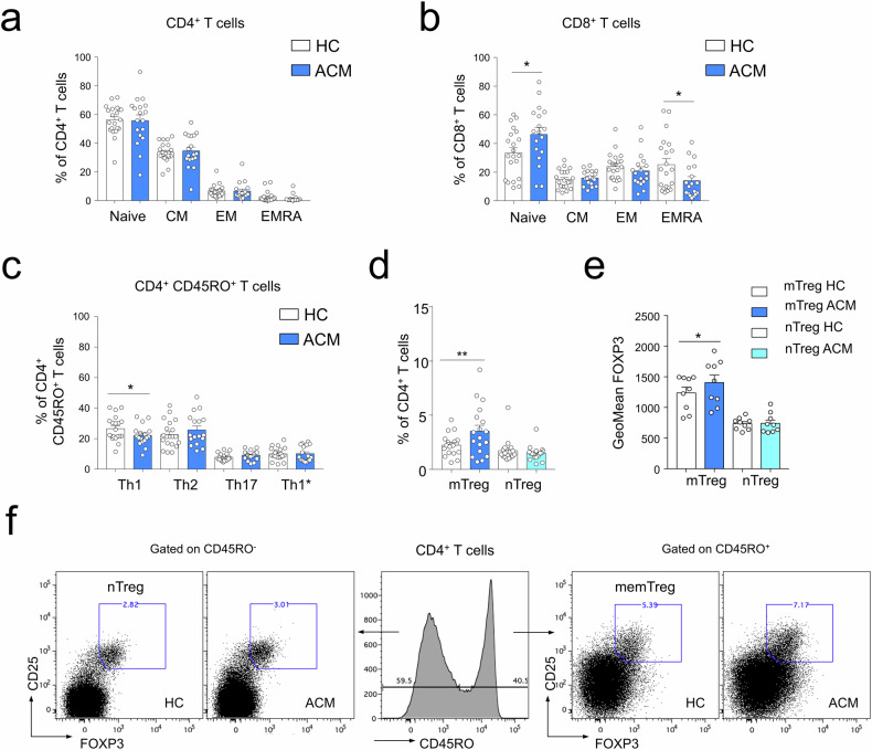
Arrhythmogenic cardiomyopathy (ACM) is an inherited cardiac disorder that causes sudden cardiac death and progressive heart failure. Besides fibro-fatty replacement and myocyte degenerative changes, inflammatory patchy infiltrates are found in myocardial histological analysis of ACM patients. Inflammatory cells could actively participate in ACM pathogenesis, contributing to the alteration of cardiac microenvironment homeostasis, thus triggering disease evolution. In order to characterize the immune-derived mediators involved in ACM pathogenesis, peripheral blood mononuclear cells from ACM patients were characterized and compared to healthy controls' ones. Flow cytometry analysis revealed a lower frequency of CD4+ T helper type 1 cells, NK cells, and terminally differentiated CD8+ EMRA+ T cells in ACM patients compared to age-matched controls. In contrast, a higher proportion of effector/memory FOXP3+ CCR4+ CD45RO+ regulatory CD4+ T cells (Treg) were found in ACM patients. Single-cell RNA-seq performed on isolated memory Treg cells (mTreg) from ACM patients and healthy controls identified 6 clusters characterized by specific gene signatures related to tissue repair and immunosuppressive pathways. Notably, interleukin 32 (IL-32) was the most differentially expressed gene in ACM patients mTreg with respect to healthy controls. Treatment of human cardiac mesenchymal stromal cells with recombinant IL-32 in vitro promoted lipid droplet accumulation and collagen deposition, thus identifying IL-32 as a new potential player in the immune-mediated trigger of cardiac fibro-fatty replacement in ACM. Overall, we here provide the first complete characterization of circulating ACM immune cells, revealing an abundance of Treg. The high expression of IL-32 in ACM Treg may contribute to accelerated cardiac remodeling in ACM patients' hearts.


Conflict of interest statement

Competing interests: The authors declare no competing interests.

Figures

Fig. 1
Fig. 1. Analysis of immune cell populations from PBMC of ACM patients revealed decreased NK cell frequency and phenotypic changes of the classical monocyte subset.
a Frequencies of major blood leukocyte subsets of HC (n = 20) and ACM patients (n = 17) analyzed by flow cytometry. *p < 0.05, **p < 0.01, and ns not significant using Unpaired t-test or Mann–Whitney t-test. b Blood monocyte subsets of controls and ACM patients were characterized by flow cytometry using CD14 and CD16 staining. c Histograms showing monocyte subset frequencies percentages of monocytes positive for d CCR2, e HLA-DR, and f CD86 for the monocyte subsets of healthy controls (HC; n = 24) and ACM patients (ACM; n = 20). g Representative fluorescence overlay histograms show CCR2, HLA-DR, and CD86 expression by classical monocytes of HC and ACM patients compared to isotype mAb staining (gray). *p < 0.05, **p < 0.01, and ns not significant using Unpaired t-test or Mann–Whitney t-test.
Fig. 2
Fig. 2. The analysis of CD4+ T cell subsets of ACM patients revealed blood effector/memory CD4+ T regulatory cell increase and effector-memory CD8+ cell decrease.
a, b Percentages of naive, CM, EM, and EMRA subsets in CD4+ T cells (a) or in CD8+ T cells (b) from controls (n = 21) and ACM patients (n = 18) as histograms. c Percentages of memory CD45RO+ CD4+ T helper subsets Th1-like, Th2-like, Th17-like and Th1*-like from ACM patients and age-matched controls (HC; n = 17 pairs). d, e Percentages (n = 18 per group) of mTreg and naive (nTreg) in CD4+ T cells (d) and geometric mean fluorescence intensity (n = 9 per group) of FOXP3 (e). f Analysis of mTreg and nTreg CD4+ T regulatory cell subsets of HC and ACM patients by flow cytometry based on CD45RO expression. Data are shown as mean ± SEM. **p < 0.01, *p < 0.05 and ns not significant using unpaired T-test (a, b) or Wilcoxon matched-pairs signed rank test (ce).
Fig. 3
Fig. 3. Single cell RNA-seq analysis of effector/memory CD4+ T cell reveal different subsets enrichment in ACM patients vs. HC.
at-SNE and UMAP projections of single-cell transcriptomes of cell-sorted CD45RO+ CD25+ CCR4+ CD4+ mTreg from PBMC of ACM patients with PKP2* mutation and controls (n = 5 per group) show the presence of 6 clusters bt-SNE projections of HC or ACM cells (separately) and c their respective contribution (frequency and cell numbers) to the 6 clusters. d Pseudotemporal gene-expression profiles of DICE selected mTreg defined three trajectories on a UMAP plot with cells colored according to its pseudotime. e Clustering of significant genes based on their expression pattern over pseudotime trajectories (|area between the curves|>0.5 with p-value < 0.01 in at least a pairwise comparison), defined 18 groups of genes. f Profiles of marker genes differentially expressed across the trajectories and pathway analysis related to groups of genes with similar expression patterns.
Fig. 4
Fig. 4. Differential expression analysis of Treg of ACM patients vs. HC revealed dysregulation of specific pathways and higher expression of IL-32.
a, c Pathway analysis of differentially expressed genes between HC and ACM mTreg (a) and for each cluster (c), according to NCI-Nature Pathway Database. b Heatmap showing log-fold change expression of genes in each of the 6 clusters with significant ANOVA test (p > 0.01) between ACM and control identifying IL-32.
Fig. 5
Fig. 5. A higher expression of IL-32 characterizes plasma and right ventricle of ACM patients compared to HC.
a, b Relative mRNA expression of total (a) and γ isoform (b) of the IL-32 gene in PBMC from ACM patients or healthy donors (HC). Data were expressed using the 2ΔΔCt method over the housekeeping gene GAPDH. c Percentages of IL-32+ mTreg in CD4+ T cells from PBMC of ACM patients or healthy donors determined by flow cytometry after intracellular staining. d IL-32 concentration (pg/ml) in the plasma of ACM or HC (n = 24 each). e, f Representative images of right ventricular tissue sections from HC and ACM showing IL-32 staining by immunofluorescence (e) and histogram quantification (f), data are expressed as the total area of IL-32 positivity on nuclei area. Data are shown as mean +/− SEM. **p < 0.01, *p < 0.05 using T-test. Scale bar: 50 µm.
Fig. 6
Fig. 6. Adipogenic and pro-fibrotic effects of IL-32 on cardiac mesenchymal stromal cells (C-MSC) of ACM or HC.
a, b Nile red staining showing lipid accumulation in C-MSC in basal conditions and after treatment with IL-32 20 ng/ml for 3 days (a), and its quantification (b) showed by sum nile red intensity per number of nuclei. c, d Immunohistochemistry showing collagen accumulation in C-MSC in basal conditions or after treatment with IL-32 20 ng/ml for 3 days (c) and its quantification (d) showed by sum 488 intensity per number of nuclei. Data are shown as mean ± SEM. ***p < 0.001, **p < 0.01, *p < 0.05 using two-way ANOVA test followed by Tukey’s multiple comparisons test. Scale bars: 50 µm.

References

    1. Chen S, Chen L, Duru F, Hu S. Heart failure in patients with arrhythmogenic Cardiomyopathy. J Clin Med. 2021;10:4782. - PMC - PubMed
    1. Huynh K. Increased degradation of desmosomal proteins in ACM. Nat Rev Cardiol. 2023;20:371. - PubMed
    1. Corrado D, Basso C, Judge DP. Arrhythmogenic cardiomyopathy. Circ Res. 2017;121:784–802. - PubMed
    1. Sommariva E, Brambilla S, Carbucicchio C, Gambini E, Meraviglia V, Dello Russo A, et al. Cardiac mesenchymal stromal cells are a source of adipocytes in arrhythmogenic cardiomyopathy. Eur Heart J. 2016;37:1835–46. - PMC - PubMed
    1. Maione AS, Stadiotti I, Pilato CA, Perrucci GL, Saverio V, Catto V, et al. Excess TGF-β1 drives cardiac mesenchymal stromal cells to a pro-fibrotic commitment in arrhythmogenic cardiomyopathy. Int J Mol Sci. 2021;22:2673. - PMC - PubMed

MeSH terms