. 2025 Feb 11;16(1):88.
doi: 10.1038/s41419-025-07421-6.

E3 ligase TRIM8 suppresses lung cancer metastasis by targeting MYOF degradation through K48-linked polyubiquitination

Affiliations

E3 ligase TRIM8 suppresses lung cancer metastasis by targeting MYOF degradation through K48-linked polyubiquitination

Chi-Hsuan Wei et al. Cell Death Dis. .

Abstract

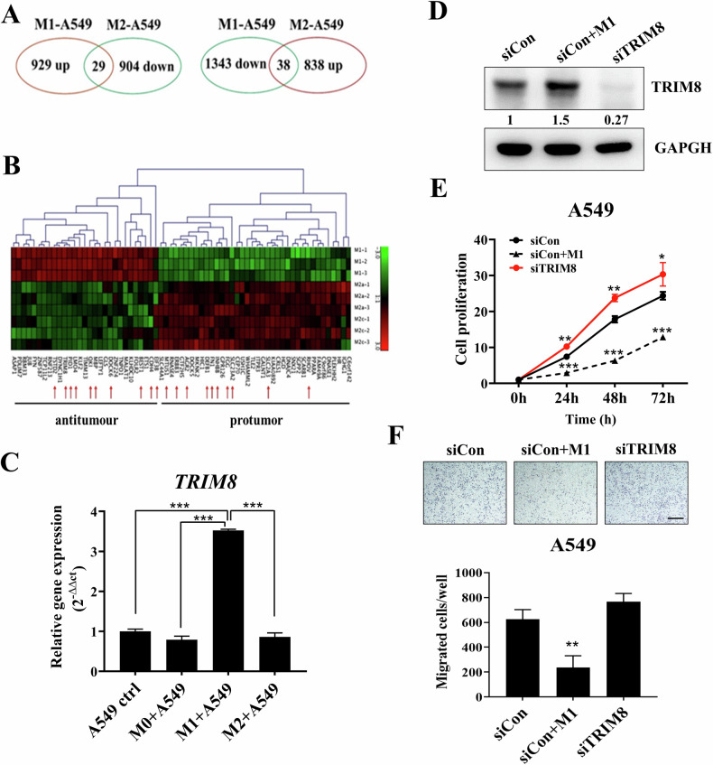
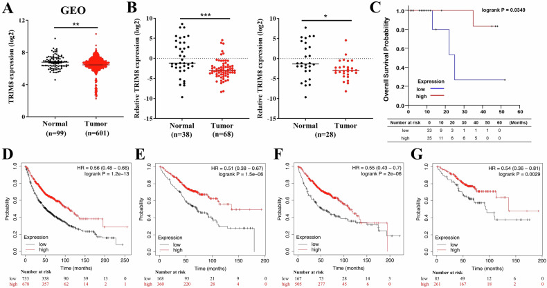
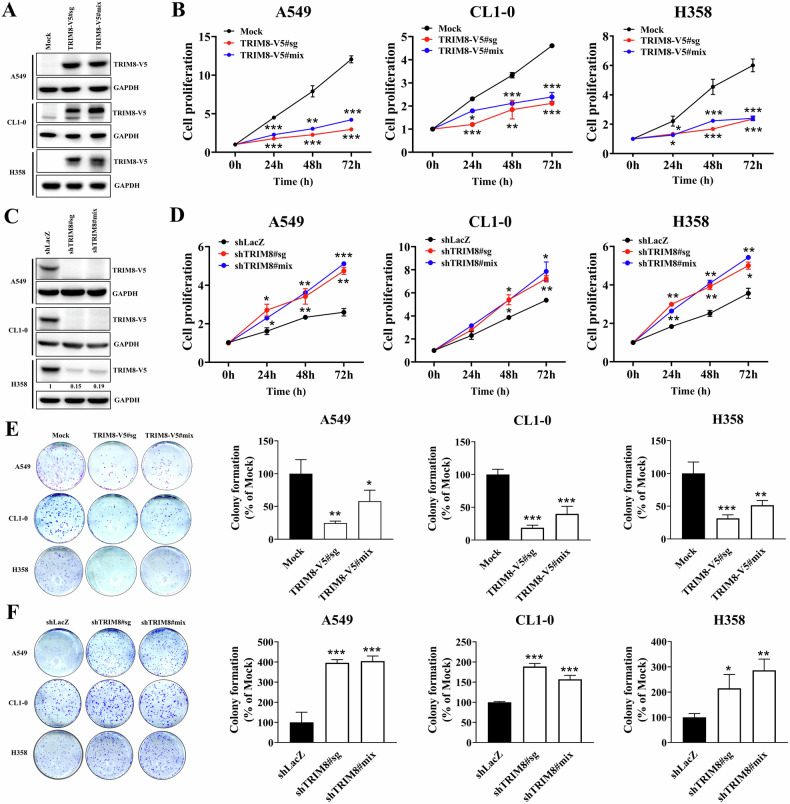
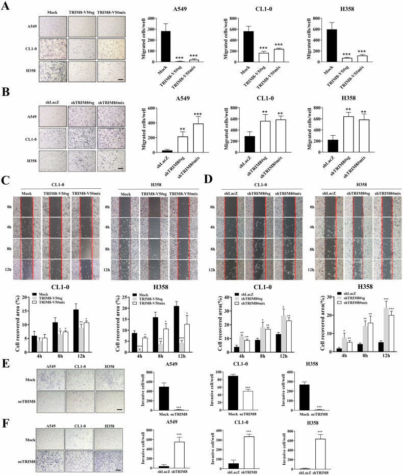
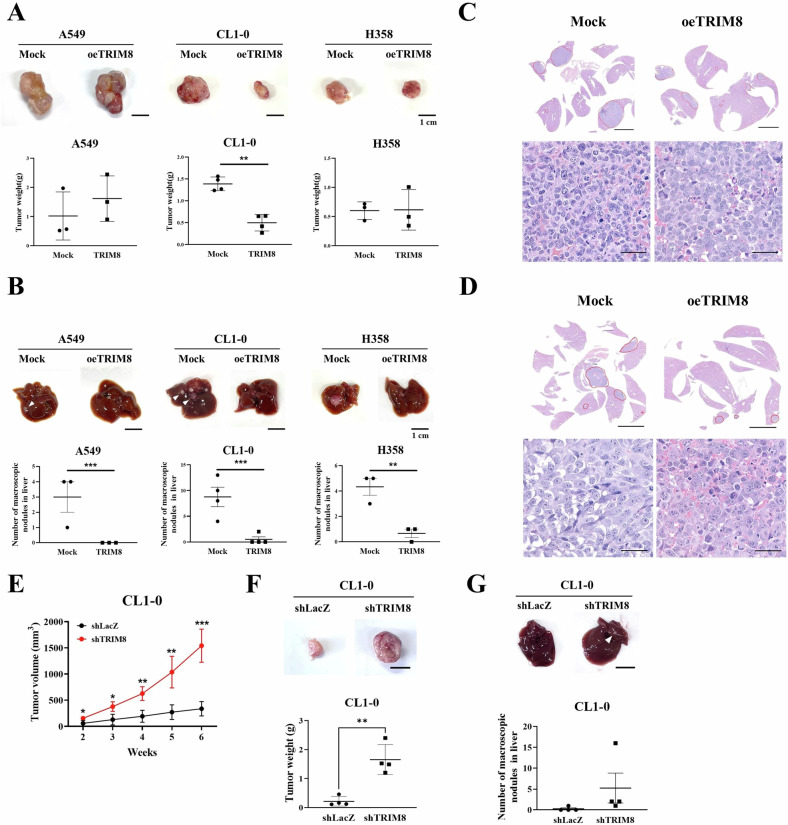
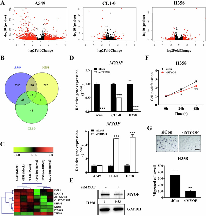
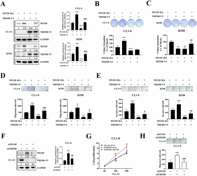
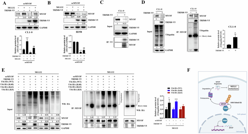
Ubiquitination is a posttranslational modification that regulates tumour progression-associated proteins through the ubiquitin‒proteasome system, making E3 ligases potential antitumour targets. Here, we report that TRIM8, a member of the TRIM family and an E3 ligase, can act as a tumour suppressor in non-small cell lung cancer (NSCLC). Both gain- and loss-of-function experiments revealed that TRIM8 inhibits the proliferation, colony formation, migration and invasion of NSCLC cells. Experiments with a xenograft model showed that TRIM8 expression suppresses tumour metastasis in vivo. Moreover, low expression of TRIM8 was associated with poor overall survival in both the Taiwanese and GEO lung cancer cohorts. TRIM8 overexpression in lung cancer cells reduced MYOF expression, and restoring MYOF rescued cell migration in TRIM8-overexpressing cells. TRIM8 targeted MYOF for K48-linked ubiquitination, facilitating proteasome-mediated degradation and subsequently suppressing the extracellular secretion of MMPs. Our results provide new insights into the contribution of TRIM8 to lung cancer progression, suggesting that TRIM8 is a new biomarker and a novel therapeutic target for lung cancer.


Conflict of interest statement

Competing interests: The authors declare no competing interests. Ethics approval: The study on human lung adenocarcinoma specimens has been approved by the Institutional Review Board of Chung Shan Medical University Hospital (IRB CSMUH No: CS2-20206). Informed consent was obtained from all patients included in this study. The animal study was approved by the Institutional Animal Care and Utilization Committee of National Chung Hsing University, Taichung, Taiwan (Approval No: 109-111 R).

Figures

Fig. 1
Fig. 1. TRIM8 is induced by M1 macrophages.
A Venn diagram of the overlapping genes of A549 cells treated with polarized macrophage-conditioned medium (CM). A549 cells were cultured with M1 or M2 macrophage CM for 48 hours. Those genes that are upregulated by M1 macrophages and downregulated by M2 macrophages are classified as tumour suppressors (left panel). Those genes that are downregulated by M1 macrophages and upregulated by M2 macrophages are classified as oncogenes (right panel). B Heatmap of differentially expressed genes in macrophage-treated A549 cells. The red arrow indicates the top 20 genes. C Evaluation of TRIM8 mRNA expression after culture in macrophage CM by RT‒qPCR. TATA-binding protein (TBP) was used as an internal control. D The expression of TRIM8 after M1 macrophage coculture and control or TRIM8 siRNA transfection, as detected by western blotting. The TRIM8 protein levels were quantified using ImageJ, with GAPDH serving as a control. E The viability of the cells in the silencing control (siCon), siCon with M1 macrophage coculture (siCon+M1), and TRIM8-silenced (siTRIM8) groups was evaluated via PrestoBlue™ reagent assays at the indicated times. F The cell migration of the siCon control, siCon with M1 macrophage coculture, and siTRIM8 cells, as determined by Transwell assay. Scale bar = 50 μm. The data are shown as the means ± SDs of at least three independent experiments, and the significance levels are indicated by *P < 0.05, **P < 0.01, and ***P < 0.001. The data were analysed with one-way ANOVA (C, E, F).
Fig. 2
Fig. 2. Correlation between TRIM8 expression and clinical outcome in patients with NSCLC.
A TRIM8 mRNA expression levels in 601 tumour tissues and 99 normal tissues from lung cancer patients in the GEO datasets (GSE19188, GSE30219 and GSE31210). The data were quantified by the log2 method. The data were analysed with t tests. B TRIM8 mRNA expression in a Taiwanese lung cancer cohort comprising 68 lung cancer tissues and 38 adjacent normal tissues, as determined by real-time qPCR (left panel). The right panel shows TRIM8 mRNA expression in 28 pairs of tumour tissue and normal tissue samples. The data were quantified by the log2 transformation of 2−Δct values and are presented as the means ± SDs; *P < 0.05, *P < 0.01, and ***P < 0.001. The data were analysed with t tests (left panel) or paired t tests (right panel). C Kaplan‒Meier curves for overall survival in 68 Taiwanese lung adenocarcinoma patients. Survival curves were also estimated and visualized by using published cohorts in the Kaplan‒Meier Plotter database. D Overall survival curve and univariate Cox regression analysis of 1411 NSCLC patients in the published cohort. E Overall survival curve and univariate Cox regression analysis of 528 patients with stage I NSCLC in the published cohort. F Overall survival curve and univariate Cox regression analysis of 672 lung adenocarcinoma patients in the published cohort. G Overall survival curve and univariate Cox regression analysis of 346 patients with stage I lung adenocarcinoma in the published cohort. The log-rank test was used to assess statistical significance, with a significance level set at P < 0.05.
Fig. 3
Fig. 3. Effect of TRIM8 expression on lung cancer cell proliferation and colony formation.
A Expression of the TRIM8-V5 protein in the transfectants, as measured through western blot analysis with an antibody against V5. The control cells (Mock) were transfected with the pcDNA3.1 vector. B The viability of mock, TRIM8-V5 single clone (#sg) and mixed clone (#mix) cells was evaluated by PrestoBlue™ reagent exclusion assays at the indicated times. C The silencing efficacy of the TRIM8-specific shRNA was determined by western blot analysis using an anti-V5 antibody. The exogenous TRIM8-V5 protein levels were quantified using ImageJ, with GAPDH serving as a control. D The viability of cells infected with the shLacZ or shTRIM8 virus was evaluated by PrestoBlue™ reagent exclusion assays at the indicated times. E Anchorage-dependent colony formation assay of TRIM8 transfectants and mock transfectants. The colon-covered area in the experimental group was normalized to that in the mock control group. F Anchorage-dependent colony formation assay of shTRIM8 transfectants and shLacZ transfectants. The colony-covered area in the experimental group was normalized to that in the shLacZ control group. The data are presented as the mean ± SD of three independent experiments. *P < 0.05, **P < 0.01, and ***P < 0.001 compared with the mock or shLacZ control groups. The data were analysed with one-way ANOVA (B, D, E, F).
Fig. 4
Fig. 4. Effect of TRIM8 expression on lung cancer cell invasion and migration.
A The vertical migration of mock and TRIM8-overexpressing transfectants was evaluated via Transwell assays. B The vertical migration of the shLacZ control transfectant and shTRIM8 transfectants was evaluated via Transwell assays. C The horizontal migration of the mock and TRIM8-overexpressing transfectants was assessed by a scratch wound assay, and wound closure was monitored at 0, 4, 8 and 12 h. Graphic quantification showed that TRIM8 overexpression significantly decreased wound closure. D The horizontal migration of the shLacZ control and shTRIM8 transfectants was assessed by a scratch wound assay, and wound closure was monitored at 0, 4, 8 and 12 h. E The invasion of the TRIM8 transfectants and mock transfectants was evaluated via Transwell assays. Scale bar = 50 μm. The data are shown as the means ± SDs of at least three independent experiments, and the level of significance was identified as *P < 0.05, **P < 0.01, and ***P < 0.001. The data were analysed with one-way ANOVA (AD) or t tests (E, F).
Fig. 5
Fig. 5. Overexpression of TRIM8 suppressed tumour metastasis in a xenograft model.
A In vivo experiment in which A549, CL1-0 or H358 lung cancer cells were subcutaneously injected into a xenograft model (upper panels). The number of mice in each group was as follows: n = 3 for A549 and H358 cells and n = 4 for CL1-0 cells. Tumour weights of the mock control group and TRIM8-overexpressing group (oeTRIM8) after 49 days (A549 cells) or 56 days (CL1-0 and H358 cells) are shown in the lower panels. Scale bar = 1 cm. B Representative images of livers harvested from the mock control and oeTRIM8 animal groups (upper panels). Quantitative analysis of the total number of tumour nodules that metastasized from subcutaneous tumours to all liver regions was performed. Arrowheads indicate metastatic nodules. Scale bar = 1 cm. C Histopathological findings of the liver via subcutaneous injection of CL1-0 lung cancer cells in mice. Scale bar (upper panel) = 5 mm; scale bar (lower panel) = 50 μm. D Histopathological findings of the liver after subcutaneous injection of H358 lung cancer cells in mice. Circles show the tumour area. Scale bar (upper panel) = 5 mm; scale bar (lower panel) = 50 μm. H&E staining, ×0.4 and ×20. E Quantification of tumour volumes in solid tumour-bearing mice from the shLacZ and shTRIM8 groups over a 6-week period. The number of mice in each group was 4. F In vivo experiments were performed by subcutaneously injecting CL1-0 lung cancer cells into a xenograft model (upper panel). Quantification of tumour volumes in solid tumour-bearing mice from the shLacZ and shTRIM8 groups over a 6-week period (lower panel). Scale bars = 1 cm. G Representative images of livers harvested from animals in the shLacZ control and shTRIM8 groups (upper panel). A quantitative analysis of the total number of tumour nodules that metastasized from subcutaneous tumours to all liver regions was performed. Arrowheads indicate metastatic nodules (lower panel). Scale bars = 1 cm. The data are presented as the means ± SDs. **P < 0.01 and ***P < 0.001. The data were analysed with t tests.
Fig. 6
Fig. 6. Downstream target genes regulated by TRIM8.
A Volcano plot showing 2918, 92 and 353 differentially expressed genes in A549, CL1-0 and H358 cells (mock control vs. oeTRIM8#mix transfectant), respectively. B Venn diagram showing the overlapping differentially expressed genes in A549, CL1-0 and H358 cells. C A heatmap showing differential gene expression between mock and TRIM8 transfectants in three NSCLC cell lines. D MYOF mRNA expression in different transfectants was measured via RT‒qPCR. E Silencing of MYOF expression in H358 cells with a control siRNA or MYOF-specific siRNA. The protein level was detected by western blotting with the indicated antibody. The MYOF protein levels were quantified using ImageJ, with GAPDH serving as a control. F The viability of the silencing control (siCon) and MYOF-silenced H358 cells (siMYOF) was evaluated by PrestoBlue™ reagent exclusion assays at the indicated times. G Transwell migration assays were performed to evaluate the migration of siCon- and siMYOF-transfected H358 cells. Scale bar = 50 μm. The data are shown as the means ± SDs of at least three independent experiments. TBP was used as an internal control. The data were analysed with t tests; *P < 0.05, **P < 0.01, and ***P < 0.001.
Fig. 7
Fig. 7. TRIM8 exerts its tumour-suppressive function by reducing MYOF expression.
A CL1-0 and H358 cells stably expressing TRIM8-V5 were transfected with the MYOF-HA plasmid, and the expression levels of TRIM8-V5 and MYOF were detected by western blotting using the indicated antibodies. The protein expression levels were quantified using ImageJ, with GAPDH serving as a control. The relative levels of MYOF protein were normalized to GAPDH expression (right panel). B, C A colony formation assay was performed to detect the proliferation of CL1-0 and H358 cells. The colony-covered area was normalized to that in the mock control group. D A Transwell migration assay was performed to evaluate the migration of CL1-0 and H358 cells. E A Transwell invasion assay was performed to evaluate the invasion of CL1-0 and H358 cells. F Transfection of TRIM8-silenced CL1-0 transfectants with the control siRNA or MYOF siRNA. The expression levels of TRIM8-V5 and MYOF were detected via western blotting using the indicated antibodies. The protein expression levels were quantified using ImageJ, with GAPDH serving as a control. The relative levels of MYOF protein were normalized to GAPDH expression (right panel). G Viability of CL1-0 cells with or without double silencing of TRIM8 and MYOF, as determined using the PrestoBlue™ reagent. H Transwell migration assays were performed to detect the migration capability of CL1-0 cells with or without double silencing of TRIM8 and MYOF. Scale bar = 50 μm. The data are shown as the means ± SDs of at least three independent experiments. *P < 0.05, **P < 0.01, ***P < 0.001 compared with the mock or shLacZ control groups. #P < 0.05, ##P < 0.01, and ###P < 0.001 compared with the TRIM8-overexpressing or TRIM8 knockdown groups. The data were analysed with one-way ANOVA.
Fig. 8
Fig. 8. TRIM8 mediated MYOF degradation through the K48-ubiquitin‒proteasome pathway.
A, B C1-0 and H358 mock and oeTRIM8 transfectants were transfected with the MYOF-HA plasmid and then treated with MG132 (10 μM) for 6 h before collection. The protein expression levels were quantified using ImageJ, with GAPDH serving as a control. The relative levels of MYOF protein were normalized to GAPDH expression (lower panel). C western blot analysis of the interactions between TRIM8 and MYOF in CL1-0 cells by coimmunoprecipitation with anti-V5 antibodies. The MYOF protein levels in the input groups were quantified using ImageJ, with GAPDH serving as a control. D Western blot analysis of the expression of endogenous MYOF and TRIM8-V5 (left panel). To measure the ubiquitination of endogenous MYOF, cell lysates were analysed via immunoprecipitation with ubiquitin antibodies followed by western blotting with anti-MYOF antibodies. The relative levels of ubiquitin protein were normalized to MYOF expression (right panel). E Cells were transfected with the MYOF-HA plasmid, wild-type (WT), or K48- or K63 or K0-ubiquitin plasmid and then analysed via immunoprecipitation with anti-MYOF antibodies followed by western blotting with antibodies against the HA tag to measure the polyubiquitination of MYOF. The cells were treated with MG132 (10 μM) for 6 h before collection. The ubiquitin-HA and MYOF protein levels in input were quantified using ImageJ, with GAPDH serving as a control (left panel). The relative levels of ubiquitin-HA protein in the IP group were quantified and normalized to MYOF expression (right panel). The above data were obtained from three independent experiments. F A schematic model of TRIM8-mediated inhibition of NSCLC metastasis. The dashed line represents an unclear mechanism.

Similar articles

References

    1. Halliday PR, Blakely CM, Bivona TG. Emerging targeted therapies for the treatment of non-small cell lung cancer. Curr Oncol Rep. 2019;21:21. - PubMed
    1. Herbst RS, Morgensztern D, Boshoff C. The biology and management of non-small cell lung cancer. Nature. 2018;553:446–54. - PubMed
    1. Altorki NK, Markowitz GJ, Gao D, Port JL, Saxena A, Stiles B, et al. The lung microenvironment: an important regulator of tumour growth and metastasis. Nat Rev Cancer. 2019;19:9–31. - PMC - PubMed
    1. Fridman WH, Zitvogel L, Sautes-Fridman C, Kroemer G. The immune contexture in cancer prognosis and treatment. Nat Rev Clin Oncol. 2017;14:717–34. - PubMed
    1. Arneth B. Tumor microenvironment. Medicina (Kaunas). 2019;56:15. - PMC - PubMed

MeSH terms