Review
. 2025 Feb;27(2):1098612X241309176.
doi: 10.1177/1098612X241309176.

2025 iCatCare consensus guidelines on the diagnosis and management of lower urinary tract diseases in cats

Affiliations
Review

2025 iCatCare consensus guidelines on the diagnosis and management of lower urinary tract diseases in cats

Samantha Taylor et al. J Feline Med Surg. 2025 Feb.

Abstract

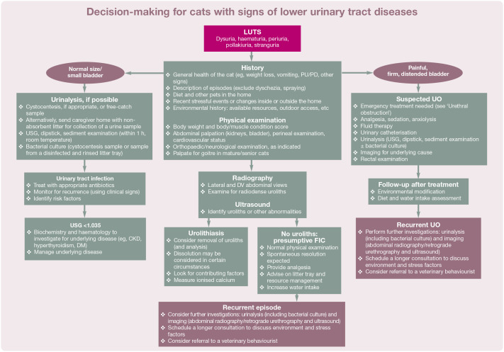
Practical relevance: Lower urinary tract signs (LUTS) such as dysuria, haematuria, periuria, pollakiuria and stranguria can occur as the result of a variety of underlying conditions and diagnostic investigation is required to uncover the underlying cause and select appropriate treatment.

Aim: The '2025 iCatCare consensus guidelines on the diagnosis and management of lower urinary tract diseases in cats' provide an overview of the common presenting signs caused by underlying feline lower urinary tract (LUT) diseases in cats, which often are indistinguishable between different underlying causes. The Guidelines set out a diagnostic approach to affected cats before focusing on the most common causes of LUTS: feline idiopathic cystitis (FIC), urolithiasis, urinary tract infection and urethral obstruction. The aim is to provide practitioners with practical information on these problematic conditions.

Clinical challenges: The fact that LUTS are similar despite different underlying causes creates a diagnostic challenge. The most common cause of LUTS, FIC, is challenging to manage due to a complex pathogenesis involving organs outside the LUT. Urethral obstruction is a life-threatening complication of various underlying LUT diseases and recurrent LUTS can lead to relinquishment or euthanasia of affected cats.

Evidence base: These Guidelines have been created by a panel of experts brought together by International Cat Care (iCatCare) Veterinary Society (formerly the International Society of Feline Medicine [ISFM]). Information is based on the available literature, expert opinion and the panel members' experience.

Keywords: Urolithiasis; catheterisation; cystitis; stress; urethral obstruction; urinary tract infection; urine.


Conflict of interest statement

Conflict of interestMembers of the panel have received financial remuneration for providing educational material, speaking at conferences

Figures

Figure 1
Figure 1
Lower urinary tract signs in cats include dysuria, haematuria, periuria, pollakiuria and stranguria in various combinations, and are caused by a variety of underlying pathologies. Image courtesy of Samantha Taylor
Figure 2
Figure 2
Young Bengal cat adopting a standing position, with erect quivering tail, typical of urine spraying as a problem behaviour. Image courtesy of Linda Ryan
Figure 3
Figure 3
Overgrooming of the abdomen in a cat with idiopathic cystitis. Image courtesy of Samantha Taylor
Figure 4
Figure 4
Algorithm for the approach to cats with signs of lower urinary tract (LUT) diseases. CKD = chronic kidney disease; DM = diabetes mellitus; DV = dorsoventral; FIC = feline idiopathic cystitis; LUTS = lower urinary tract signs; PU/PD = polyuria/polydipsia; UO = urethral obstruction; USG = urine specific gravity. See relevant sections of the Guidelines for further information on many aspects listed in the algorithm, including emergency treatment of UO
Figure 5
Figure 5
Microscopic appearance of (a) haematuria, (b) struvite crystalluria, (c) calcium oxalate dihydrate crystalluria and (d) a bilirubin crystal. Images courtesy of (a-c) Francesco Cian and (d) lona Mayer (d)
Figure 6
Figure 6
Gentle, cat friendly interactions can facilitate conscious cystocentesis. Image courtesy of Richard Murgatroyd
Figure 7
Figure 7
Lateral abdominal radiograph showing radiopaque uroliths in the bladder and urethra of a cat. Image courtesy of Rachel Korman
Figure 8
Figure 8
Lateral radiograph of the caudal abdomen of a cat, with a region of the penis magnified. The faint long tubular thin mineral opacity (arrow) at the level of the penis is consistent with the os penis. Image courtesy of Lumbry Park Veterinary Specialists
Figure 9
Figure 9
A urinary catheter with attached syringe filled with contrast material placed in the distal penis. The catheter can be held in place with forceps; a swab placed between the forceps and prepuce can limit trauma. Image courtesy of Samantha Taylor
Figure 10
Figure 10
(a) A normal retrograde study and (b) a retrograde urethrogram revealing a stricture and urolith, in two male cats. Narrowing of the urethra, as indicated by the arrow in image (a), is a normal finding and should not be mistaken for a focal stenosis. In image (b), there is a stricture (white arrow) and a filling defect in a dilated section of the intrapelvic urethra (black arrow), consistent with a urolith. This cat later underwent a perineal urethrostomy and several calcium oxalate uroliths were removed. Images courtesy of (a) Lumbry Park Veterinary Specialists and (b) London Vet Specialists
Figure 11
Figure 11
Ultrasound image showing a cystolith with acoustic shadowing, along with a thickened bladder wall. Image courtesy of Samantha Taylor
Figure 12
Figure 12
Sagittal CT image from a 4-year-old cat presenting with incontinence. The green arrow shows a mineral material plug in the penile urethra that was causing a partial obstruction. Image courtesy of Samantha Taylor
Figure 13
Figure 13
Schematic diagram of chronic pain associated with feline idiopathic cystitis (FIC). FIC has long been thought to result from some toxins in the urine and/or a urothelial permeability defect, which leads to 'bottom-up' nociceptive input to the brain (solid arrows). 'Top-down' (dotted) arrows show how this activation of the central threat response system affects the bladder, causing signs of FIC. More recent studies have found that environmental threats can also result in signs of FIC via top-down input to the central threat response system. Adapted from Koban et al, courtesy of Tony Buffington
Figure 14
Figure 14
Calcium oxalate cystoliths removed from a Ragdoll via cystotomy. Image courtesy of Samantha Taylor
Figure 15
Figure 15
Bacteriuria can be identified on sediment examination but treatment should take into account the presence or absence of clinical signs. Image courtesy of Francesco Cian
Figure 16
Figure 16
Algorithm for the approach to cats with suspected urethral obstruction. CBC = complete blood count; FIC = feline idiopathic cystitis; GA = general anaesthesia; LUTS = lower urinary tract signs; MEMO = multimodal environmental modification; PCV = packed cell volume; POCUS = point-of-care ultrasound; TS = total solids; UTI = urinary tract infection
Figure 17
Figure 17
Ultrasound images demonstrating findings and changes relating to the caudal vena cava (CVC) in cats with differing volume status. (a,b) Hypovolaemic cats will have a flat/collapsed CVC (white arrows) that may or may not change between (a) expiration and (b) inspiration, depending on the severity of hypovolaemia. In this example the CVC does change significantly between expiration and inspiration. (c,d) Hypervolaemic cats will have a distended CVC (white arrows) that changes very little between (c) expiration and (d) inspiration. (e,f) Cats who are euvolaemic should have at least a 20% change in the diameter of the CVC between (e) expiration and (f) inspiration. In this example the change is around 40%. Images courtesy of Soren Boysen
Figure 18
Figure 18
Algorithm for the management of hyperkalaemia in cats with urethral obstruction. The inset box shows changes on an electrocardiogram (ECG) that are typical in cases of feline hyperkalaemia, including decreased to absent P waves, widened QRS complexes, shortened QT intervals and tall tented or enlarged reversed T waves. IM = intramuscularly; IV = intravenously. ECG image courtesy of Samantha Taylor
Figure 19
Figure 19
Sacrococcygeal epidural administration of local anaesthetics can provide an adequate means of pain management in cats with urethral obstruction. Leakage of spinal fluid is less likely at this location but can occur, as shown in this image. Image courtesy of Paulo Steagall
Figure 20
Figure 20
Gentle massage of the tip of the penis to remove urethral plugs and distal uroliths. Image courtesy of Rachel Korman
Figure 21
Figure 21
(a-d) Photographs and accompanying diagrams to illustrate the importance of moving the penis (and thus urethra) dorsally and caudally to straighten the naturally occurring 'S' bend in the urethra (a,b) and allow passage of the catheter (c,d). This is performed by gently moving the prepuce (arrow). Images courtesy of Soren Boysen
Figure 22
Figure 22
A lubricated tomcat catheter with attached syringe of saline is inserted into the cleansed urethral orifice of an obstructed cat. Using pulsatile flushes, the catheter is slowly advanced. Image courtesy of Rachel Korman
Figure 23
Figure 23
(a; top to bottom) Polypropylene catheter, polypropylene catheter with adjustable suture wings, Jackson cat catheter with stylet, polytetrafluoroethylene catheter, and (insert) polytetrafluoroethylene catheter used both for unblocking and as an indwelling catheter. (b) Polyvinyl catheter. (c) Olive tip catheters. Images courtesy of (a) Danielle Gunn-Moore and (b,c) Serge Chalhoub
Figure 24
Figure 24
An intravenous catheter (with stylet removed) can be useful for flushing the distal urethra. Image courtesy of Rachel Korman
Figure 25
Figure 25
(a) A red rubber catheter that has been advanced into the urinary bladder. Two stay sutures have been placed, one on each side of the perineum ('earring method'), and butterfly tape has been used to fix the catheter to the stay sutures. Two simple interrupted sutures secure the tape to the stay sutures. (b) An alternative method for securing a urinary catheter. In this case a 'Slippery Sam'-type catheter is secured with sutures to the prepuce and connected to a closed urine collection set using a 'Little Herbert' Luer lock connector. See text for discussion regarding choice of indwelling catheter. Images courtesy of (a) Serge Chalhoub and (b) Rachel Korman between the catheter and prepuce (or vagina) should be wiped with 0.05% chlorhexidine every 8 h or whenever contamination is seen. The following should be monitored:
Figure 26
Figure 26
Finger trap suture on a red rubber urinary catheter. The suture was started at the butterfly tape end, and continued downwards along the catheter for about 1-2 cm. Image courtesy Chantal McMillan
Figure 27
Figure 27
Urinary catheters (red rubber [a] and Mila [b]), each attached to a closed urinary collection system, have been secured to the cat's tail to avoid tension on the prepuce. Images courtesy of (a) Chantal McMillan and (b) Laura Jones
Figure 28
Figure 28
An indwelling catheter connected to the collection tubing with a swivel device, allowing the tubing to twist and avoid traction on the prepuce. Image courtesy of Rachel Korman
Figure 29
Figure 29
(a) A sterilised intravenous drip bag is used as an economic closed collection system. Note it is not in direct contact with the floor and disinfectant is available to wipe connections clean when emptying or disconnecting the system. The cage front is covered with a towel to offer the cat a hiding opportunity. (b) A commercially available urine collection bag that allows easy measurement of urine output and is likewise kept off the floor. Images courtesy of (a) Rachel Korman and (b) Lumbry Park Veterinary Specialists
Figure 30
Figure 30
A cystostomy tube placed in a cat with a proximal urethral rupture to provide urinary diversion. Image courtesy of Helen Kirkpatrick
Figure 31
Figure 31
Ultrasound image of a cat's bladder, showing a mass that was later confirmed to be an invasive urothelial carcinoma. Image courtesy of Rachel Korman
Figure 32
Figure 32
Intravenous urography revealing ectopic ureters in a cat with incontinence. Image courtesy of Rachel Korman
Figure 33
Figure 33
Urination outside the litter tray is a common problem behaviour that may or may not be due to lower urinary tract disease. Environmental changes are likely to be beneficial; for example, the litter tray in this image may be inadequate in size and an open tray could be trialled. Image courtesy of Rachel Korman
Figure 34
Figure 34
Summary of areas of focus for caregivers of cats with lower urinary tract disease. Adapted, with permission from Mikel Delgado
Figure 35
Figure 35
Options to allow resting and perching in elevated locations should be provided. Image courtesy of Mikel Delgado
Figure 36
Figure 36
A large litter tray made from a storage box with soft litter. Several such resources, placed in quiet locations and scooped daily, can encourage use. Image courtesy of Mikel Delgado
Figure 37
Figure 37
Outdoor latrine in a quiet area with a rakeable surface to encourage regular voiding.
Figure 38
Figure 38
Water intake should be encouraged for cats with any type of lower urinary tract disease; for example, using a water fountain. Image courtesy of Rachel Korman
Figure 39
Figure 39
Puzzle feeders can provide environmental enrichment and some types, such as the one pictured here, can be used with wet food for cats with lower urinary tract diseases. Image courtesy of Samantha Taylor
None
None
None
None
None
None
None
None
None
None
None
None
None
None
None
None
None
None
None
None
None

References

    1. O'Neill DG, Gunn-Moore D, Sorrell S, et al.. Commonly diagnosed disorders in domestic cats in the UK and their associations with sex and age. J Feline Med Surg 2023; 25. DOI: 10.1177/1098612X231155016. - DOI - PMC - PubMed
    1. Robinson NJ, Dean RS, Cobb M, et al.. Investigating common clinical presentations in first opinion small animal consultations using direct observation. Vet Rec 2015; 176: 463. DOI: 10.1136/vr.102751. - DOI - PMC - PubMed
    1. Lekcharoensuk C, Osborne CA, Lulich JP. Epidemiologic study of risk factors for lower urinary tract diseases in cats. J Am Vet Med Assoc 2001; 218: 1429-1435. - PubMed
    1. Westropp JL, Delgado M, Buffington CAT. Chronic lower urinary tract signs in cats: current understanding of patho-physiology and management. Vet Clin North Am Small Anim Pract 2019; 49: 187-209. - PubMed
    1. Osbaldiston GW, Taussig RA. Clinical report on 46 cases of feline urological syndrome. Vet Med Small Anim Clin 1970;65: 461-468. - PubMed

LinkOut - more resources