. 2025 Apr 2;33(4):1566-1583.
doi: 10.1016/j.ymthe.2025.02.013. Epub 2025 Feb 11.

A BPTF-specific PROTAC degrader enhances NK cell-based cancer immunotherapy

Affiliations

A BPTF-specific PROTAC degrader enhances NK cell-based cancer immunotherapy

Yunjia Li et al. Mol Ther. .

Abstract

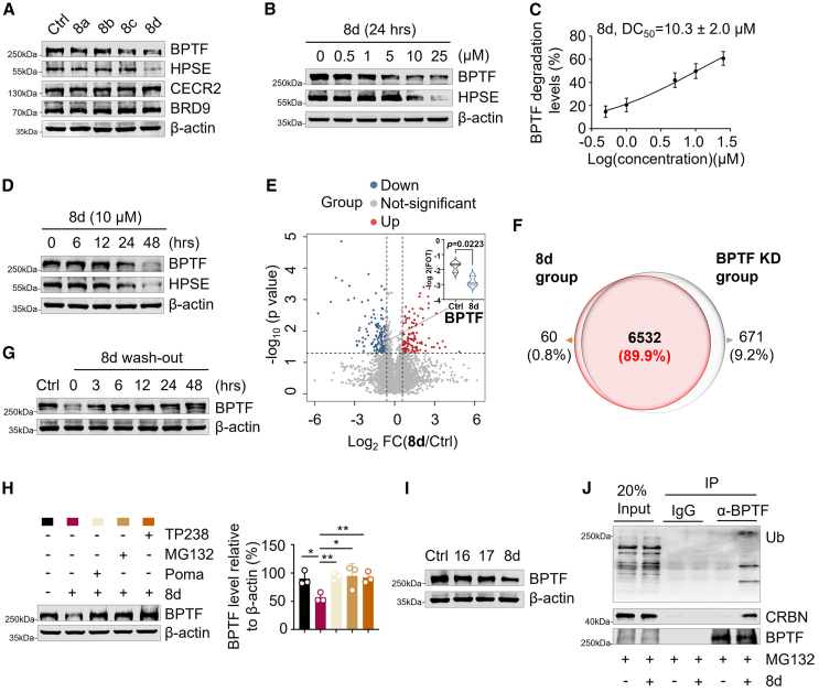
Natural killer (NK) cell-based immunotherapy shows promise in cancer treatment, but its efficacy remains limited, necessitating the development of novel strategies. In this study, we demonstrate that the epigenetic factor bromodomain PHD-finger containing transcription factor (BPTF) hinders hepatocellular carcinoma (HCC) recognition by NK cells through its PHD finger's interpretation of H3K4me3. We have generated a small-molecule proteolysis-targeting chimera (PROTAC) that selectively degrades human and murine BPTF. The degradation of BPTF using PROTACs directly enhances the abundance of natural cytotoxicity receptor ligands on HCC cells, facilitating their recognition by NK cells and thereby augmenting NK cell cytotoxicity against HCC both in vitro and in vivo. Through multidisciplinary techniques, our findings establish targeting BPTF with PROTACs as a promising approach to overcome immune evasion of HCC from NK cells and provide a new strategy to enhance NK cell-based cancer immunotherapy.

Keywords: PROTACs; epigenetic modification; hepatocellular carcinoma; immunotherapy; natural killer cells.


Conflict of interest statement

Declaration of interests The authors declare that they have no competing interests.

Figures

None
Graphical abstract
Figure 1
Figure 1
HPSE expression is regulated through BPTF’s recognition of H3K4me3 (A) Comparisons of the protein abundance of BPTF between adjacent normal tissue (N) samples and tumor (T) (paired t test) in the HCC patients’ cohort (n = 159). The line represents mean with SEM and upper and lower quartiles, respectively. The p value is depicted in the figure. (B) Volcano plot illustrating differentially expressed gene sets that are significantly down-regulated or up-regulated in Huh7 cells in response to BPTF silencing according to RNA-seq data; comparison between two biological replicates (n = 2). (C) RT-qPCR analysis showing changes in HPSE mRNA levels in Huh7 cells with or without KD of BPTF. The RT-qPCR signal was normalized by GAPDH (n = 3). (D) Genome browser view of CUT&Tag sequencing signal on the chromosome 4 segment, showing the localization of BPTF (depicted in red) and H3K4me3 (shown in blue) binding peaks at the promoter region of HPSE in Huh7 cells; and H3K4me3 (shown in pink) binding peaks at the promoter region of HPSE in BPTF KD Huh7 cells. (E) IP assay using anti-BPTF antibody and IgG control to confirm the endogenous association between BPTF with H3K4me3 in Huh7 cells. The displayed image is representative of three biological replicates. (F) CUT&Tag qPCR analysis using anti-BPTF antibody and IgG control to detect the enrichment of BPTF at the promoter region of HPSE in Huh7 cells (n = 3). (G) CUT&Tag qPCR analysis using anti-H3K4me3 antibody and IgG control to detect the enrichment of H3K4me3 at the promoter region of HPSE in Huh7 cells (n = 3). The data in (C), (F), and (G) were presented as mean ± SD; ∗p < 0.05, ∗∗p < 0.01, ∗∗∗p < 0.001, ∗∗∗∗p < 0.0001; unpaired t test.
Figure 2
Figure 2
Design and synthesis of BPTF-targeting PROTACs (A) The chemical structure of BPTF PROTACs. The BPTF PROTAC degraders (8a-d) are designed with a structure that involves the conjugation of a BPTF inhibitor (TP238) to an E3 ligase binder (Poma) via linkers of varying PEG lengths. The TP238, E3 ligase binder, and linkers are highlighted in blue, red, and black respectively. (B) Binding energy of four different linker systems. T, E, and P symbolize the target protein, E3 ligase, and PROTAC molecule, respectively. The error bar was calculated as the standard deviation between different frames in a single molecular dynamics simulation system. (C) CRBN-PROTAC-BPTF ternary structure of the best-ranked 8d, in which the chemical structure of PROTAC was highlighted. CRBN was shown cyan and BPTF was shown green. The highlighted PROTAC molecule was represented using multiple colors, with TP238 in yellow, Poma in blue, and linker in orange. (D) Scheme of the action model for the designed BPTF PROTAC degrader. The PROTAC degrader recruits E3 ligase to induce ubiquitination and subsequent proteasomal degradation of BPTF.
Figure 3
Figure 3
8d selectively degrades BPTF in a proteasome-dependent manner (A) Immunoblotting of BPTF, HPSE, CECR2 and BRD9 in the total cell lysates. The lysates were isolated from Huh7 cells treated with BPTF PROTAC degraders (8a-d) at 10 μM for 24 h, respectively. β-Actin was used as the loading control. The displayed image is representative of three biological replicates. (B) Immunoblotting of BPTF and HPSE in the total cell lysates. The lysates were isolated from Huh7 cells treated with 8d at the indicated doses (0, 0.5, 1, 5, 10, and 25 μM) for 24 h β-actin was used as the loading control. The displayed image is representative of three biological replicates. (C) Degradation curves of BPTF in 8d-treated Huh7 cells. The expression level of BPTF was normalized to the level of β-actin. n = 3. (D) Immunoblotting of BPTF and HPSE in the total cell lysates. The lysates were isolated from Huh7 cells treated with 8d at 10 μM for the indicated times (0, 6, 12, 24, and 48 h). β-Actin was used as the loading control. The displayed image is representative of three biological replicates. (E) Volcanic plot illustrating the altered proteins between control and 8d-treated Huh7 cells. The significantly downregulated and upregulated proteins are represented by blue and red dots, respectively. (Two-sided paired t test, p < 0.05, FC > 1.5). (F) Venn diagram showing the similarities and differences between 8d-treated and BPTF KD Huh7 cell groups. (G) Immunoblotting of BPTF in the total cell lysates. The lysates were isolated from Huh7 cells that were pretreated with 8d (10 μM) for 24 h followed by wash-out for the indicated times. β-Actin was used as the loading control. The displayed image is representative of three biological replicates. (H) Immunoblot analysis (left) and quantification (right, n = 3) of BPTF in the total cell lysates. The lysates were isolated from Huh7 cells that were respectively pretreated with Poma (1 μM), MG132 (1 μM), and TP238 (10 μM) for 1 h before 8d treatment (10 μM for 24 h). β-Actin was used as the loading control. The displayed image is representative of three biological replicates. The data was presented as mean ± SD; ∗p < 0.05, ∗∗p < 0.01, ∗∗∗p < 0.001, ∗∗∗∗p < 0.0001; Unpaired t test. (I) Immunoblotting of BPTF in the total cell lysates. The lysates were isolated from Huh7 cells that were treated with the deactivated degraders 16, 17 and PROTAC degrader 8d (10 μM for 24 h), respectively. β-Actin was used as the loading control. The displayed image is representative of three biological replicates. (J) IP assay using anti-BPTF and IgG control to detect the endogenous association of BPTF with CRBN (E3 ligase) and the ubiquitination level of BPTF in Huh7 cells pretreated with MG132 (1 μM) for 3 h before 8d treatment (10 μM for 24 h). The displayed image is representative of three biological replicates.
Figure 4
Figure 4
The effector function of human primary NK cells toward Huh7 cells is enhanced by 8d treatment (A) Flow cytometry analysis (left) and quantification (right, n = 3) showing the percentage of Annexin V+ Huh7 cells (target cells) in a co-culture with human primary NK cells (effector cells) for 4 h at an effector/target ratio of 1:20. The control Huh7 cells, 16-treated Huh7 cells, 17-treated Huh7 cells, TP238-treated Huh7 cells and 8d-treated Huh7 cells were included in the experiment. (B) Immunoblotting of HSPG in the total cell lysates. The lysates were obtained from Huh7 cells treated with TP238 (10 μM) or 8d (10 μM) for 24 h β-actin served as the loading control. The presented image is representative of three independent experiments. (C–E) Flow cytometry analysis (left) and quantification (right, n = 3) showing the expression of NKp30 (C), NKp46 (D), and NKG2D (E) in primary human NK cells. The NK cells were co-cultured with control Huh7 cells, TP238-treated Huh7cells, and 8d-treated Huh7 cells for a duration of 4 h. (F–I) Flow cytometry analysis (left) and quantification (right, n = 3) showing the proportion of CD69+ NK cells (F), Perforin+ GZMB+ NK cells (G), IFN-γ+ CD107a+ NK cells (H), and TNF-α+ CD107a+ NK cells (I) within the total population of primary human NK cells. The NK cells were co-cultured with control Huh7 cells, TP238-treated Huh7cells and 8d-treated Huh7 cells for a duration of 4 h. The data in (A) and (C–I) were presented as mean ± SD; ∗p < 0.05, ∗∗p < 0.01, ∗∗∗p < 0.001, ∗∗∗∗p < 0.0001; unpaired t test.
Figure 5
Figure 5
The effector function of primary human NK cells toward primary HCC cells isolated from the tumor tissue of HCC patients is enhanced by 8d treatment (A) Immunoblotting of the expression of BPTF and HPSE in the lysates extracted from primary HCC cells isolated from the tumor tissue of HCC patients. The primary HCC cells were treated with or without 8d (10 μM for 24 h) before lysate extraction. β-Actin was used as the loading control. The presented image is representative of three biological replicates. (B and C) Quantification of the expression level of BPTF (B) and HPSE (C) depicted in (A). The expression levels of BPTF and HPSE were normalized to the levels of β-actin. n = 3. (D) Immunoblotting of the expression of HSPG (left) and quantification (right, n = 4) in the lysates extracted from primary HCC cells isolated from tumor tissue of HCC patients. The primary HCC cells were subjected to treatment with or without 8d (10 μM for 24 h) before lysate extraction. β-Actin was used as the loading control. The image presented is representative of four biological replicates. (E) Flow cytometry analysis (left) and quantification (right, n = 3) showing the percentage of AnnexinV+ primary HCC cells (target cells) in a co-culture with primary human NK cells (effector cells) for 4 h, with a ratio of effector/target at 1:20. The control and 8d-treated primary HCC cells were included in the experiment. (F–H) Flow cytometry analysis (left) and quantification (right, n = 4) showing the expression of NKp30 (F), NKp46 (G), and NKG2D (H) on primary human NK cells following a 4-h co-culture with either control or 8d-treated primary HCC cells. (I–L) Flow cytometry analysis (left) and quantification (right, n = 4) showing demonstrated the proportion of CD69+ NK cells (I), Perforin+GZMB+ NK cells (J), IFN-γ+CD107a+ NK cells (K), and TNF-α+CD107a+ NK cells (L) within the total population of primary human NK cells after a 4-h co-culture with either control or 8d-treated primary HCC cells. The data in (B–L) are presented as mean ± SD; ∗p < 0.05, ∗∗p < 0.01, ∗∗∗p < 0.001, ∗∗∗∗p < 0.0001; paired t test.
Figure 6
Figure 6
Proteomic profiling reveals the effect of 8d on primary HCC cells isolated from tumor tissues of HCC patients (A) Heatmap displays the normalized protein expression levels in primary HCC cells treated with control (yellow) or 8d (pink), (n = 3). Each column represents one individual sample. (B and C) The pathway enrichment analysis comparing control and 8d-treated primary HCC cells, which is enriched with up-regulated (B) or down-regulated (C) proteins. EGF, epidermal growth factor; FGF, fibroblast growth factor; HIF, hypoxia-inducible growth factor; IGF, insulin-like growth factor; PDGF, platelet-derived growth factor; VEGF, vascular endothelial growth factor.
Figure 7
Figure 7
8d treatment prevents HCC outgrowth in vivo (A) Treatment schedule of 8d in the orthotopic HCC mouse model, both in the presence of NK cells and after their depletion by applying anti-mouse NK1.1 antibody. (B) Representative images of HCC on day 21 from four different groups: HCC mice with NK cell depletion treated with vehicle (first row), HCC mice with NK cell depletion treated with 8d (second row), control HCC mice (third row), and HCC mice treated with 8d (fourth row). n = 5 for each group. The arrowheads indicate the area where the tumor is growing. (C) Quantification of liver/body weight ratio in mice from the four groups shown in (B) at autopsy on day 21. n = 5. (D–G) Immunoblot analysis and quantification (n = 4) of the expression of BPTF (D and E), HPSE (D and F), and HSPG (D and G) in the lysates extracted from the liver tissues of HCC mice in the control and 8d groups. β-Actin was used as the loading control. (H) Representative images (left) and quantification (right, n = 4) of immunohistochemistry (IHC) staining of BPTF and HPSE in the liver tissue sections of HCC mice from the four groups shown in B at autopsy on day 21. Scale bars, 50 μm. I-N. Flow cytometry analysis (left) and quantification (right, n = 5) of the expression of NKG2D (I), NKp46 (J), GZMB (K), Perforin (L), IFN-γ+ TNF-α+ (M), and CD107a+ (N) on NK cells isolated from the liver tissues of HCC mice treated with control or 8d. The data in (C) and (E–N) are presented as mean ± SD; ∗p < 0.05, ∗∗p < 0.01, ∗∗∗p < 0.001, ∗∗∗∗p < 0.0001; unpaired t test.

References

    1. Llovet J.M., Castet F., Heikenwalder M., Maini M.K., Mazzaferro V., Pinato D.J., Pikarsky E., Zhu A.X., Finn R.S. Immunotherapies for hepatocellular carcinoma. Nat. Rev. Clin. Oncol. 2022;19:151–172. doi: 10.1038/s41571-021-00573-2. - DOI - PubMed
    1. Sangro B., Sarobe P., Hervás-Stubbs S., Melero I. Advances in immunotherapy for hepatocellular carcinoma. Nat. Rev. Gastroenterol. Hepatol. 2021;18:525–543. doi: 10.1038/s41575-021-00438-0. - DOI - PMC - PubMed
    1. Riley R.S., June C.H., Langer R., Mitchell M.J. Delivery technologies for cancer immunotherapy. Nat. Rev. Drug Discov. 2019;18:175–196. doi: 10.1038/s41573-018-0006-z. - DOI - PMC - PubMed
    1. Sun C., Sun H.Y., Xiao W.H., Zhang C., Tian Z.G. Natural killer cell dysfunction in hepatocellular carcinoma and NK cell-based immunotherapy. Acta Pharmacol. Sin. 2015;36:1191–1199. doi: 10.1038/aps.2015.41. - DOI - PMC - PubMed
    1. Kyrysyuk O., Wucherpfennig K.W. Designing Cancer Immunotherapies That Engage T Cells and NK Cells. Annu. Rev. Immunol. 2023;41:17–38. doi: 10.1146/annurev-immunol-101921-044122. - DOI - PMC - PubMed

MeSH terms

Substances

LinkOut - more resources