. 2025 Jan 10;28(2):111790.
doi: 10.1016/j.isci.2025.111790. eCollection 2025 Feb 21.

Spatial transcriptomic analysis identifies epithelium-macrophage crosstalk in endometriotic lesions

Affiliations

Spatial transcriptomic analysis identifies epithelium-macrophage crosstalk in endometriotic lesions

Gregory W Burns et al. iScience. .

Abstract

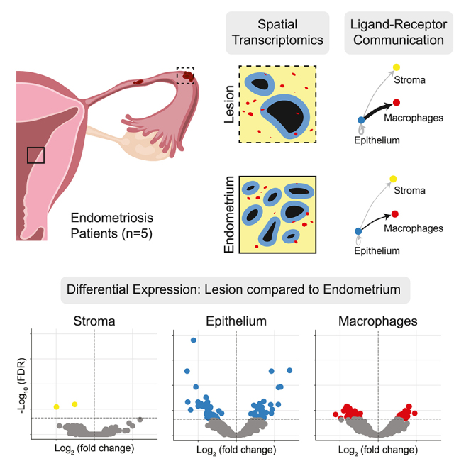
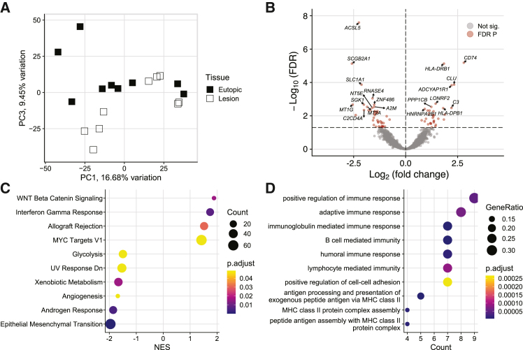
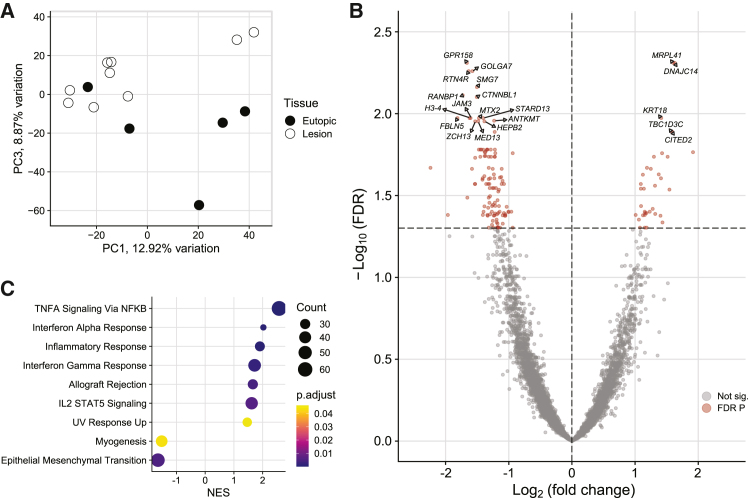
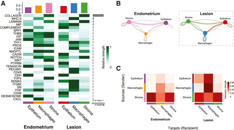
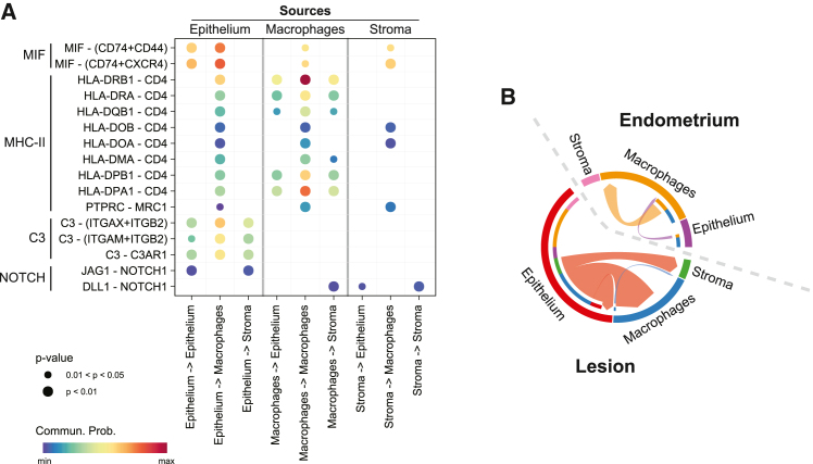
The mechanisms underlying the pathophysiology of endometriosis, characterized by the presence of endometrium-like tissue outside the uterus, remain poorly understood. This study aimed to identify cell type-specific gene expression changes in superficial peritoneal endometriotic lesions and elucidate the crosstalk among the stroma, epithelium, and macrophages compared to patient-matched eutopic endometrium. Surprisingly, comparison between lesions and eutopic endometrium revealed transcriptional similarities, indicating minimal alterations in the sub-epithelial stroma and epithelium of lesions. Spatial transcriptomics highlighted increased signaling between the lesion epithelium and macrophages, emphasizing the role of the epithelium in driving lesion inflammation. We propose that the superficial endometriotic lesion epithelium orchestrates inflammatory signaling and promotes a pro-repair phenotype in macrophages, providing a new role for complement 3 in lesion pathobiology. This study underscores the significance of considering spatial context and cellular interactions in uncovering mechanisms governing disease in endometriotic lesions.

Keywords: integrative aspects of cell biology; organizational aspects of cell biology; reproductive medicine; transcriptomics.


Conflict of interest statement

The authors declare no competing interests.

Figures

None
Graphical abstract
Figure 1
Figure 1
Segmentation strategy for spatial transcriptomics (A) Superficial peritoneal endometriotic lesions and matched endometrium (n = 10) were visualized with trichrome staining to confirm the presence of epithelial and stromal compartments. Representative images of selected samples are shown. (B) Tissues were immunostained for pan-cytokeratin, CD68, and ACTA2 for segmentation and spatial transcriptome collection on the GeoMx platform. Representative regions of interest (ROI) are shown for each tissue type. Duplicate ROI were collected for each tissue and stroma, epithelium, and macrophages were segmented from each for a total of 20 ROI and 60 segments. Scale bar, 100 μm.
Figure 2
Figure 2
Spatial transcriptomics results from eutopic endometrium superficial peritoneal endometriotic lesions (A) Bar plot of all 60 segments separated by gene detection rate. Segments with a detection rate of less than 10% were removed. (B) Confirmation of gene and negative probe count separation by segment in a scatterplot and by distribution for each cell type in histograms. Separation after gene and segment filtering is required for effective Q3 normalization. (C) Bar plot of detectable genes per segment used for filtering genes found in less than 10% of segments leaving 7,945 genes for further analysis. (D) Dot plot of marker genes for epithelium (n = 9), stroma (n = 6), and macrophages (n = 6) confirming transcriptomic separation between cell types. See also Figures S1 and S2.
Figure 3
Figure 3
Endometriotic sub-epithelial stroma is minimally altered compared to eutopic endometrium (A) Principal component (PC) plot of lesion stroma compared to eutopic endometrium showed group separation most clearly on PC3. (B) Volcano plot of two down-regulated genes in the endometriotic stroma. (C) Gene set enrichment analysis of hallmark pathways found four increased inflammatory gene sets and two decreased.
Figure 4
Figure 4
Immune response genes are increased in the endometriotic lesion epithelium (A) Principal component (PC) plot of lesion epithelium compared to eutopic endometrium showed group separation most clearly on PC3. (B) Volcano plot of 76 differentially expressed genes (DEG) in the endometriotic epithelium with 33 increased and 43 decreased. Of note, complement 3, C3, was one of the top increased DEG and several others were major histocompatibility complex class II related genes, like CD74 and HLA-DRB1. (C) Gene set enrichment analysis of hallmark pathways found four increased gene sets and six decreased, indicating increasedinflammation and proliferation. (D) Nine of the top 10 enriched gene ontology biological process terms were immune related.
Figure 5
Figure 5
Macrophages are altered in endometriotic lesions (A) Principal component (PC) plot of lesion macrophages compared to eutopic endometrium showed group separation most clearly on PC3. (B) Volcano plot of 148 differentially expressed genes (DEG) with 35 increased and 113 decreased in the endometriotic macrophages. (C) Gene set enrichment analysis of hallmark pathways found seven increased gene sets and two decreased, indicating increased inflammation likely reflective of the presence of monocyte-derived macrophages in the lesion tissue. See also Figure S5.
Figure 6
Figure 6
CellChat ligand-receptor analysis identified increased communication from the lesion epithelium to macrophages (A) Signaling pathway heatmap of all 29 significant pathways identified by CellChat sorted by signaling strength, indicated by the gray bar. Interactions, combined incoming and outgoing, totaled for each cell type in the bar plot at the top of the heatmap and scaled per row. (B) Circle plots of communication networks including 24 pathways in the endometrium and 26 in lesions where line thickness corresponds to strength. Stroma to epithelium communication was strong in the eutopic endometrium and similarly present in the lesions. However, epithelium to macrophage communication was increased 3.7-fold in lesions. (C) Interaction strength heatmaps highlight the gain of epithelium to macrophage signals in lesions and strong macrophage-macrophage interactions.
Figure 7
Figure 7
Inflammatory ligands originate in the endometriotic lesion epithelium (A) Ligand—receptor dot plot of inflammatory pathways in endometriotic lesions. Pathway names are indicated on the left side of the plot followed by specific ligand and receptor interactions. All signals originating from the lesion epithelium are in the left three columns, macrophages in the center, and stroma on the right. Complement and major histocompatibility complex class II (MHC-II) pathways originated in the lesion epithelium and not the stroma. Macrophage migration inhibitory factor (MIF) signaling was strongest in the epithelium, but also sent from the stroma and macrophage cell types. (B) Complement signaling chord diagram of the weighted complement signaling pathway indicating increased signaling from the lesion epithelium compared to the eutopic endometrium epithelium.

Update of

References

    1. Eskenazi B., Warner M.L. Epidemiology of endometriosis. Obstet. Gynecol. Clin. North Am. 1997;24:235–258. - PubMed
    1. Bulun S.E. Endometriosis. N. Engl. J. Med. 2009;360:268–279. doi: 10.1056/NEJMra0804690. - DOI - PubMed
    1. Giudice L.C. Clinical practice. Endometriosis. N. Engl. J. Med. 2010;362:2389–2398. doi: 10.1056/NEJMcp1000274. - DOI - PMC - PubMed
    1. Practice Committee of American Society for Reproductive Medicine Treatment of pelvic pain associated with endometriosis. Fertil. Steril. 2008;90:S260–S269. doi: 10.1016/j.fertnstert.2008.08.057. - DOI - PubMed
    1. Practice Committee of the American Society for Reproductive Medicine Endometriosis and infertility: a committee opinion. Fertil. Steril. 2012;98:591–598. doi: 10.1016/j.fertnstert.2012.05.031. - DOI - PubMed

LinkOut - more resources