Review
. 2025 Jan 28:12:1478549.
doi: 10.3389/fcell.2024.1478549. eCollection 2024.

Synthetic embryology of the human heart

Affiliations
Review

Synthetic embryology of the human heart

Maria Belen Paredes-Espinosa et al. Front Cell Dev Biol. .

Abstract

The evolution of stem cell-based heart models from cells and tissues to organoids and assembloids and recently synthetic embryology gastruloids, is poised to revolutionize our understanding of cardiac development, congenital to adult diseases, and patient customized therapies. Human induced pluripotent stem cell-derived cardiomyocytes (hiPSC-CMs) have already been integrated into transplantable patches and are in preclinical efforts to reverse fibrotic scarring from myocardial infarctions. To inform on the complexity of heart diseases, multi-tissue morphogenic heart models are needed that replicate fundamental components of heart function to heart organogenesis in vitro and which require a deep understanding of heart development. Organoid and assembloid models capture selected multicellular cardiac processes, such as chamber formation and priming events for vascularization. Gastruloid heart models offer deeper insights as synthetic embryology to mimic multi-staged developmental events of in vivo heart organogenesis including established heart fields, crescent formation and heart tube development along with vascular systemic foundation and even further steps. The human Elongating Multi-Lineage Organized Cardiac (EMLOC) gastruloid model captures these stages and additional events including chamber genesis, patterned vascularization, and extrinsic central and intrinsic cardiac nervous system (CNS-ICNS) integration guided by spatiotemporal and morphogenic processes with neural crest cells. Gastruloid synthetic embryology heart models offer new insights into previously hidden processes of development and provide powerful platforms for addressing heart disease that extends beyond cardiomyocytes, such as arrhythmogenic diseases, congenital defects, and systemic injury interactions, as in spinal cord injuries. The holistic view that is emerging will reveal heart development and disease in unprecedented detail to drive transformative state-of-the-art innovative applications for heart health.

Keywords: ICNS; assembloid; cardiogenesis; gastruloid; hiPSC; neurons; organogenesis; organoid.


Conflict of interest statement

The research was conducted as part of the aforementioned CATN2 award between the Paluh lab and Cytocybernetics, Inc a New York business with expertise that includes cardiac electrophysiology.

Figures
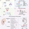
FIGURE 1
FIGURE 1
(A) Formation of the cardiac crescent from the cardiogenic mesoderm, including the first heart field (FHF) and second heart field (SHF). (B) Formation of the heart tube from the arterial pole to the venous pole, including the primitive ventricle, atria, and the proepicardial organ, with the endocardium, cardiac jelly, and myocardium layers. (C) Process of heart looping. (D) Septation and the formation of heart chambers, including the outflow tract (OFT) cushions, atrioventricular (AVC) cushions, right ventricle (RV), left ventricle (LV), and trabecula. (E) Differentiation of cardiac neural crest cells (cNCCs), proepicardium, and cardiac mesoderm into various cardiac cells and their neural integration. Created with BioRender.
FIGURE 2
FIGURE 2
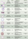
Overview of cardiac organoid and gastruloid models. In vitro 3D heterogenous cell culture systems include most advanced gastruloids as models of synthetic embryology, and partial organogenesis models depicted in organoids, and assembloids. Created with BioRender.
FIGURE 3
FIGURE 3
Stages of human heart development alongside corresponding advanced cardiac stem cell models including synthetic embryology approaches. (A) heart tube formation in human heart development, and corresponding, cardiac stem cell models by Rossi et al. (2021) (mouse) a synthetic embryology approach and Volmert et al. (2023) depicting endocardium-like structures, gut tube-like formations, and vascular networks that mimic heart tube-like structures. (B) Heart looping, septation, and chamber formation in human heart development, and stem cell models by Drakhlis et al. (2021) and Lewis-Israeli et al. (2021) showing mesenchymal cells, septum-transversum-like cells, endocardial-like cells, and chamber structures. (C) Cardiac cell differentiation and neural integration in human heart development, showing the differentiation of cardiac neural crest cells (cNCCs) and their integration with cardiac mesoderm and proepicardium, forming various cardiac cells and nerves and corresponding neuronal integration models by Olmsted and Paluh (2022) exemplifying synthetic embryology through neuro-cardiac integration and sympathetic innervation and Zeltner et al. (2024) highlighting neural rosettes, and sympathetic innervation in cardiac chambers. Created with BioRender.

References

    1. Acharya A., Nemade H., Papadopoulos S., Hescheler J., Neumaier F., Schneider T., et al. (2022). Microgravity-induced stress mechanisms in human stem cell-derived cardiomyocytes. iScience 25, 104577. 10.1016/j.isci.2022.104577 - DOI - PMC - PubMed
    1. Ameen M., Sundaram L., Shen M., Banerjee A., Kundu S., Nair S., et al. (2022). Integrative single-cell analysis of cardiogenesis identifies developmental trajectories and non-coding mutations in congenital heart disease. Cell. 185, 4937–4953.e23. 10.1016/j.cell.2022.11.028 - DOI - PMC - PubMed
    1. Arnold S. J., Robertson E. J. (2009). Making a commitment: cell lineage allocation and axis patterning in the early mouse embryo. Nat. Rev. Mol. Cell. Biol. 10, 91–103. 10.1038/nrm2618 - DOI - PubMed
    1. Asp M., Giacomello S., Larsson L., Wu C., Fürth D., Qian X., et al. (2019). A spatiotemporal organ-wide gene expression and cell atlas of the developing human heart. Cell 179, 1647–1660.e19. 10.1016/j.cell.2019.11.025 - DOI - PubMed
    1. Blinova K., Dang Q., Millard D., Smith G., Pierson J., Guo L., et al. (2018). International multisite study of human-induced pluripotent stem cell-derived cardiomyocytes for drug proarrhythmic potential assessment. Cell. Rep. 24, 3582–3592. 10.1016/j.celrep.2018.08.079 - DOI - PMC - PubMed

LinkOut - more resources