Review
. 2025 Jan 28:13:1532050.
doi: 10.3389/fcell.2025.1532050. eCollection 2025.

Mechanisms and roles of membrane-anchored ATG8s

Affiliations
Review

Mechanisms and roles of membrane-anchored ATG8s

Soo-Kyeong Lee et al. Front Cell Dev Biol. .

Abstract

Autophagy-related protein 8 (ATG8) family proteins, including LC3 and GABARAP subfamilies, are pivotal in canonical autophagy, driving autophagosome formation, cargo selection, and lysosomal fusion. However, recent studies have identified non-canonical roles for lipidated ATG8 in processes such as LC3-associated phagocytosis (LAP), LC3-associated endocytosis (LANDO), and lipidated ATG8-mediated secretory autophagy. These pathways expand ATG8's functional repertoire in immune regulation, membrane repair, and pathogen clearance, as ATG8 becomes conjugated to single-membrane structures (e.g., phagosomes and lysosomes). This review examines the molecular mechanisms of ATG8 lipidation, focusing on its selective conjugation to phosphatidylethanolamine (PE) in autophagy and phosphatidylserine (PS) in CASM. We highlight LIR-based probes and LC3/GABARAP-specific deconjugases as critical tools that allow precise tracking and manipulation of ATG8 in autophagic and non-autophagic contexts. These advancements hold therapeutic promise for treating autophagy-related diseases, including cancer and neurodegenerative disorders, by targeting ATG8-driven pathways that maintain cellular homeostasis.

Keywords: LAP; LC3/GABARAP; LIR motif; Lando; autophagy; deconjugase; non-canonical autophagy; probe.


Conflict of interest statement

The authors declare that the research was conducted in the absence of any commercial or financial relationships that could be construed as a potential conflict of interest.

Figures
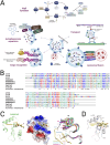
FIGURE 1
FIGURE 1
Function, classification and structure of mATG8 family proteins in autophagy. (A) A schematic illustration of the mATG8 lipidation process and the functions of lipidated mATG8 proteins in autophagosomal membranes. Pro-mATG8 proteins are cleaved by ATG4 enzymes to form cytosolic mATG8 (Type I), which is then conjugated to phosphatidylethanolamine (PE) through a series of enzyme actions: E1 (ATG7), E2 (ATG3), and E3 (the ATG5–ATG12–ATG16L1 complex), converting it into lipidated mATG8 (Type II). Lipidated LC3/GABARAP proteins perform multiple functions, including autophagosome expansion, transport, cargo recognition, and lysosomal fusion, by providing accessible sites on the autophagosomal membrane for autophagy machinery proteins. They bind to LIR-containing receptors on cargo destined for degradation. (B) Sequence alignment of LC3/GABARAP and fungal Atg8 proteins. (C) The crystal structure of the LIR (TP)-GABARAP fusion was prepared by combining GABARAP and LIR from different fusion molecules interacting within the crystal. The structure displays a ribbon model on the left and electrostatic surface potentials with a ribbon model of LIR peptides in the center, highlighting interacting LIR residues. On the right, a structural comparison of the α2 helix across LC3/GABARAP proteins is shown, using Cα atoms for superimposition. The following PDB IDs were used: LC3A, 5CX3; LC3B, 5D94; LC3C, 3VVW; GABARAP, 1GNU; GABARAP-L1, 6HOI; and GABARAP-L2, 4CO7. (D) A modeled structure of the LIR [TP(T)]-LC3C complex is also shown, with potential electrostatic or hydrogen-bond interactions represented by a stick model.
FIGURE 2
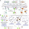
FIGURE 2
Canonical and Non-Canonical Autophagy Pathways: Canonical Autophagy and CASM. (A) Autophagy initiation requires the ULK1 kinase complex, regulated by AMPK and mTOR as activator and inhibitor, respectively. During the expansion phase, AMPK phosphorylates ULK1, which includes FIP200, ATG13, and ATG101, thereby activating the class III PI3K complex composed of BECN1, AMBRA1, ATG14L, VPS15, and VPS34. This complex generates phosphatidylinositol 3-phosphate (PI3P), promoting phagophore elongation. Within the phagophore, mATG8 proteins recruit cargo for degradation. mATG8 is cleaved by ATG4 and lipidated by the combined actions of ATG7, ATG3, and the ATG5-12-16L1 complex, anchoring it to autophagosomal membranes. The mature, closed autophagosomes are transported via SNARE proteins to actin filaments and ultimately fuse with lysosomes through mATG8s, enabling degradation. (B) LC3-Associated Phagocytosis (LAP) process is initiated in murine macrophages through interactions between extracellular components and receptors, including Dectin (a phagocytic receptor), Fc receptors (FcRs), and Toll-like receptors (TLRs). Unlike canonical autophagy, LAP formation does not require the ULK1 complex. Upon phagocytosis, PI3P is generated by the VPS34 complex on the single phagosomal membrane, activating the NADPH oxidase complex (NOX). NOX activation leads to ROS production within the phagosomal lumen, raising pH levels. This high pH then promotes the assembly of V-ATPase on the phagosomal membrane. V-ATPase interacts with the WD40 domain of ATG16L1, subsequently recruiting the ATG5-ATG12-ATG16L1 complex, which facilitates the conjugation of LC3 or GABARAP to phosphatidylserine (PS) and phosphatidylethanolamine (PE) on the phagosomal membrane. The mature mATG8-conjugated phagosome is ultimately degraded through lysosomal fusion. (C) LC3-Associated Endocytosis (LANDO) is linked with recycling membrane receptors, such as TLRs, TREM, and CD36, and plays a role in regulating inflammation within the immune system. Similar to LAP, LANDO does not require the ULK1 complex. Cytosolic endosomes marked by Rab5 interact with the VPS34 complex to generate PI3P on the single-membrane endosome. PI3P enables the conjugation of mATG8 proteins, such as LC3 and GABARAP, through the ATG5-ATG12-ATG16L1 complex. This process facilitates endosome fusion with lysosomes for degradation or recycling via the ATG8-mediated endosomal pathway. (D) LC3-Dependent EV Loading and Secretion (LDELS) is a regulated pathway for degrading proteins, RNAs, and lipids through extracellular vesicles (EVs). EVs, derived from multivesicular bodies (MVBs), carry cargos recognized by mATG8 proteins, which also facilitate the secretion of LC3-dependent EVs.
FIGURE 3
FIGURE 3
Mechanism of LIR-Based Probes and LC3/GABARAP Deconjugases, and Their Applications in Research and Therapy. (A) Schematic diagram of LIR based probes and deconjugases: The HyD-LIR probe illustrated on the left effectively detects mATG8s present on autophagosomal membranes through the synergy of a short hydrophobic domain and LIR. The MT-LIR probe shown in the middle of the picture contains the MT domain of RavZ, which binds PI3P, and selective LIRs on both terminals, allowing it to specifically bind to specific mATG8. The deconjugase on the right consists of the catalytic domain of RavZ and selective LIRs that irreversibly cleave specific mATG8 on autophagosomal membranes. LIR-based probes are commonly used to visualize autophagosomes by weak and transient expression. On the other hand, continuous and overexpression of the probes inhibits autophagy by blocking the binding of mATG8 to other autophagy-related proteins in the autophagosomal membranes. The deconjugases can be expressed to remove specific mATG8 proteins from the autophagosomal membranes to form special situations where LC3s or GABARAPs are lacking, which are different from mATG8 knock-out cells. (B) Considerations for the use of LIR-based probes and deconjugases. Probes and deconjugases are used across the entire process of conventional and selective autophagy. The utilisation of probes and deconjugases in the extended functions of mATG8 proteins conjugated to a single membrane (LAP, LANDO and LDELS) is useful for studying the contribution and importance of mATG8 proteins in each pathway, and further enables new studies of mATG8.

References

    1. Alemu E. A., Lamark T., Torgersen K. M., Birgisdottir A. B., Larsen K. B., Jain A., et al. (2012). ATG8 family proteins act as scaffolds for assembly of the ULK complex: sequence requirements for LC3-interacting region (LIR) motifs. J. Biol. Chem. 287 (47), 39275–39290. 10.1074/jbc.M112.378109 - DOI - PMC - PubMed
    1. Birgisdottir Å. B., Mouilleron S., Bhujabal Z., Wirth M., Sjøttem E., Evjen G., et al. (2019). Members of the autophagy class III phosphatidylinositol 3-kinase complex I interact with GABARAP and GABARAPL1 via LIR motifs. Autophagy 15 (8), 1333–1355. 10.1080/15548627.2019.1581009 - DOI - PMC - PubMed
    1. Boyle K. B., Ellison C. J., Elliott P. R., Schuschnig M., Grimes K., Dionne M. S., et al. (2023). TECPR1 conjugates LC3 to damaged endomembranes upon detection of sphingomyelin exposure. EMBO J. 42 (17), e113012. 10.15252/embj.2022113012 - DOI - PMC - PubMed
    1. Carosi J. M., Nguyen T. N., Lazarou M., Kumar S., Sargeant T. J. (2021). ATG8ylation of proteins: a way to cope with cell stress? J. Cell Biol. 220 (11), e202108120. 10.1083/jcb.202108120 - DOI - PMC - PubMed
    1. Chen J. J., Nathaniel D. L., Raghavan P., Nelson M., Tian R., Tse E., et al. (2019). Compromised function of the ESCRT pathway promotes endolysosomal escape of tau seeds and propagation of tau aggregation. J. Biol. Chem. 294 (50), 18952–18966. 10.1074/jbc.RA119.009432 - DOI - PMC - PubMed

LinkOut - more resources