. 2025 Feb 12;45(2):BSR20241133.
doi: 10.1042/BSR20241133.

Dynamical organization of vimentin intermediate filaments in living cells revealed by MoNaLISA nanoscopy

Affiliations

Dynamical organization of vimentin intermediate filaments in living cells revealed by MoNaLISA nanoscopy

Mariano Smoler et al. Biosci Rep. .

Abstract

Intermediate filaments are intimately involved in the mechanical behavior of cells. Unfortunately, the resolution of optical microscopy limits our understanding of their organization. Here, we combined nanoscopy, single-filament tracking, and numerical simulations to inspect the dynamical organization of vimentin intermediate filaments in live cells. We show that a higher proportion of peripheral versus perinuclear vimentin pools are constrained in their lateral motion in the seconds time window, probably due to their cross-linking to other cytoskeletal networks. In a longer time scale, active forces become evident and affect similarly both pools of filaments. Our results provide a detailed description of the dynamical organization of the vimentin network in live cells and give some cues on its response to mechanical stimuli.

Keywords: MoNaLISA nanoscopy; cytoskeleton; vimentin filaments.


Conflict of interest statement

The authors declare no competing interests.

Figures
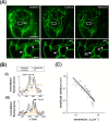
Figure 1:
Figure 1:. Determination of the apparent persistence length of vimentin filaments in living U2OS cells.
(A) Representative images of U2OS cells endogenously expressing rsEGFP2-vimentin acquired with widefield, confocal and MoNaLISA setups (upper panels). Zoom-in images of the regions included in the rectangles delimited in the whole-cell images (bottom panels). Scale bars: 10 µm and 1 µm, respectively. (B) Intensity profiles perpendicular to the main axis of close-by vimentin filaments (arrows); the filaments width (~ 80 nm) in the MoNaLISA image was determined by fitting a Lorentzian function (orange lines) to the intensity profiles (black dots). (C) Analysis of filament´s curvatures by a Fourier decomposition method. The continuous line corresponds to the fitting of equation 4 to the Fourier data.
Figure 2:
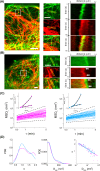
Figure 2:. The lateral motion of vimentin filaments followed a constrained diffusion model in a time window of seconds.
(A) Vimentin IFs were classified as perinuclear and peripheral (dotted squares:1 and 2, respectively) based on their location relative to the contour of the cell nucleus (magenta line) as described in the text. Scale bar: 10 µm. Zoom-in regions of the image, to facilitate the visualization of individual filaments. Scale bar: 2 µm. (B) Procedure for the quantitative analysis of single filament trajectories. The spatial coordinates of the segments were recovered in each frame of the movie to obtain the trajectory of its center of mass (YCM) and the MSDL. Scale bar: 2 µm. (C) MSDL curves obtained for perinuclear (magenta) and peripheral (light blue) vimentin filaments showing corralled motion. The data were individually fitted with equation 6 for τt < τtcorral (black line). (D) Proportion of corralled motion observed in each subcellular region.
Figure 3:
Figure 3:. The lateral motion of vimentin filaments followed anomalous diffusion in a time window of minutes.
(A-B) Representative Airyscan images of U2OS cells co-transfected with mCherry-vimentin (red) and EMTBx3GFP (A, green) or EGFP-actin (B, green). Scale bar: 2 µm. Zoom in regions (white squares, left panel) and kymographs (right panel). The arrows point contact sites between filaments. Scale bar: 2 µm. (C) MSDL curves presenting anomalous diffusion obtained for perinuclear (magenta) and peripheral (light blue) filament segments in log-log scales. Dotted lines display the expected behavior for sub diffusion with α=0.5 (bottom) and ballistic motion (α=2, top). DI values determined for these tracking data were < 10%. Inset: representative MSDL data fitted with equation 8 (continuous line). (D) Distributions of α and Dapp obtained by fitting individual MSDL data (left and middle panels) and their relation in log-log scale (right panel). Continuous lines correspond to the fitting of an allometric function, α=ADappB, where A and B are constants.
Figure 4:
Figure 4:. Numerical simulations of the lateral dynamics of vimentin filaments.
(A) Cartoon illustrating the simulated model. In a time scale of seconds, the absence of net external forces acting on vimentin filaments (green) generates displacements in a confined space (Δx1=Δx2) due to its connections to static cytoskeletal polymers (red). Active forces transmitted by the cytoskeleton become appreciable at longer periods of time and indirectly affect the mobility of the vimentin filaments (Δx1§gt;Δx2). The black arrow illustrates a net displacement of a cytoskeletal polymer in the horizontal direction. (B) Relation of α and Dapp parameters recovered from the simulations (green). Continuous lines represent the fitting of the simulated (green) and peripheral (light blue, Figure 3D) data with an allometric function α=ADappB, where A and B are constants.

References

    1. Mizuno D., Tardin C., Schmidt C.F., Mackintosh F.C. Nonequilibrium mechanics of active cytoskeletal networks. Science. 2007;315:370–373. doi: 10.1126/science.1134404. - DOI - PubMed
    1. Pegoraro A.F., Janmey P., Weitz D.A. Mechanical properties of the cytoskeleton and cells. Cold Spring Harb. Perspect. Biol. 2017;9:a022038. doi: 10.1101/cshperspect.a022038. - DOI - PMC - PubMed
    1. Chung B.M., Rotty J.D., Coulombe P.A. Networking galore: intermediate filaments and cell migration. Curr. Opin. Cell Biol. 2013;25:600–612. doi: 10.1016/j.ceb.2013.06.008. - DOI - PMC - PubMed
    1. Leduc C., Etienne-Manneville S. Intermediate filaments in cell migration and invasion: the unusual suspects. Curr. Opin. Cell Biol. 2015;32:102–112. doi: 10.1016/j.ceb.2015.01.005. - DOI - PubMed
    1. De Pascalis C., Pérez-González C., Seetharaman S., Boëda B., Vianay B., Burute M., et al. Intermediate filaments control collective migration by restricting traction forces and sustaining cell-cell contacts. J. Cell Biol. 2018;217:3031–3044. doi: 10.1083/jcb.201801162. - DOI - PMC - PubMed

Publication types

MeSH terms

LinkOut - more resources