. 2025 Feb 18;122(7):e2415287122.
doi: 10.1073/pnas.2415287122. Epub 2025 Feb 12.

Substance P receptor signaling contributes to host maladaptive responses during enteric bacterial infection

Affiliations

Substance P receptor signaling contributes to host maladaptive responses during enteric bacterial infection

Michael Cremin et al. Proc Natl Acad Sci U S A. .

Abstract

Immune responses in the intestine are intricately balanced to prevent pathogen entry without inducing immunopathology. The nervous system is well established to interface with the immune system to fine-tune immunity in various organ systems including the gastrointestinal tract. Specialized sensory neurons can detect bacteria, bacterial products, and the resulting inflammation, to coordinate the immune response in the gastrointestinal tract. These sensory neurons release peptide neurotransmitters such as Substance P (SP), to induce both neuronal signaling and localized responses in nonneuronal cells. With this in mind, we assessed the immunoregulatory roles of SP receptor signaling during enteric bacterial infection with the noninvasive pathogen Citrobacter rodentium. Pharmacological antagonism of the SP receptor significantly reduced bacterial burden and prevented colonic crypt hyperplasia. Mice with SP receptor signaling blockade had significantly reduced inflammation and recruitment of T cells in the colon. Reduced colonic T cell recruitment is due to reduced expression of adhesion molecules on colonic endothelial cells in SP receptor antagonist-treated mice. Using SP receptor T cell conditional knockout mice, we further confirmed SP receptor signaling enhanced select aspects of T cell responses. Our data demonstrate that SP receptor signaling can significantly reduce inflammation and prevent host-maladaptive responses without impinging upon host protection.

Keywords: Citrobacter rodentium; immunopathology; inflammation; mucosal Immunology; neuroimmunology.


Conflict of interest statement

Competing interests statement:The authors declare no competing interest.

Figures
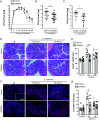
Fig. 1.
Fig. 1.
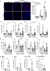
Antagonism of TACR1 signaling reduces C. rodentium burden and induced pathology. Administration of the highly selective TACR1 antagonist CP96345 (2.5 mg/kg, orogastric gavage, once daily) significantly reduces fecal C. rodentium over the course of infection (A). Reduced fecal (B) and colonic adherent (C) C. rodentium were observed in CP96345 compared to vehicle-treated mice at 10 dpi. Crypt length was measured on H&E-stained histology sections (D) and quantified (E) from vehicle or CP96345-treated uninfected or infected mice at 10 and 29 dpi. Immunofluorescent staining and confocal imaging were performed to identify (F) and enumerate (G) proliferating (DAPI+ Ki67+ CDH1+) IEC in uninfected or infected mice treated with vehicle or CP96345. Results are from individual mice, mean ± SEM. For (AC) *P ≤ 0.05, **P ≤ 0.01, ***P ≤ 0.001. For (DG), *P ≤ 0.05, **P ≤ 0.01, ***P ≤ 0.001 compared to uninfected controls of the same treatment and #P ≤ 0.05, ##P ≤ 0.01, ###P ≤ 0.001 compared between treatment groups. Two-way ANOVA with Tukey’s post hoc test. (Scale bar, 100 µm.) Luria broth (LB), postinfection (p.i.), IEC.
Fig. 2.
Fig. 2.
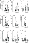
TACR1 signaling is required for recruitment of IFNγ and IL-17A producing T cells during enteric infection. Colonic recruitment of T cells (DAPI+ CD3+) in uninfected (LB) or C. rodentium–infected mice treated with vehicle or CP96345 was assessed in tissues sections obtained 10 and 29 dpi by immunofluorescence and confocal microscopy (A) and enumerated (B). LB or C. rodentium–infected mice treated with vehicle or CP96345 10 dpi were quantified via flow cytometry for T cell recruitment by frequency of live CD3+ T cells (C) as well as intracellular cytokine staining for CD4+ T cells that express IFNγ (D), IL-17A (E), IL-22 (F), and FoxP3 (G). In a separate cohort, colonic BEC (CD45−, CD31+ gp38−) from uninfected (LB) and C. rodentium–infected mice treated with vehicle or CP96345 10 dpi were analyzed for their expression of MAdCAM-1 (H), ICAM-1 (I), and VCAM-1 (J). Tacr1 mRNA expression was assessed in sorted colonic BEC (K), lymphatic endothelial cells (LEC) (L), T cells (M), and distal IEC (N) in naïve (LB) or 10 dpi C. rodentium infected WT mice. Each data point represents an individual mouse. Mean ± SEM, *P ≤ 0.05, **P ≤ 0.01, ***P ≤ 0.001 compared to uninfected controls of the same treatment and #P ≤ 0.05, ##P ≤ 0.01, ###P ≤ 0.001 compared between treatment groups. One-way ANOVA with Tukey’s post hoc test was used. (Scale bar, 100 µm.) Luria broth (LB), postinfection (p.i.), C. rodentium (C. rod).
Fig. 3.
Fig. 3.
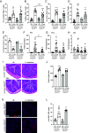
TACR1 blockade attenuates expression of specific proinflammatory genes in colonic tissues during C. rodentium infection. Colonic tissue from uninfected (LB) and infected mice treated with vehicle or CP96354 were assessed for expression of Ifnγ (A), Il17a (B), Il22 (C) at 10 and 29 dpi. General inflammatory responses were assayed by measuring colonic expression of Nos2 (D), Regiiiy (E), Chi3l3 (F), Tnfa (G), Il1β (H), and Il6 (I). Results are from individual mice, mean ± SEM, *P ≤ 0.05, **P ≤ 0.01, ***P ≤ 0.001 compared to uninfected controls of the same treatment and #P ≤ 0.05, ##P ≤ 0.01, ###P ≤ 0.001 compared between treatment groups. Two-way ANOVA with Tukey’s post hoc test was used. Postinfection (p.i.).
Fig. 4.
Fig. 4.
T cell intrinsic TACR1 signaling impacts IFNγ production during enteric infection. Colonic lamina propria lymphocytes were quantified in Lck.Cre+ TACR1f/f mice and their Lck.Cre− TACR1f/f littermates. Frequency of live CD3+ CD4+ T cells in the colonic lamina propria of C. rodentium–infected mice 10 dpi (A). Intracellular cytokine staining of IFNγ (B), IL-17A (C), IL-22 (D), and FoxP3 (E) producing CD4+ T cells 10 d post–C. rodentium infection. Colonic tissue from Lck.Cre+ TACR1f/f mice and their Lck.Cre− TACR1f/f littermates was assessed for mRNA expression of Ifnγ (F), Il17a (G), Il22 (H) at 10 dpi. Colonic crypt length was measured in H&E-stained sections of LB or 10 dpi C. rodentium–infected Lck.Cre+ TACR1f/f mice or Lck.Cre− TACR1f/f littermates (I and J). DAPI+ Ki67+ CDH1+ IEC were stained (K) and enumerated (L) via immunofluorescent staining and confocal imaging in uninfected or infected Lck.Cre+ TACR1f/f mice and their Lck.Cre− TACR1f/f littermates 10 dpi. Results are from individual mice, mean ± SEM, *P ≤ 0.05, **P ≤ 0.01, ***P ≤ 0.001 compared to uninfected controls of the same treatment and #P ≤ 0.05, ##P ≤ 0.01, ###P ≤ 0.001 compared between treatment groups. One-way ANOVA with Tukey’s post hoc test was used. (Scale bar, 100 µm.) Luria broth (LB), C. rodentium (C. rod).
Fig. 5.
Fig. 5.
TACR1 signaling exacerbates inflammation, pathology, and bacterial burden. TACR1 signaling on BEC upregulates MAdCAM-1 which increases recruitment of T cells to the lamina propria during C. rodentium infection. TACR1 signaling on T cells enhances the expression of IFNγ which exacerbates crypt hyperplasia pathology and bacterial burden. Treatment with the TACR1 antagonist CP96345 reduces the recruitment of T cells by reducing the expression of MAdCAM-1 while also reducing IFNγ expression in T cells once in the lamina propria. These reductions lead to reduced crypt hyperplasia pathology and bacterial burden throughout the course of the infection.

Update of

References

    1. Ramirez V., Swain S., Murray K., Reardon C., Neural immune communication in the control of host-bacterial pathogen interactions in the gastrointestinal tract. Infect. Immun. 88, e00928-19 (2020), 10.1128/iai.00928-00919. - DOI - PMC - PubMed
    1. Chiu I. M., von Hehn C. A., Woolf C. J., Neurogenic inflammation and the peripheral nervous system in host defense and immunopathology. Nat. Neurosci. 15, 1063–1067 (2012), 10.1038/nn.3144. - DOI - PMC - PubMed
    1. Dantzer R., Neuroimmune interactions: From the brain to the immune system and vice versa. Physiol. Rev. 98, 477–504 (2018), 10.1152/physrev.00039.2016. - DOI - PMC - PubMed
    1. Reardon C., Murray K., Lomax A. E., Neuroimmune communication in health and disease. Physiol. Rev. 98, 2287–2316 (2018), 10.1152/physrev.00035.2017. - DOI - PMC - PubMed
    1. Spencer N. J., Hu H., Enteric nervous system: Sensory transduction, neural circuits and gastrointestinal motility. Nat. Rev. Gastroenterol. Hepatol. 17, 338–351 (2020), 10.1038/s41575-020-0271-2. - DOI - PMC - PubMed