. 2025 Feb 14;11(7):eadp5958.
doi: 10.1126/sciadv.adp5958. Epub 2025 Feb 12.

Targeted approach to determine the impact of cancer-associated protease variants

Affiliations

Targeted approach to determine the impact of cancer-associated protease variants

Kira Bickenbach et al. Sci Adv. .

Abstract

Several steps of cancer progression, from tumor onset to metastasis, critically involve proteolytic activity. To elucidate the role of proteases in cancer, it is particularly important to consider single-nucleotide variants (SNVs) that affect the active site of proteases, thereby influencing cleavage specificity, substrate processing, and thus cancer cell behavior. To facilitate systematic studies, we here present a targeted approach to determine the impact of cancer-associated protease variants (TACAP). Starting with the semiautomated identification of potential specificity-modulating SNVs, our workflow comprises mass spectrometry-based cleavage specificity profiling and substrate identification, localization, and inhibitor studies, followed by functional analyses investigating cancer cell properties. To demonstrate the feasibility of TACAP, we analyzed the meprin β R238Q variant. This amino acid exchange R238Q leads to a loss of meprin β's characteristic cleavage preference for acidic amino acids at P1' position, accompanied with changes in substrate pool and inhibitor affinity compared to meprin β wild type.


Figures
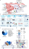
Fig. 1.
Fig. 1.. TACAP is used as tool for the identification and characterization of potential specificity-modulating cancer-associated SNVs in protease genes.
(A) Various proteases are involved in cancer progression by influencing different processes (stated in the boxes). TNFα, tumor necrosis factor–α; VEGF-A, vascular endothelial growth factor A; ECM, extracellular matrix. (B) Overview of presented targeted approach to determine the impact of cancer-associated protease variants (TACAP). (C) Distribution of SNVs in MEP1B annotated in COSMIC for different cancer entities. (D) Identified cancer-associated SNVs affecting different domains of meprin β. SP, signal peptide; PRO, prodomain; CAT, catalytic domain; MAM, meprin A5 protein tyrosine phosphatase μ domain with disulphide bridge; TRAF, tumor necrosis factor receptor–associated factor domain; I, inserted domain; EGF, epidermal growth factor like domain; TM, transmembrane region; C, cytosolic region. [(C and D) status as of 7 October 2024]. (E) The positively charged R238 is located within the active site at S1′ position and interacts via ionic interactions with the negatively charged amino acid D36 within the propeptide of meprin β.
Fig. 2.
Fig. 2.. PICS analysis and quenched fluorogenic peptide cleavage assays revealed a loss of cleavage specificity for acidic amino acids in P1′ position for meprin β R238Q.
(A) Workflow of PICS analysis to compare the cleavage preference of meprin β WT and R238Q. (B and C) Cleavage specificity of meprin β WT (left) and R238Q (right) is presented in specificity logos (including P4 to P4′ positions). All semi-peptides identified in Lys-C and CNBR library, which did not contain K or R at the P1 position, were analyzed. For (B), only peptides with a log2(diff) greater than 2 and for (C) only peptides that were unique to meprin β WT or meprin β R238Q–treated samples were included. (D) Principle of quenched fluorogenic peptide cleavage assays to analyze cleavage specificity of meprin β WT or R238Q: To measure meprin β activity, the quenched fluorogenic peptide EDEDED, flanked with a fluorogenic group [mca = (7-methoxycoumarin-4-yl)acetyl] and a quencher (dnp = 2,4-dinitrophenyl), was used. Cleavage of the peptide results in detectable fluorescence intensity at 405 nm with an excitation at 320 nm. (E) Exemplary peptide cleavage assay using HEK 293T cells transfected with meprin β variants. Relative cell surface activity of HEK 293T cells transfected with meprin β variants using the (F) (mca)-EDEDED-(dnp), (G) (mca)-EEEEE-(dnp), (H) (mca)-EEEEE-(dnp), (I) (mca)-DDDDDD-(dnp), (J) (mca)-DDDDDD-(dnp), (K) (mca)-EDAEDA-(dnp), and (L) (mca)-EDAEDA-(dnp) substrate (each n = 3). (M) Activity of purified meprin β WT, meprin β R238Q, and meprin α toward the (mca)-EDAEDA-(dnp) substrate (n = 3) [means ± SD; one-way ANOVA, followed by a Tukey posttest (** ≙ P < 0.01; *** ≙ P < 0.001)]. IP, immunoprecipitated.
Fig. 3.
Fig. 3.. Meprin β WT and meprin β R238Q do not differ in cellular localization and shedding.
(A) For cell surface biotinylation assays, membrane proteins on intact cells were labeled with thiol-cleavable amine–reactive biotin and pulled down from lysates with magnetic streptavidin beads. (B) Cell surface biotinylation assay of HEK 293T cells overexpressing meprin β variants, analyzed by Western blot. Pan-cadherin and glyceraldehyde-3-phosphate dehydrogenase (GAPDH) served as controls for cell surface and intracellular proteins, respectively. (C and D) Immunofluorescence microscopy images of HEK 293T transfected with the indicated meprin β variants, stained with anti–meprin β antibody (hEcto1, green) (C) or anti–flag tag antibody (green) (D). Antibody against PDIA6 (red) served as ER marker (D) and nuclei were stained with DAPI (blue). (E) Inactive pro–meprin β can be shed from the cell surface by ADAM10/17 and MT1-MMP. Soluble pro–meprin β can be activated by trypsin. (F) HEK 293T WT/ADAM10/17−/− cells were transfected with meprin β variants and ADAM10. Lysates and ultracentrifuged supernatants were analyzed by Western blot. GAPDH served as loading control. (G) Activity of shed meprin β variants in ultracentrifuged cell supernatants [untreated or activated with trypsin, measured using (mca)-EDAEDA-(dnp)] (n = 3) [means ± SD; two-way ANOVA, followed by a Tukey posttest (* ≙ P < 0.05; ** ≙ P < 0.01; *** ≙ P < 0.001)].
Fig. 4.
Fig. 4.. Meprin β R238Q and WT share the same activation mechanisms but differ in inhibition properties.
(A) Meprin β reaches the cell surface as a zymogen and requires tryptic activation, for example, by trypsin or MT-2. (B) Activation of meprin β variants transfected in HEK 293T ADAM10/17−/− cells by trypsin treatment or co-transfected with MT-2 was analyzed by a cell surface activity assay with the (mca)-EDAEDA-(dnp) substrate. (C) Lysate controls from (B) were blotted against meprin β, MT-2 (myc), and GAPDH. (D) HEK 293T cells transfected with meprin β variants with strep tag N-terminal of the propeptide were stained with an anti–strep tag and anti–meprin β antibody (binding to the ectodomain) and analyzed via flow cytometry. (E) Normalized ratio of strep tag to meprin β median fluorescence intensity, representing levels of pro–meprin β to activated meprin β variants on the cell surface [n = 3, means ± SD; one-way ANOVA, followed by a Tukey posttest (* ≙ P < 0.05)]. (F) Chemical structure of the hydroxamate inhibitor actinonin, which inhibits meprin β by chelating the Zn2+ ion within the active site with the hydroxamic moiety. A pentanyl side chain targets the P1′ position. (G) Chemical structure of the F2-inhibitor, a phosphinic acid–based peptide inhibitor, designed to inhibit meprin β WT based on the assumption that the non-prime site aspartate builds ionic interactions with R238 of the S1′-subpocket. (H) Efficacy of inhibitors for meprin β WT (actinonin and F2-inhibitor) toward meprin β R238Q compared also to meprin α. For activity measurements of meprin β WT and meprin α, (mca)-EDAEDA-(dnp) and (mca)-HVANDPIW-(dnp) were used, respectively. Relative activity of inhibitor treated samples is normalized to the respective untreated control [n = 3, means ± SD; two-way ANOVA, followed by a Tukey posttest (*** ≙ P < 0.001)].
Fig. 5.
Fig. 5.. N-terminomics indicates differential protein substrate cleavage of meprin β WT compared to that of meprin β R238Q.
(A) Principle of HYTANE analysis using lysates and supernatants of transfected U251MG cells. (B) Native protein substrate cleavage specificities of meprin β WT (left) and meprin β R238Q (right) analyzed from HYTANE analysis of cell supernatants presented in specificity logos. (C to I) Volcano plots showing all identified proteolytic events detected by N-terminomics of U251MG supernatants comparing the indicated conditions. Gray lines represent threshold values (±0.58 for log2 difference, P = 0.05). Selected cleavage events within cancer-associated proteins are highlighted.
Fig. 6.
Fig. 6.. APP, syndecan-1, CD99, and IL-6R are differentially cleaved by meprin β R238Q compared to those by meprin β WT.
(A) Cleavage of APP by meprin β WT, BACE-1, ADAM10/17, and γ-secretase. (B) Cleavage sites of meprin β within APP (46, 48, 49). (C) HEK 293T cells were co-transfected with APP and different meprin β variants or BACE-1. APP cleavage products were analyzed by Western blot. (D) Densitometric analysis of sAPPβ + Asp from Western blots (n = 7; nd, not detectable). (E) Serum-free supernatants from (C) were analyzed for Aβ concentrations via Aβx-40 ELISA (n = 5). (F) Cleavage sites within syndecan-1 for meprin β WT (black) and R238Q (red), identified via N-terminomics of transfected U251MG cells, are annotated. (G) Western blot analysis of HEK 293T cells transiently transfected with syndecan-1 and meprin β variants. (H) Densitometric analysis of a cleavage fragment labeled with * from Western blots (normalized to GAPDH, n = 5). (I) Cleavage of CD99 by meprin β WT within the ectodomain generates two C-terminal fragments (CTFI/II), which can be further processed by γ-secretase. Cleavage sites within CD99 for meprin β WT (black) and R238Q (red), identified via N-terminomics of transfected U251MG cells, are shown. (J) Hela cells were transiently transfected with CD99 and meprin β variants and treated with the γ-secretase inhibitor DAPT. Cell lysates were analyzed via Western blot. (K) Densitometric analysis of CTFII (*) from Western blots (normalized to GAPDH, n = 3). (L) Cleavage site within the IL-6R identified for meprin β WT in (19). (M) HEK 293T ADAM10/17−/− cells were transiently transfected with IL-6R and different meprin β variants and analyzed via Western blot. (N) Densitometric analysis of sIL-6R (*) from Western blots (n = 3) [(D, E, H, K, and N) means ± SD; one-way ANOVA, followed by a Tukey posttest (* ≙ P < 0.05; ** ≙ P < 0.01)].
Fig. 7.
Fig. 7.. Meprin β increases migration of glioma cells.
(A) Workflow of transendothelial migration (TEM) assay with U251MG cells expressing meprin β variants. (B) For evaluation of TEM assay, transmigrated glioma cells were imaged by fluorescence microscopy and counted. (C) Quantification of migrated U251MG cells expressing different meprin β variants (n = 7). (D) Principle of tumor spheroid invasion assay through a basement membrane–like matrix. (E) Representative pictures of spheroid invasion for meprin β variants expressing cells at different time points. (F) Quantification of invasion area of spheroids (n = 4). (G) Analysis of migration of glioma cells within murine organotypic brain slice cultures. (H) Representative images of migration of transfected GL261 cells injected into organotypic brain slices as spheroids. Highlighted migration area of glioma cells outside the spheroid core was quantified. (I) Quantification of area of cells migrated from the spheroids into the organotypic brain slice (n = 3). (J) Workflow of invasion assay through Matrigel- or fibronectin-coated transwell inserts. (K) Representative images of migration of transfected U251MG cells through Matrigel-coated transwells. (L and M) Quantification of invaded U251MG cells expressing different meprin β and treated with different inhibitors (F2-inhibitor or actinonin) for Matrigel-fibronectin (L) and fibronectin-coated (M) inserts (n = 4) [means ± SD; for (C) and (I), one-way ANOVA, followed by a Tukey posttest; for (F), (L), and (M), two-way ANOVA, followed by a Tukey posttest (* ≙ P < 0.05; ** ≙ P < 0.01; *** ≙ P < 0.001)].

References

    1. Bond J. S., Proteases: History, discovery, and roles in health and disease. J. Biol. Chem. 294, 1643–1651 (2019). - PMC - PubMed
    1. Eatemadi A., Aiyelabegan H. T., Negahdari B., Mazlomi M. A., Daraee H., Daraee N., Eatemadi R., Sadroddiny E., Role of protease and protease inhibitors in cancer pathogenesis and treatment. Biomed. Pharmacother. 86, 221–231 (2017). - PubMed
    1. Jackson H. W., Defamie V., Waterhouse P., Khokha R., TIMPs: Versatile extracellular regulators in cancer. Nat. Rev. Cancer 17, 38–53 (2017). - PubMed
    1. Vizovisek M., Ristanovic D., Menghini S., Christiansen M. G., Schuerle S., The tumor proteolytic landscape: A challenging frontier in cancer diagnosis and therapy. Int. J. Mol. Sci. 22, 2514 (2021). - PMC - PubMed
    1. Gnosa S. P., Blasco L. P., Piotrowski K. B., Freiberg M. L., Savickas S., Madsen D. H., Auf dem Keller U., Kronqvist P., Kveiborg M., ADAM17-mediated EGFR ligand shedding directs macrophage-promoted cancer cell invasion. JCI Insight 7, e155296 (2022). - PMC - PubMed

LinkOut - more resources