. 2025 Apr 1;26(7):e202401014.
doi: 10.1002/cbic.202401014. Epub 2025 Feb 26.

Design of Zn-Binding Peptide(s) from Protein Fragments

Affiliations

Design of Zn-Binding Peptide(s) from Protein Fragments

Ján Michael Kormaník et al. Chembiochem. .

Abstract

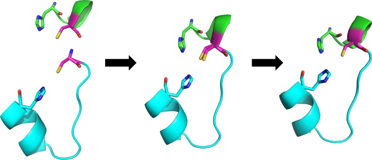
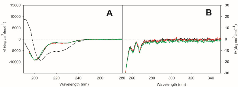
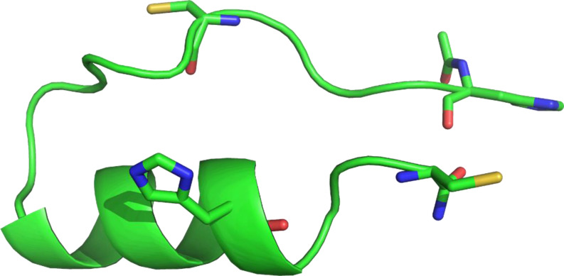
We designed a minimalistic zinc(II)-binding peptide featuring the Cys2His2 zinc-finger motif. To this aim, several tens of thousands of (His/Cys)-Xn-(His/Cys) protein fragments (n=2-20) were first extracted from the 3D protein structures deposited in Protein Data Bank (PDB). Based on geometrical constraints positioning two Cys (C) and two His (H) side chains at the vertices of a tetrahedron, approximately 22 000 sequences of the (H/C)-Xi-(H/C)-Xj-(H/C)-Xk-(H/C) type, satisfying Nmetal-binding H=Nmetal-binding C=2, were processed. Several other criteria, such as the secondary structure content and predicted fold stability, were then used to select the best candidates. To prove the viability of the computational design experimentally, three peptides were synthesized and subjected to isothermal calorimetry (ITC) measurements to determine the binding constants with Zn2+, including the entropy and enthalpy terms. For the strongest Zn2+ ions binding peptide, P1, the dissociation constant was shown to be in the nanomolar range (KD=~220 nM; corresponding to ΔGbind=-9.1 kcal mol-1). In addition, ITC showed that the [P1 : Zn2+] complex forms in 1 : 1 stoichiometry and two protons are released upon binding, which suggests that the zinc coordination involves both cysteines. NMR experiments also indicated that the structure of the [P1 : Zn2+] complex might be quite similar to the computationally predicted one. In summary, our proof-of-principle study highlights the usefulness of our computational protocol for designing novel metal-binding peptides.

Keywords: Computer design; Isothermal calorimetry; Metal-binding peptide; NMR; QM modeling; Zinc(II).


Conflict of interest statement

The authors declare no conflict of interest.

Figures

Figure 1
Figure 1
Merging two fragments into a larger peptide. When we find two compatible fragments (left), the terminal AA of the first fragment is overlaid onto the first AA of the second fragment (center). We only consider the 10 best alignments according to the clustering by the RMSD. These fragments are then merged into a new peptide and only the C‐terminal AA from the first fragment is kept in the new peptide (right). Similar process is then repeated to obtain the final design. The two fragments are shown in cartoon in lime and cyan, respectively, the shared cysteine is shown in magenta. All terminal binding AAs are shown as sticks, hydrogens are omitted for clarity.
Figure 2
Figure 2
Source proteins for peptide P1. Each fragment is colored according to the source PDB with the proposed binding residues shared by two fragments in magenta. Only parts of the proteins from PDB are shown. Proteins are shown by cartoon with binding residues as sticks, hydrogens are omitted for clarity.
Figure 3
Figure 3
(A) An example of isothermal titration of ZnCl2 to peptide P1 performed in 20 mM HEPES, pH 7.0 at 298.15 K. Experimentally observed binding enthalpies (ΔH obs) from titration of ZnCl2 to peptide P1 (B) and P1C (C) in four different buffers, plotted against enthalpy of ionization of each buffer (ΔH ion).
Figure 4
Figure 4
ECD spectra of peptide P1 in buffer solution (solid black curve) and 25 % (v/v) TFE (dashed black curve) and of [P1 : Zn2+] complex in water in stoichiometry 1 : 1 (red curve) and 1 : 2 (green curve) in far‐UV (A) and near‐UV (B) spectral region.
Figure 5
Figure 5
ECD spectra of peptide P1C in buffer solution (solid black curve) and 25 % (v/v) TFE (dashed black curve) and of [P1C : Zn2+] complex in water in stoichiometry 1 : 1 (red curve) and 1 : 2 (green curve) in far‐UV (A) and near‐UV (B) spectral region.
Figure 6
Figure 6
Comparison of the NMR models (lime) with the designed peptide P1 (cyan; shown without zinc). Using the restraints from NMR and ITC, 20 models of the peptide were generated. Here we show the best model (left, all‐atom RMSD=2.9 Å) and the median of the 20 models (right, all‐atom RMSD=4.1 Å) according to the all‐atom RMSD. Peptides are shown by cartoon with binding residues shown as sticks, hydrogens are omitted for clarity.
Figure 7
Figure 7
(A) The average distances between the metal‐binding residues and the zinc ion during MD run. (B) Selected frames from the MD study of [P1 : Zn2+] (at 138 ns, yellow, and 434 ns, teal). While the overall shape of the binding site is very similar, the conformations of the binding residues vary, which can be seen on both the terminal cysteine residue (at the top of the figure) and relative orientation of the rings of both histidine residues. Peptide is shown by cartoon with binding residues shown as sticks and zinc ions shown as spheres, hydrogens are omitted for clarity.
Figure 8
Figure 8
The final structure of the free P1C from the MD run at 450 ns. The peptide folded into a more compact conformation, where the N‐terminal section is seemingly held in place by the arginines on the C‐terminal α‐helix (residues not shown). The proposed binding residues (shown in sticks) are unable to form a tetrahedral binding site in this conformation. Peptide is shown by cartoon with presumed binding residues shown in sticks, hydrogens are omitted for clarity.

References

    1. Zheng H., Cooper D. R., Porebski P. J., Shabalin I. G., Handing K. B., Minor W., Acta Crystallogr. Sect. Struct. Biol. 2017, 73 (Pt 3), 223–233. - PMC - PubMed
    1. Sánchez-Aparicio J.-E., Tiessler-Sala L., Velasco-Carneros L., Roldán-Martín L., Sciortino G., Maréchal J.-D., J. Chem. Inf. Model. 2021, 61 (1), 311–323. - PubMed
    1. Putignano V., Rosato A., Banci L., Andreini C., Nucleic Acids Res. 2018, 46 (D1), D459–D464. - PMC - PubMed
    1. Bertini I., Gray H. B., Stiefel E. I., Valentine J. S., Biological Inorganic Chemistry. Structure and Reactivity, University Science Books, 2007, ISBN 978-1-891389-43-6.
    1. Liu J., Chakraborty S., Hosseinzadeh P., Yu Y., Tian S., Petrik I., Bhagi A., Lu Y., Chem. Rev. 2014, 114 (8), 4366–4469. - PMC - PubMed

LinkOut - more resources