. 2025 Mar;22(2):e00532.
doi: 10.1016/j.neurot.2025.e00532. Epub 2025 Feb 12.

The dual role of VEGF-A in a complex in vitro model of oxaliplatin-induced neurotoxicity: Pain-related and neuroprotective effects

Affiliations

The dual role of VEGF-A in a complex in vitro model of oxaliplatin-induced neurotoxicity: Pain-related and neuroprotective effects

A Toti et al. Neurotherapeutics. 2025 Mar.

Abstract

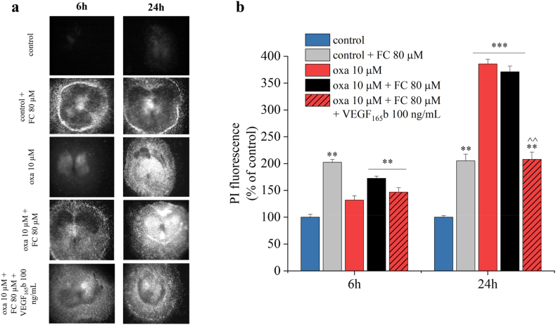
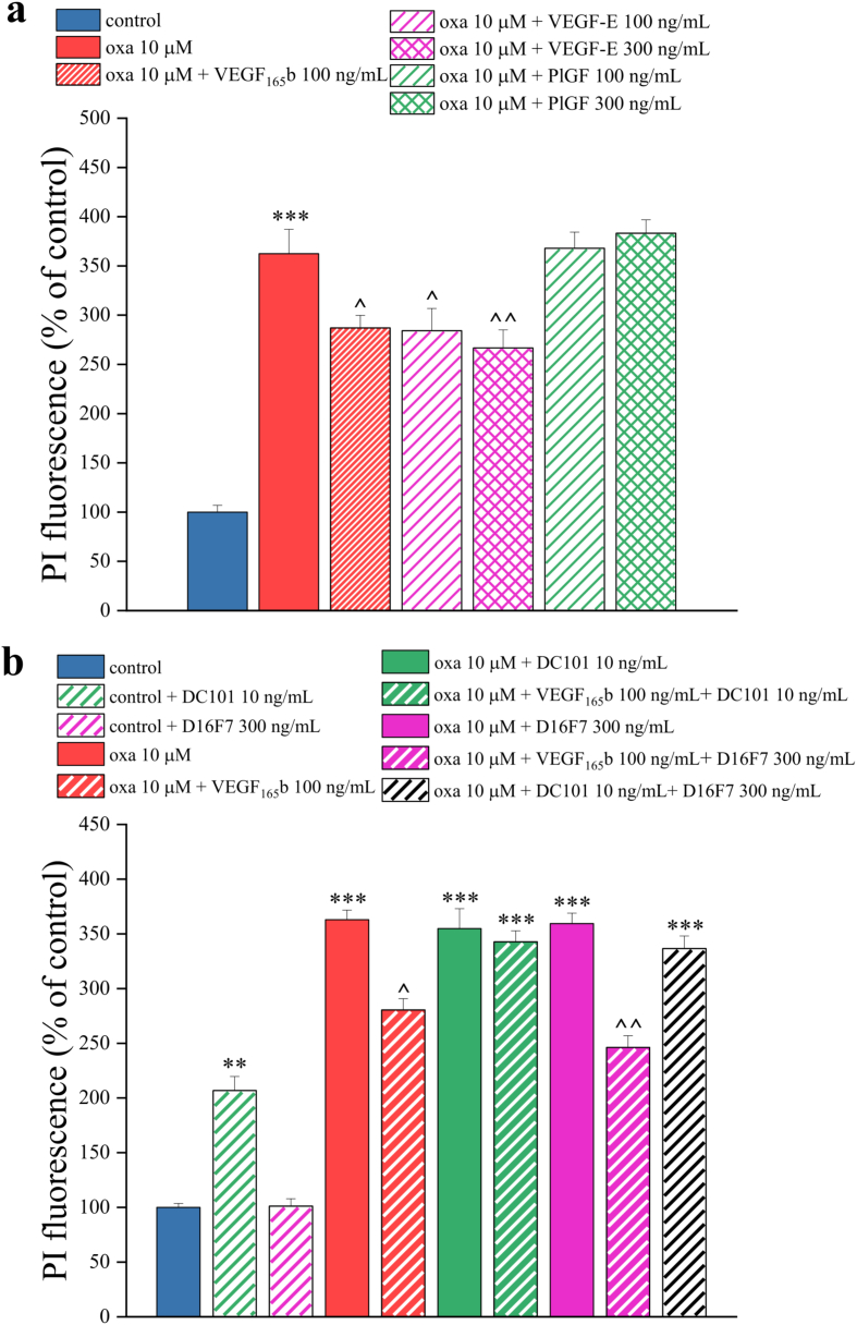
Vascular endothelial growth factor (VEGF)-A is a main player in the development of neuropathic pain induced by chemotherapy and the pharmacological blockade of VEGF receptor (VEGFR) subtype 1 is a pain killer strategy. Interestingly, VEGF-A has been demonstrated to have also neuroprotective properties. The aim of the study was to investigate the neuroprotective role of VEGF-A against oxaliplatin neurotoxicity, attempting to discriminate pain-related and restorative signaling pathways. We used rat organotypic spinal cord slices treated with oxaliplatin, as an in vitro model to study chemotherapy-induced toxicity. In this model, 10 ​μM oxaliplatin caused a time-dependent release of VEGF-A, which was reduced by the astrocyte inhibitor fluorocitrate. Moreover, glia inhibition exacerbated oxaliplatin-induced cytotoxicity in a VEGF-A sensitive manner. Treatment with VEGF165b, the main isoform of VEGF-A, prevented the oxaliplatin-induced neuronal damage (indicated by NeuN staining) and astrocyte activation (indicated by GFAP staining). In addition, the blockade of VEGFR-2 by the selective antibody DC101 blunted the protective action of VEGF165b. In the same model, VEGF165b increased the release of molecules relevant in pain signaling, like substance P and CGRP, as well as the mRNA expression of glutamate transporters (EAAT1 and EAAT2), similarly to oxaliplatin and these effects were prevented by the selective VEGFR-1 blocker antibody D16F7. In conclusion, VEGF-A plays a dichotomic role in an in vitro model of chemotherapy-induced toxicity, either promoting neuroprotection or triggering pain mediators release, depending on which of its two receptors is activated. The selective management of VEGF-A signaling is suggested as a therapeutic approach.

Keywords: Chemotherapy-induced neurotoxicity; Glial cells; Neuroprotection; Spinal cord organotypic slices; VEGF-A; VEGFRs.


Conflict of interest statement

Declaration of competing interest The authors declare that they have no competing interests.

Figures

Image 1
Graphical abstract
Fig. 1
Fig. 1
Structural organization and vascular network in organotypic spinal cord slices. (a) Immunohistochemical analysis of GFAP (astrocyte marker, red) and NeuN (neuron marker, green) performed after 14 days of slice maturation (image obtained by scanning with 20X magnification of each field of view; 63× magnification for GFAP insert and 40× magnification for NeuN inserts). Scale bar ​= ​100 ​μM. (b) Immunohistochemical analysis and representative images of RECA-1 (blood vessels marker, red), performed after 7 and 14 days of slices maturation (40X magnification). Scale bar ​= ​100 ​μM ∗∗P ​< ​0.01 vs slice 7 days.
Fig. 2
Fig. 2
Oxaliplatin induces cytotoxicity in spinal cord slices. Qualitative (a) and quantitative (b) analyses of PI (5 ​μg/mL) fluorescence intensity were performed to investigate the toxicity of oxaliplatin at different concentrations (1, 10 and 100 ​μM) for 6 and 24 ​h. Each value represents the mean ​± ​SEM of three independent experiments, with a total of 15 slices for condition. ∗∗∗P ​< ​0.001 vs control.
Fig. 3
Fig. 3
Alterations of astrocytes and neurons induced by oxaliplatin. (a) Representative images of GFAP (red) and NeuN (green) staining in organotypic spinal cord slices, treated with 10 ​μM oxaliplatin for 24 ​h in comparison to untreated slices. Quantitative analyses were expressed as mean of GFAP fluorescence intensity (b), NeuN fluorescence intensity (c) and NeuN positive cells per optic field (d) (20X magnification). Scale bar = 100 μM. Each value represents the mean ​± ​SEM of three independent experiments, with a total of 12 slices for condition. ∗∗P ​< ​0.01 vs control.
Fig. 4
Fig. 4
Fluorocitrate inhibits oxaliplatin-induced release of VEGF-A. Amount of VEGF-A measured by ELISA into the culture medium of the organotypic slices, after treatment with 80 ​μM fluorocitrate (FC) alone or in combination with 10 ​μM oxaliplatin (oxa) for 6 and 24 ​h. VEGF-A levels were normalized to cell protein concentrations. Each value represents the mean ​± ​SEM of three independent experiments, with two wells per condition. ∗P < 0.05 and ∗∗∗P < 0.001 vs control; ˆ P < 0.05 and ˆ ˆ P < 0.01 vs oxaliplatin treatment.
Fig. 5
Fig. 5
Inhibition of astrocytic VEGF-A by fluorocitrate enhances oxaliplatin cytotoxicity. Qualitative (a) and quantitative (b) analysis of PI (5 ​μg/mL) fluorescence intensity in spinal cord slices to evaluate the effect of 80 ​μM fluorocitrate (FC) alone or in combination with 100 ​ng/mL VEGF165b on 10 ​μM oxaliplatin-induced toxicity applied for 6 and 24 ​h. Each value represents the mean ​± ​SEM of three independent experiments, with a total of 15 slices for condition. ∗∗P ​< ​0.01 and ∗∗∗P ​< ​0.001 vs control; ˆ ˆ P ​< ​0.01 vs oxaliplatin treatment.
Fig. 6
Fig. 6
VEGF165b prevents the cytotoxicity induced by oxaliplatin. Qualitative (a) and quantitative (b) analyses of PI (5 ​μg/mL) fluorescence intensity were performed to study the effect of VEGF165b treatment (30, 100 and 300 ​ng/mL) on 10 μM oxaliplatin-induced toxicity at 24 ​h. Each value represents the mean ​± ​SEM of three independent experiments, with a total of 15 slices for condition. ∗∗∗P ​< ​0.001 vs control; ˆ ˆ P ​< ​0.01 vs oxaliplatin treatment.
Fig. 7
Fig. 7
VEGF165b reduces the alterations in astrocytes and neurons caused by oxaliplatin. Representative images of GFAP (red) and NeuN (green) staining on organotypic spinal cord slices, treated with oxaliplatin (10 ​μM) alone or in combination with VEGF165b (100 ​ng/mL) for 24 ​h in comparison to untreated slices (a). Quantitative analyses were expressed as mean of GFAP fluorescence intensity (b), NeuN fluorescence intensity (c) and NeuN positive cells per optic fields (d). (20X magnification). Scale bar = 100 μM. Each value represents the mean ​± ​SEM of three independent experiments, with a total of 12 slices for condition. ∗P ​< ​0.05 and ∗∗P ​< ​0.01; ˆ P ​< ​0.05 vs oxaliplatin treatment.
Fig. 8
Fig. 8
VEGFR-2 mediates the VEGF-A neuroprotective effects. Quantitative analyses of PI (5 ​μg/mL) fluorescence intensity performed to evaluate the effect of 24 ​h treatment with the VEGFR-2 ligand, VEGF-E (100 and 300 ​ng/mL), and the VEGFR-1 ligand, PlGF (100 and 300 ​ng/mL), (a) and with the selective VEGFR-1 antagonist, D16F7 (300 ​ng/mL) or the selective VEGFR-2 antagonist, DC101 (10 ​ng/mL) mAbs (b) on oxaliplatin-induced toxicity (10 ​μM, 24 ​h), in combination with VEGF165b (100 ​ng/mL). Each value represents the mean ​± ​SEM of three independent experiments. ∗∗P ​< ​0.01; ∗∗∗P ​< ​0.001 vs control; ˆ P ​< ​0.05; ˆ ˆ P ​< ​0.01 vs oxaliplatin treatment.
Fig. 9
Fig. 9
VEGFR-1-mediates the release of CGRP and SP evoked by both oxaliplatin and VEGF165b. The quantity of SP and CGRP released into the medium of the organotypic slices was measured by ELISA, after treatment with oxaliplatin (10 ​μM) or VEGF165b (100 ​ng/mL) for 6 ​h, alone or in the presence of the selective VEGFR-1 antagonist, D16F7 (300 ​ng/mL), or the selective VEGFR-2 antagonist, DC101 (10 ​ng/mL). Levels of the neuropeptides were normalized to cell protein concentration. Each value represents the mean ​± ​SEM of three independent experiments. ∗∗∗P ​< ​0.001 vs control; ˆ ˆ P ​< ​0.01 vs oxaliplatin treatment; $$P ​< ​0.01 vs VEGF165b treatment.
Fig. 10
Fig. 10
VEGFR-1 mediates the downregulation of glutamate transporters evoked by both oxaliplatin and VEGF165b. The mRNA expression of EAAT1 and EAAT2 genes was measured by RT-PCR, after treatment of organotypic spinal cord slices with oxaliplatin (10 ​μM) or VEGF165b (100 ​ng/mL) for 6 ​h, alone and in combination with the VEGFR-1 antagonist D16F7 (300 ​ng/mL) or the VEGFR-2 antagonist DC101 (10 ​ng/mL). Each value represents the mean ​± ​SEM of three independent experiments. ∗∗P ​< ​0.01 vs control; ˆ ˆ P ​< ​0.01 vs oxaliplatin treatment; $$P ​< ​0.01 and $$$P ​< ​0.001 vs VEGF165b treatment.
Suppl. File 1
Suppl. File 1
Rat organotypic spinal cord slices obtaining. The experimental protocol to obtain the organotypic spinal cord culture, starting from rat pups 4–6 post-natal days (a). Representative pictures of spinal cord slices during the 14 days of maturation, showing that structural integrity is maintained (b).
Suppl. File 2
Suppl. File 2
Representative pictures of the qualitative analyses of PI (5 ​μg/mL) fluorescence intensity, performed to evaluate the effect of the VEGFR-2 ligand, VEGF-E (100 and 300 ​ng/mL) and the VEGFR-1 ligand, PlGF (100 and 300 ​ng/mL) treatment on 10 ​μM oxaliplatin-induced toxicity.
Suppl. File 3
Suppl. File 3
Representative pictures of the qualitative analyses of PI (5 ​μg/mL) fluorescence intensity, performed to evaluate the effect of the selective VEGFR-1 antagonist, D16F7 (300 ​ng/mL) or the selective VEGFR-2 antagonist, DC101 (10 ​ng/mL) mAbs treatment on 10 ​μM oxaliplatin-induced toxicity, in combination with VEGF165b (100 ​ng/mL).

References

    1. Campbell J.N., Meyer R.A. Mechanisms of neuropathic pain. Neuron. 2006 Oct 5;52(1):77–92. - PMC - PubMed
    1. Seretny M., Currie G.L., Sena E.S., Ramnarine S., Grant R., MacLeod M.R., et al. Incidence, prevalence, and predictors of chemotherapy-induced peripheral neuropathy: a systematic review and meta-analysis. Pain. 2014 Dec;155(12):2461–2470. - PubMed
    1. Aldskogius H., Kozlova E.N. Central neuron-glial and glial-glial interactions following axon injury. Prog Neurobiol. 1998 May;55(1):1–26. - PubMed
    1. Volterra A., Meldolesi J. Astrocytes, from brain glue to communication elements: the revolution continues. Nat Rev Neurosci. 2005 Aug;6(8):626–640. - PubMed
    1. Haydon P.G., Carmignoto G. Astrocyte control of synaptic transmission and neurovascular coupling. Physiol Rev. 2006;86(3):1009–1031. - PubMed

MeSH terms

Substances