. 2025 Feb 12;10(1):10.
doi: 10.1038/s41525-024-00460-6.

LMX1B haploinsufficiency due to variants in the 5'UTR as a cause of Nail-Patella syndrome

Affiliations

LMX1B haploinsufficiency due to variants in the 5'UTR as a cause of Nail-Patella syndrome

Serena Cappato et al. NPJ Genom Med. .

Abstract

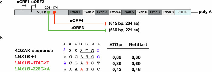
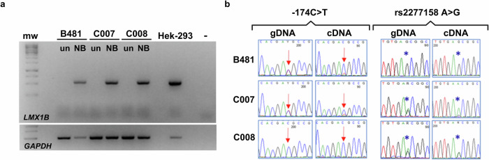
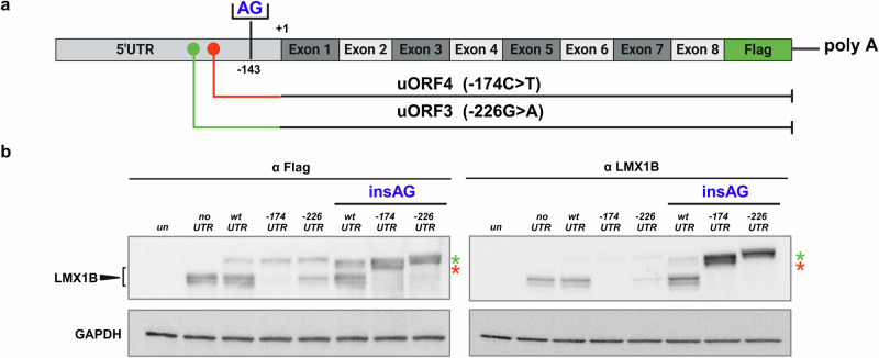
Nail-Patella syndrome (NPS) is a rare autosomal dominant condition due to haploinsufficiency of LMX1B, caused by loss-of-function variants affecting the coding sequence, or partial/whole deletions of the gene. In here, we describe two familial cases of NPS, carrying novel variants of the LMX1B 5'UTR region (-174C>T and -226G>A). To verify their pathogenic role, we carried out a functional characterization, both by reporter gene assays in heterologous systems and in patient's derived cells. We demonstrated that both variants impair LMX1B expression at post-transcriptional level. They introduce two upstream open reading frames (uORFs), out-of-frame with the main LMX1B coding sequence, generating transcripts detected by the non-sense mediated decay (NMD). We also demonstrated that the escape of the altered mRNA from NMD, if any, may lead to the synthesis of an aberrant LMX1B protein.


Conflict of interest statement

Competing interests: The authors declare no competing interests.

Figures

Fig. 1
Fig. 1. Pedigrees and genetic analysis.
a Pedigrees of the two reported families with recurrence of NPS. Black filled symbols represent individuals affected with NPS, the arrows indicate the two probands (B481 and E477). The individual indicated by the gray symbol in Family 2 is affected by a genetic condition not related to NPS (see text for details). b Chromatograms obtained by Sanger sequencing of the 5’UTR region spanning the two identified variants of the LMX1B gene, the −174C>T in Family 1 (absent in the C008 unaffected mother) and the −226G>A in the proband of Family 2 (E477) and in her affected brother (II-3, 0032). Image created with BioRender.com.
Fig. 2
Fig. 2. In silico analysis of the two LMX1B 5’UTR variants.
a Schematic representation of the LMX1B mRNA. The coding exons are indicated by the numbered rectangles. The light gray rectangle indicates the 5’UTR sequence harboring the wild type uORF1 and 2. The position of the identified variants is indicated by the green and red dots, respectively, and the newly generated uORF3 and 4 are also schematically represented. b Comparison between the canonical Kozak sequence around ATG with that surrounding the main LMX1B starting codon, and those in which each newly generated ATG is embedded (LMX1B −174C>T; LMX1B −226G>A). The left panel shows the sequence alignment with variant positions indicated in red. The right panel provides the values indicating the strength of each translation starting codon as predicted by ATGpr and NetStart tools. Values range from 0 to 1 and when greater than 0.5 predict a probable translation start site. Image created with BioRender.com.
Fig. 3
Fig. 3. Analysis of LMX1B cDNA.
a Sodium butyrate induces LMX1B gene expression in lymphoblastoid cell lines derived from B481 proband and his parents (C007 and C008). Indeed, an RT-PCR product of 537 bp spanning from the 5’UTR region to exon 2 of LMX1B is well detectable in lymphoblasts treated for 24 h with 10 mM Sodium butyrate and in Hek-293 used as positive control, but not in untreated cells. The housekeeping gene GAPDH was used to check the retro-transcription efficiency. b The variant −174C>T likely impairs the expression of the carrying transcript as assessed by comparing Sanger sequencing of PCR products obtained from genomic versus cDNA. The position of the variant is highlighted by red arrows and that of the A>G polymorphism by blue stars (see text for details). Mw molecular weight, un untreated cells, NB Sodium butyrate, – PCR reaction negative control (no template). Image created with BioRender.com.
Fig. 4
Fig. 4. The identified LMX1B 5’UTR variants impair gene expression at post-transcriptional level.
a Workflow for the functional study of LMX1B 5’UTR variants at post transcriptional level as assessed by reporter gene assay (see text for details). b Histograms represent the luciferase activity measured after transient transfection of the different pGL3.PV expression constructs, schematically reported on the left part of the panel. The luciferase values were normalized for transfection efficiency by co-transfection with a Renilla reporter control construct (see Methods for details) and are represented as the fold activation compared to the vector carrying the WT 5’UTR of LMX1B (value 1). Values represent the mean +/−the standard deviation of at least n = 4 biological replicates performed in technical triplicate or quadruplicate (p < 0.001***). EV, luciferase reporter vector without the 5’UTR sequence; WT, wild-type LMX1B 5’UTR. c Workflow for the functional study of LMX1B 5’UTR variants at post transcriptional level as assessed by western blot analysis (see text for details). d Hek-293 cells were transfected with pRP containing the LMX1B cDNA with or without the 5’UTR sequence, wild type or mutated. The expression level of the protein was evaluated by Western Blot, by using anti-Flag and anti-LMX1B primary antibody. Arrows indicate the band corresponding to LMX1B protein (60 kDa) and a non-specific band appearing only in cells transfected with the 5’UTR constructs. Densitometric analysis of LMX1B protein levels was obtained by the anti-Flag and anti-LMX1B normalized on the signal derived from anti-GAPDH antibody. Histograms represent the mean +/− the standard deviation of 6 independent experiments and represent the values compared to the vector without the 5’UTR (value 1). p < 0.01§, p < 0.001# (Fig. 5d right panel). un, untransfected cells; no UTR, vector containing the LMX1B cDNA without 5’UTR sequence; UTRwt, expression vector containing the LMX1B cDNA with the wild type 5’UTR sequence; UTR−174, expression vector carrying the 5’UTR −174C > T variant; UTR−226, expression vector with the 5’UTR −226G>A substitution. Image created with BioRender.com.
Fig. 5
Fig. 5. Inhibition of the NMD restores the expression of the mutated allele of LMX1B in the affected members of Family 1 cells.
a Lymphoblastoid cell lines derived from B481 proband and his parents (C007 and C008) were treated 24 h with 10 mM of Sodium butyrate and overnight with 200 µg/ml of Puromycin. cDNA was obtained after retro-transcription of 2 µg of RNA and a fragment including the 5’UTR region and the first 4 exons of LMX1B was amplified by PCR. The housekeeping gene GAPDH was used to check the retro-transcription efficiency. b Details of the electropherograms showing the variant −174C>T with (NB/P) or without (NB) the cotreatment with Puromycin. The position of the variant is highlighted by red arrows and the polymorphism rs2277158 A>G by blue stars. un untreated cells, NB Sodium butyrate treated cells, NB/P cells treated with Sodium butyrate and Puromycin, mw molecular weight, – PCR no template control. Image created with BioRender.com.
Fig. 6
Fig. 6. Functional evaluation of the newly generated uORF3 and uORF4.
a Schematic representation of the strategy we applied to investigate the expression of the new uORFs. We inserted two nucleotides (AG) in position −143 upstream the main LMX1B ATG, to put the new uORFs in frame with the main one in pRP-CMV/5’UTR (hLMX1B) expression vectors, WT and mutated versions. b After transfection of the different plasmids in Hek-293 cells, the expression of the different proteins was evaluated both with anti-LMX1B antibody (left panel) and an anti-FLAG antibody (right panel). The black arrowhead indicates the LMX1B protein and the red and green stars the products of the uORF3 and uORF4 translation when they are placed in frame with the principal ATG. The blot image is representative of three independent experiments. un, untransfected cells; noUTR, vector containing the LMX1B cDNA from ATG; UTRwt, vector containing the WT 5’UTR region and the cDNA of LMX1B; UTR−174, vector containing the −174C>T variant; and the cDNA of LMX1B; UTR−226, vector carrying −226G>A substitution; insAG, expression vectors with the AG insertion in position −143. Image created with BioRender.com.

References

    1. Bongers, E. M. et al. Genotype-phenotype studies in nail-patella syndrome show that LMX1B mutation location is involved in the risk of developing nephropathy. Eur. J. Hum. Genet.13, 935–946 (2005). - PubMed
    1. McIntosh, I. et al. Nail patella syndrome revisited: 50 years after linkage. Ann. Hum. Genet.69, 349–363 (2005). - PubMed
    1. Sweeney, E., Fryer, A., Mountford, R., Green, A. & McIntosh, I. Nail patella syndrome: a review of the phenotype aided by developmental biology. J. Med. Genet.40, 153–162 (2003). - PMC - PubMed
    1. Ghoumid, J. et al. Nail-Patella Syndrome: clinical and molecular data in 55 families raising the hypothesis of a genetic heterogeneity. Eur. J. Hum. Genet.24, 44–50 (2016). - PMC - PubMed
    1. Dunston, J. A., Lin, S., Park, J. W., Malbroux, M. & McIntosh, I. Phenotype severity and genetic variation at the disease locus: an investigation of nail dysplasia in the nail patella syndrome. Ann. Hum. Genet.69, 1–8 (2005). - PubMed

LinkOut - more resources