. 2025 Feb 12;15(1):5204.
doi: 10.1038/s41598-025-89882-5.

Inhibiting CXCR4 reduces immunosuppressive effects of myeloid cells in breast cancer immunotherapy

Affiliations

Inhibiting CXCR4 reduces immunosuppressive effects of myeloid cells in breast cancer immunotherapy

Nicholas G Ciavattone et al. Sci Rep. .

Abstract

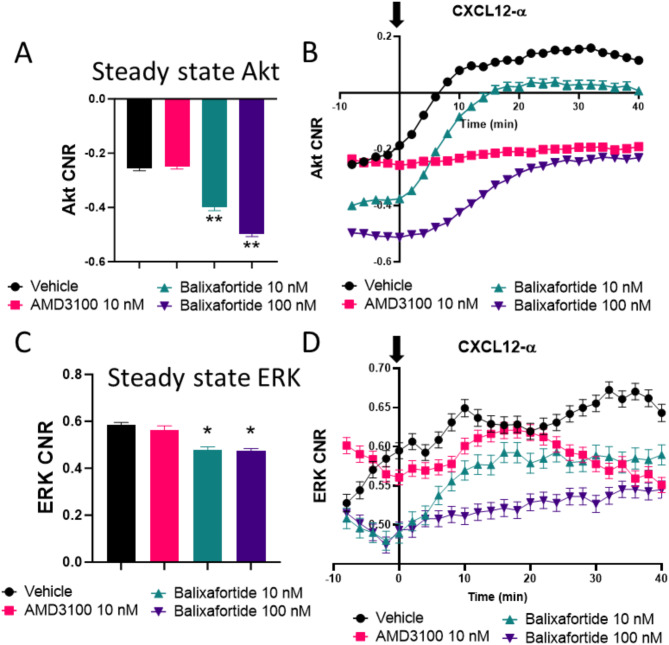
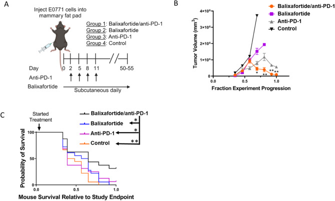
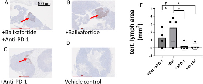
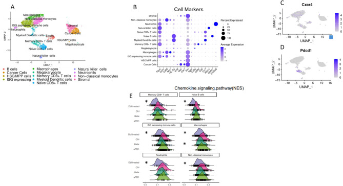
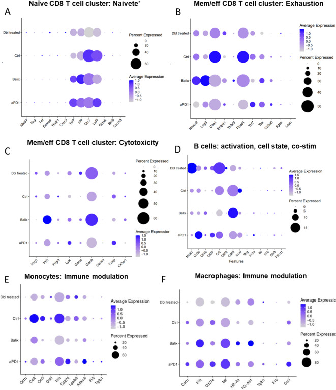
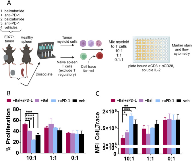
Patients with triple negative breast cancer (TNBC) show only modest response rates to immune checkpoint inhibitor immunotherapy, motivating ongoing efforts to identify approaches to boost efficacy. Using an immunocompetent mouse model of TNBC, we investigated combination therapy with an anti-PD-1 immunotherapy antibody plus balixafortide, a cyclic peptide inhibitor of CXCR4. Cell-based assays demonstrated that balixafortide functions as an inverse agonist, establishing a mode of action distinct from most compounds targeting CXCR4. Combination anti-PD-1 plus balixafortide significantly reduced growth of orthotopic tumors and extended overall survival relative to single agent therapy or vehicle. Adding balixafortide to anti-PD-1 increased numbers of tertiary lymphoid structures, a marker of local tumor immune responses associated with favorable response to immunotherapy in TNBC. Single cell RNA sequencing revealed that combination anti-PD-1 plus balixafortide reduced T cell exhaustion and increased markers of effector T cell activity. Combination therapy also reduced signatures of immunosuppressive myeloid derived suppressor cells (MDSCs) in tumors. MDSCs isolated from mice treated with anti-PD-1 plus balixafortide showed reduced inhibition of T cell proliferation following ex vivo stimulation. These studies demonstrate that combining inhibition of CXCR4 with anti-PD-1 to enhances responses to checkpoint inhibitor immunotherapy in TNBC, supporting future clinical trials.

Keywords: Breast cancer; CXCR4; Checkpoint inhibitor immunotherapy; Myeloid derived suppressor cells.


Conflict of interest statement

Declarations. Competing interests: Spexis (formerly Polyphor LLC) provided partial funding to G.D.L. for this research. No other authors have a competing interest.

Figures

Fig. 1
Fig. 1
Balixafortide functions as an inverse agonist for CXCR4. We measured levels of (A) AKT and C) ERK using kinase translocation reporters (KTRs) in MDA-MB-231 breast cancer cells after treatment with different CXCR4 antagonists as indicated. Bars show mean + SEM values for kinase activity after 30 min of treatment with the listed compounds. Data represent the log2 of cytoplasmic/nuclear fluorescence intensities for each reporter (n > 400 cells per condition). We quantified activation of (B) AKT and D) ERK by KTRs after treatment with 100 ng/ml CXCL12-α added at time 0 (arrow). Data represent mean values for log2 of cytoplasmic/nuclear fluorescence intensities at each time point. We tracked > 300 cells over time for each condition. Error bars denote SEM where visible beyond the symbol. Data for all panels are representative of two independent experiments. *, p < 0.05, **, p < 0.01.
Fig. 2
Fig. 2
Balixafortide improves anti-PD-1 immunotherapy to reduce tumor growth in mice with TNBC. (A) We implanted mice with E0771 breast cancer cells and randomly assigned mice to listed treatment groups on day 2. Groups 1 and 3 received 4 doses of anti-PD-1 antibody at 5 mg/kg per dose every 3 days for 4 cycles. Groups 1 and 2 received 20 mg/kg balixafortide through subcutaneous injection in the rear flank daily. Control mice received matched vehicles only (n = 10 /group). (B) We calculated tumor volumes by caliper measurements. To account for differences in days required to reach pre-defined study endpoints, we normalized the reported duration of each experiment to fraction experiment progression. Presented data are mean values + SEM combined from 2 different experiments. We compared differences across groups at each time point by ANOVA corrected for multiple comparisons. *, p < 0.05; **, p < 0.01. (C) Graph shows overall survival of mice in different treatment groups with log-rank p-values calculated using a Mantel-Cox test. Asterisks in legend symbolize significance (* <0.05; ** <0.01). We normalized data to fraction of days to reach the study endpoint (defined as 1.0), combining two different experiments with different overall days until endpoint.
Fig. 3
Fig. 3
Balixafortide increases area of tertiary lymphatic structures in orthotopic tumors. (A-D) Representative immunohistochemistry images of tumor sections stained by for B lymphocyte marker B220 from mice treated with (A) balixafortide/anti-PD-1, (B) balixafortide, (C) anti-PD-1, and (D) vehicle. Arrows show B cell rich tertiary lymphatic structures. (E) Graph shows mean values and SEM for area occupied by tertiary lymphoid structures on each section. Results are combined data from two independent experiments with slides from 2 separate mice analyzed for each experiment (n = 4 per group). *, p < 0.05 by t-test. NS, not significant.
Fig. 4
Fig. 4
Drop-seq transcriptomic analysis reveals modulation of the tumor microenvironment in mice treated with balixafortide and anti-PD-1. We performed single-cell RNA sequencing on tumors from mice treated with balixafortide/anti-PD-1, balixafortide, anti-PD-1, or vehicle (n = 3/group). (A) Clustered UMAP plot shows annotated populations of cells recovered from tumors. (B) We tested representative marker genes for each annotated cluster using DotPlot to verify proper classification of cells through scType. (C, D) Feature plots from the UMAP in panel A show expression of genes targeted by balixafortide (CXCR4) (C) and anti-PD-1 (Pdcd1) (D) therapies showing majority targets in the immune cell block of the UMAP clusters. (E) Ridge plots represent modulation of genes in the chemokine signaling pathway by various treatments. *, p < 0.05 by Kruskal-Wallis test.
Fig. 5
Fig. 5
Balixafortide and anti-PD-1 reduce markers of T cell exhaustion and immune modulation by myeloid cells. We analyzed single cell RNA sequencing data from Fig. 4 to compare relevant genes that regulate functions of immune cells listed in each panel. Pseudocolor scale in dot plots shows LogFC expression and bubble size represents the percent of cells from the population. (A) T cell Naivete, (B) T cell Exhaustion, (C) T cell cytotoxicity, (D) B cell activation state, (E) monocyte, and (F) macrophage functions.
Fig. 6
Fig. 6
Treatment with balixafortide and anti-PD-1 reduces immunosuppression by myeloid cell populations. (A) We treated mice with E0771 tumors for two weeks as illustrated in Fig. 1 and recovered myeloid cells from tumors with CD11b + immunomagnetic beads. We isolated T cells from a healthy mouse spleen, excluding T regulatory cells with anti-CD25, and plated indicated ratios of myeloid cells to T cells (10:1, 1:1, 0:1) with 0.1ug/mL insoluble anti-CD3 for activation signal. (B) Graph shows mean values + SEM for percent proliferation measured by taking the percentage of T cells gated below the control T cell peak that received no stimulus and thus did not divide. (C) Graph displays mean value + SEM of mean fluorescence intensity (MFI) of cell trace far red as a complementary way to assess T cell division through dye dilution (lower MFI = more proliferation). *, p < 0.05; ** p < 0.01; *** p < 0.001; **** p < 0.0001. Results are representative of two independent experiments.

References

    1. Solinas, C. et al. Targeting immune checkpoints in breast cancer: An update of early results. ESMO Open2, e000255. 10.1136/esmoopen-2017-000255 (2017). - PMC - PubMed
    1. Gruosso, T. et al. Spatially distinct tumor immune microenvironments stratify triple-negative breast cancers. J. Clin. Invest.129, 1785–1800. 10.1172/jci96313 (2019). - PMC - PubMed
    1. Chan, K. K. & Bass, A. R. Autoimmune complications of immunotherapy: Pathophysiology and management. BMJ369, m736. 10.1136/bmj.m736 (2020). - PubMed
    1. Okwundu, N., Grossman, D., Hu-Lieskovan, S., Grossmann, K. F. & Swami, U. The dark side of immunotherapy. Ann. Transl. Med.9, 1041. 10.21037/atm-20-4750 (2021). - PMC - PubMed
    1. Luker, G. D. et al. At the Bench: Pre-clinical evidence for multiple functions of CXCR4 in cancer. J. Leukoc. Biol.109, 969–989. 10.1002/jlb.2bt1018-715rr (2021). - PMC - PubMed

MeSH terms

LinkOut - more resources