. 2025 Mar;639(8056):1076-1083.
doi: 10.1038/s41586-024-08553-z. Epub 2025 Feb 12.

A metagenomic 'dark matter' enzyme catalyses oxidative cellulose conversion

Affiliations

A metagenomic 'dark matter' enzyme catalyses oxidative cellulose conversion

Clelton A Santos et al. Nature. 2025 Mar.

Erratum in

  • Publisher Correction: A metagenomic 'dark matter' enzyme catalyses oxidative cellulose conversion.
    Santos CA, Morais MAB, Mandelli F, Lima EA, Miyamoto RY, Higasi PMR, Araujo EA, Paixão DAA, Junior JM, Motta ML, Streit RSA, Morão LG, Silva CBC, Wolf LD, Terrasan CRF, Bulka NR, Diogo JA, Fuzita FJ, Colombari FM, Santos CR, Rodrigues PT, Silva DB, Grisel S, Bernardes JS, Terrapon N, Lombard V, Filho AJC, Henrissat B, Bissaro B, Berrin JG, Persinoti GF, Murakami MT. Santos CA, et al. Nature. 2025 Apr;640(8058):E7. doi: 10.1038/s41586-025-08872-9. Nature. 2025. PMID: 40119088 Free PMC article. No abstract available.

Abstract

The breakdown of cellulose is one of the most important reactions in nature1,2 and is central to biomass conversion to fuels and chemicals3. However, the microfibrillar organization of cellulose and its complex interactions with other components of the plant cell wall poses a major challenge for enzymatic conversion4. Here, by mining the metagenomic 'dark matter' (unclassified DNA with unknown function) of a microbial community specialized in lignocellulose degradation, we discovered a metalloenzyme that oxidatively cleaves cellulose. This metalloenzyme acts on cellulose through an exo-type mechanism with C1 regioselectivity, resulting exclusively in cellobionic acid as a product. The crystal structure reveals a catalytic copper buried in a compact jelly-roll scaffold that features a flattened cellulose binding site. This metalloenzyme exhibits a homodimeric configuration that enables in situ hydrogen peroxide generation by one subunit while the other is productively interacting with cellulose. The secretome of an engineered strain of the fungus Trichoderma reesei expressing this metalloenzyme boosted the glucose release from pretreated lignocellulosic biomass under industrially relevant conditions, demonstrating its biotechnological potential. This discovery modifies the current understanding of bacterial redox enzymatic systems devoted to overcoming biomass recalcitrance5-7. Furthermore, it enables the conversion of agro-industrial residues into value-added bioproducts, thereby contributing to the transition to a sustainable and bio-based economy.


Conflict of interest statement

Competing interests: C.A.S., F.M., E.A.L., G.F.P. and M.T.M. are named inventors on patent application number BR10202401483 filed by the Brazilian Center for Research in Energy and Materials, covering the use of the enzyme discovered in this study for biomass conversion and related biotechnological applications. The other authors declare no competing interests.

Figures

Fig. 1
Fig. 1. Metagenome of long-term sugarcane bagasse-covered soil.
a, Sampling site indicating the area covered with sugarcane bagasse and an adjacent area where a bulk control soil sample was taken. SBS, sugarcane bagasse-covered soil. b, The alpha diversity index shows that the sugarcane bagasse-covered soil has reduced microbial diversity compared with the control soil. c, Phylogenetic tree illustrating the relationships between the 124 recovered MAGs. Previously undescribed genomes in the taxonomy are highlighted with purple stars. d, Predicted metabolic pathways, glycoside hydrolases (GHs) and the newly described CelOCE in ‘Candidatus Telluricellulosum braziliensis’, highlighting its potential role in cellulose conversion. GHs with low sequence identity (<30%) to known GHs in the CAZy database (https://www.cazy.org/), or those with activities matching their predicted family but not subfamily, are shown in red. Predicted enzymatic activities consistent with their GH family or subfamily classification are indicated by Enzyme Commission (EC) numbers in parentheses. Source Data
Fig. 2
Fig. 2. Function and sequence orthology.
a, Boosting effect on the saccharification of pretreated sugarcane bagasse, microcrystalline cellulose and amorphous cellulose when CelOCE is combined with a cellulolytic enzyme cocktail. Data are the mean ± s.d. from three independent experiments. Statistical significance was determined by one-way analysis of variance (ANOVA) with Tukey’s post hoc test (**P < 0.01). Percentages indicate the difference between treatments. b, High-performance anion-exchange chromatography with pulsed amperometric detection (HPAEC–PAD) profiles of reactions containing reductant and enzymes CelOCE (dark green line), KdgF (orange line) and BacB (blue line). Control reactions using only sugarcane bagasse (dark grey line), only ASC (grey line), sugarcane bagasse and CelOCE, no ASC (light green line), sugarcane bagasse and ASC (light grey line) and with inactivated enzyme (grey–blue line) are also shown. Standard C1-oxidized and non-oxidized cellooligosaccharides are represented by black lines. DP, degree of polymerization; ox, oxidized. c,d, Amorphous (c) and microcrystalline cellulose (d) binding isotherms comparing the enzyme in the presence or absence of a reductant (ASC). The binding isotherms for PASC were fitted using the Langmuir–Freundlich model. The fit for CelOCE without ASC yielded n = 2.5 (n, Langmuir–Freundlich coefficient) and R2 = 0.99, whereas the fit for CelOCE with ASC resulted in n = 2.1 and R2 = 0.99. Data are the mean ± s.d. from three independent experiments. e, SSN depicting three distinct isofunctional clusters of the reference proteins BacB (Protein Data Bank (PDB) ID: 3H7J), KdgF (PDB ID: 5FPZ) and CelOCE (this study). Connections between nodes indicate at least 30% sequence identity with an alignment e-value cut-off of 1 × 10–5. Source Data
Fig. 3
Fig. 3. Crystal structure, copper properties and catalytic requirements.
a, Dimeric arrangement observed in CelOCE crystal structures, highlighting the dimer interface, the location of the active site (blue region encompassing the copper atom) and the cellulose binding site (grey region). b, Octahedral copper coordination sphere in the CelOCE crystal structures, showing the copper-coordinating residues H44, H46, H84 and Q50 as sticks, the copper atom as an orange sphere and water molecules as red spheres. Dashed lines indicate distances in ångström. c, Surface representation of a CelOCE protomer, highlighting the flattened catalytic interface that enables the interaction with the cellulose. The copper-coordinating histidine and proline residues contributing to this unconventional interface are shown. The residue F33, proposed to be involved in disaccharide recognition in the active site pocket, is also shown. d, ITC data for copper binding to CelOCE. The main plot depicts the binding isotherm (green circles) with its theoretical fit (black line). Thermodynamic parameters are shown in the bottom right. Top left inset, thermogram. ΔG, Gibbs free energy change; ΔH, enthalpy change; ΔS, entropy change; T, temperature. e, EPR spectra of CelOCE in the absence (grey line) and presence (green line) of reductant (ASC). The typical EPR spectrum of a Cu2+ centre in the resting state (grey line) is abolished after reduction to Cu+ (green line). a.u., arbitrary units. f, Time-dependent analysis of cellobionic acid production by CelOCE under aerobic and anaerobic conditions and the role of an electron donor (ASC). Anaerobic reactions were conducted with ASC, either in the absence or presence of 100 µM hydrogen peroxide (H2O2). Aerobic reactions were carried out with or without ASC. Data are the mean ± s.d. of three independent experiments. Source Data
Fig. 4
Fig. 4. Cellulose recognition and proposed cleavage mechanism.
a, Proposed model for the simultaneous interaction of CelOCE with cellulose and in situ hydrogen peroxide generation. While one active site engages the non-reducing end of a cellulose chain, the other active site in the homodimer is probably available to generate hydrogen peroxide, the essential co-substrate for cellulose oxidative cleavage. b, Cellotetraose (represented as spheres and sticks) was docked and equilibrated in the CelOCE structure, demonstrating that two glucosyl residues can be accommodated in the active site pocket. The C1 carbon of the −1 glucosyl moiety is positioned favourably for oxidative attack, leading to the production of cellobionic acid as the sole product, which aligns with the biochemical data.
Fig. 5
Fig. 5. Complementary role with classical cellulases.
a, Complementary assays showing cooperative action of CelOCE with endo- and exo-acting cellulases on microcrystalline cellulose. b, Genetic engineering approach used to integrate the sequences encoding CelOCE and L.similis AA9A into the T.reesei genome. c, Saccharification efficiency of the enzyme cocktail produced by the engineered strains under industrially relevant conditions, using pretreated sugarcane bagasse as the technical substrate. Data in a and c are the mean ± s.d. of three independent experiments. In a, statistical significance was determined by one-way ANOVA with Tukey’s post hoc test (***P < 0.001). Source Data
Extended Data Fig. 1
Extended Data Fig. 1. Glycoside hydrolase (GH) annotation.
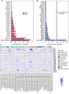
(a) Top-most abundant GH families identified in each metagenome sample. The normalized abundance of each GH family is compared between the data from Sugarcane Bagasse-covered Soil collected 20 cm below ground (SBS) and control soil sample collected from an adjacent not covered with sugarcane bagasse (Bulk). (b) Top-most abundant GH families identified in the uncultured ‘Candidatus Telluricellulosum braziliensis’ MAG. This panel provides a focused view of the GH repertoire potentially involved in plant cell wall degradation by this bacterium. (c) GH distribution in the recovered MAGs. The top 30 MAGs with the highest number of predicted GHs are shown. The heatmap color intensity indicates the relative abundance of each GH family within a given MAG, facilitating visual comparison of GH profiles across the different MAGs. Source Data
Extended Data Fig. 2
Extended Data Fig. 2. CelOCE interaction with microcrystalline cellulose.
(a) Fourier-transform infrared spectroscopy (FTIR) spectra of Avicel untreated or treated with CelOCE. The peaks at 1639 and 1520 cm−1 correspond to the –C = O and –N–H functional groups of CelOCE, respectively. (b) X-ray photoelectron spectroscopy (XPS) patterns for control (untreated) Avicel and Avicel treated with CelOCE, both before and after washing with anionic surfactant (SDS). The survey spectrum of CelOCE-treated cellulose fibers reveals a nitrogen peak (N1s), indicating the presence of N-O, C = N, and C-N groups introduced by the adsorption of CelOCE. The plotted spectra are representative curves from three independent experiments. Source Data
Extended Data Fig. 3
Extended Data Fig. 3. Distribution of CelOCE orthologs across bacterial and archaeal phyla.
Phylogenetic tree illustrates the distribution and potential evolutionary history of CelOCE-like proteins across diverse microbial taxa, encompassing 406 microbial species from 37 bacterial and archaeal phyla. The tree was constructed based on 402 complete genomes from the NCBI RefSeq database and 4 MAGs generated in this study (in red). The ‘Candidatus Telluricellulosum braziliensis’ MAG harboring the celOCE gene is denoted as SBS.bin.55 and indicated with a red asterisk. Phyla with fewer than two species (Thermosulfidibacterota, Thermodesulfobacteriota, Nanoarchaeota, Nitrospirota, and Calditrichota) are unlabeled, except when closely related to the CelOCE-containing MAG.
Extended Data Fig. 4
Extended Data Fig. 4. CelOCE structural properties.
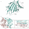
(a) CelOCE adopts a compact jelly-roll fold, which consists of two anti-parallel β-sheets, one containing 6 strands (β-sheet A, including the following strands: β2-β4, β6, β9 and β11) and the other containing 4 strands (β-sheet B, including the following strands: β5, β7, β8 and β10). (b) Cartoon representation of the two CelOCE protomers, indicating the homodimeric arrangement in a back-to-back configuration where their active sites face opposite directions. Key interactions stabilizing the dimer interface are indicated, including M1-C77, A3-S75, K4-D74, and E26-E49. These dimeric interactions primarily involve the N-terminal β-strands (β1) and distances are shown in ångström.
Extended Data Fig. 5
Extended Data Fig. 5. Copper coordination sphere in CelOCE.
(a) When glycerol, a sugar mimetic, is present (structure 2), one water molecule in the copper coordination sphere is replaced by an oxygen atom from glycerol. This causes the remaining water molecule to shift to the equatorial plane, alongside H44, H46, and Q50. (b) Conformational change in copper coordinating histidine at acidic pH. Comparison of two CelOCE crystal structures highlights a conformational change in H44 that occurs under acidic pH conditions. The structure obtained under acidic crystallization condition is shown in light orange (structure 3), while the CelOCE structure with non-flipped H44 is shown in white (structure 2) for comparison. The copper-coordinating residues are represented as sticks, the copper atom as an orange sphere, water molecules as red spheres and glycerol molecules as spheres/sticks following the protein color scheme. Distance measurements (in ångström) pertain to the structure obtained under acidic conditions, specifically showing the altered position of H44 relative to the copper.
Extended Data Fig. 6
Extended Data Fig. 6. Active site of CelOCE.
(a) Surface representation of CelOCE, emphasizing the buried nature of the catalytic copper, located approximately 5 Å from the protein surface. The copper is shown as an orange sphere. (b) Cartoon representation of CelOCE with the docked cellotetraose, demonstrating room for the accommodation of a disaccharide within the active-site pocket (−2 and −1 subsites). (c) Detailed view of the CelOCE active site, demonstrating its stereochemical compatibility with glucosyl moieties. The docked cellooligosaccharide shows the non-reducing end glucosyl (−2) residue anchored primarily by interactions with E96 and Q50, while the −1 glucosyl residue stacks against F33, productively positioning the C1 atom for oxidative attack by the catalytic copper.
Extended Data Fig. 7
Extended Data Fig. 7. The role of electron donor and co-substrate for catalysis.
(a) The role of an electron donor (ASC) in product release under aerobic conditions for CelOCE activity. (b) Cellobionic acid production by CelOCE under aerobic and anaerobic conditions in the presence of ASC as a reductant. (c) Stoichiometric ratio for co-substrate and product formation. The reactions containing 50 mM sodium acetate pH 5.0, 0.1% (w/v) Avicel, 1 mM ASC, 5 or 10 µM H2O2, and 1 µM CelOCE were incubated at 37 °C for 16 h under anaerobic conditions. For a, b and c, results are expressed as mean ± standard deviation from three independent experiments. Statistical significance was determined by one-way ANOVA with Tukey’s post hoc test (***p < 0.001). Source Data
Extended Data Fig. 8
Extended Data Fig. 8. Putative modes of substrate recognition and regioselectivity.
Cellobionic acid was the only product detected, supporting C1-carbon regioselectivity. Although molecular docking studies indicate that the enzyme likely recognizes the non-reducing end of cellulose, we cannot rule out the possibility of reducing end recognition since subsequent catalytic cycles on the same cellulose chain could also generate cellobionic acid. NR, non-reducing end; R, reducing end; Ox, oxidized.
Extended Data Fig. 9
Extended Data Fig. 9. Cellobionic acid production and saccharification efficiency by the secretome of engineered strains.
The secretome of the parental (Br_TrR03) and engineered (CelOCE::Br_TrR03) T. reesei strains were assayed on technical substrates. (a, b) Cellobionic acid production using pretreated sugarcane bagasse. Saccharification efficiency on pretreated eucalyptus chips including the comparison with Cellic® CTec2 and the (LsAA9A::Br_TrR03) strain secretome (c). Cellobionic acid production using pretreated eucalyptus chips (d, e). Increased release of cellobionic acid was observed in the saccharification assays performed with the secretome of the strain co-expressing CelOCE in both technical substrates. Cellobionic acid production was monitored at 24, 48 and 72 h and the HPAEC-PAD product profiles are shown. Results from panels a, c and d are expressed as mean ± standard deviation from three independent experiments. In a and d, statistical significance was determined by one-way ANOVA with Tukey’s post hoc test (*** p < 0.001). Source Data

References

    1. Cragg, S. M. et al. Lignocellulose degradation mechanisms across the tree of life. Curr. Opin. Chem. Biol.29, 108–119 (2015). - PMC - PubMed
    1. Bomble, Y. J. et al. Lignocellulose deconstruction in the biosphere. Curr. Opin. Chem. Biol.41, 61–70 (2017). - PubMed
    1. Lynd, L. R. et al. How biotech can transform biofuels. Nat. Biotechnol.26, 169–172 (2008). - PubMed
    1. Chundawat, S. P. S., Beckham, G. T., Himmel, M. E. & Dale, B. E. Deconstruction of lignocellulosic biomass to fuels and chemicals. Annu. Rev. Chem. Biomol. Eng.2, 121–145 (2011). - PubMed
    1. Quinlan, R. J. et al. Insights into the oxidative degradation of cellulose by a copper metalloenzyme that exploits biomass components. Proc. Natl Acad. Sci. USA108, 15079–15084 (2011). - PMC - PubMed

Supplementary concepts

LinkOut - more resources