. 2025 Jun;32(6):1167-1179.
doi: 10.1038/s41418-025-01457-z. Epub 2025 Feb 12.

STING directly interacts with PAR to promote apoptosis upon acute ionizing radiation-mediated DNA damage

Affiliations

STING directly interacts with PAR to promote apoptosis upon acute ionizing radiation-mediated DNA damage

Yirong Sun et al. Cell Death Differ. 2025 Jun.

Abstract

Acute ionizing radiation (IR) causes severe DNA damage, leading to cell cycle arrest, cell death, and activation of the innate immune system. The role and signaling pathway of stimulator of interferon genes (STING) in IR-induced tissue damage and cell death are not well understood. This study revealed that STING is crucial for promoting apoptosis in response to DNA damage caused by acute IR both in vitro and in vivo. STING binds to poly (ADP‒ribose) (PAR) produced by activated poly (ADP‒ribose) polymerase-1 (PARP1) upon IR. Compared with that in WT cells, apoptosis was suppressed in Stinggt-/gt- cells. Excessive PAR production by PARP1 due to DNA damage enhances STING phosphorylation, and inhibiting PARP1 reduces cell apoptosis after IR. In vivo, IR-induced crypt cell death was significantly lower in Stinggt-/gt- mice or with low-dose PARP1 inhibitor, PJ34, resulting in substantial resistance to abdominal irradiation. STING deficiency or inhibition of PARP1 function can reduce the expression of the proapoptotic gene PUMA, decrease the localization of Bax on the mitochondrial membrane, and thus reduce cell apoptosis. Our findings highlight crucial roles for STING and PAR in the IR-mediated induction of apoptosis, which may have therapeutic implications for controlling radiation-induced apoptosis or acute radiation symptoms. STING responds to acute ionizing radiation-mediated DNA damage by directly binding to poly (ADP-ribose) (PAR) produced by activated poly (ADP-ribose) polymerase-1 (PARP1), and mainly induces cell apoptosis through Puma-Bax interaction. STING deficiency or reduced production of PAR protected mice against Acute Radiation Syndrome.


Conflict of interest statement

Competing interests: The authors declare no competing interests.

Figures

None
STING responds to acute ionizing radiation-mediated DNA damage by directly binding to poly (ADP-ribose) (PAR) produced by activated poly (ADP-ribose) polymerase-1 (PARP1), and mainly induces cell apoptosis through Puma-Bax interaction. STING deficiency or reduced production of PAR protected mice against Acute Radiation Syndrome.
Fig. 1
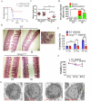
Fig. 1. STING deficiency improves survival and protects against IR-mediated tissue damage.
A Survival of WT C57BL/6 J and Stinggt-/gt- C57BL/6 J after exposure to 16.7 Gy SBI. F, female; M, male. B H&E staining of WT C57BL/6 J and Stinggt-/gt- C57BL/6 J intestines with (IR) and without (NT) abdominal radiation at 4 dpi (4 days post irradiation). C Villus height of the intestine in WT C57BL/6 J and Stinggt-/gt- C57BL/6 J mice at 4 dpi with (IR) and without (NT) abdominal radiation. The data are presented as the means ± SEMs (n = 5; two-way ANOVA; *p < 0.05; **p < 0.01; ***p < 0.001). D Crypt loss villous atrophy (percent of bowel affected) after IR in WT and Stinggt-/gt- mice with and without abdominal radiation. N. D., Not detected. ***p < 0.001. E The level of cytotoxicity of BMDMs at 6 h postirradiation (6 hpi) was measured by LDH release into the supernatant. F Survival rates of WT and Stinggt-/gt- derived BMDMs exposed to 20 and 40 Gy radiation at 6 hpi. The data are reported as the means ± standard deviations (s.d.) (n = 6; *p < 0.05; **p < 0.01; ***p < 0.001). G THP-1 and STING-/- THP-1 cell morphology was detected via transmission electron microscopy (TEM) (Hitachi HT-7800).
Fig. 2
Fig. 2. Loss of STING function suppresses the apoptotic pathway.
A TUNEL staining of the intestines of WT and Stinggt-/gt- mice at 0 and 4 dpi. B Quantification of TUNEL-positive cells in the intestine after SBI at 0 and 4 dpi. The percentages of TUNEL-stained areas in all intestine regions were statistically analyzed. C BMDM viability was measured by annexin V-FITC-propidium iodide staining of WT and Stinggt-/gt- BMDMs after exposure to the indicated dose of IR. D Quantification of Annexin V and propidium iodide labeling. E The number of apoptotic cells after exposure to the indicated dose of IR. Apoptotic cells were quantified with a Cell-APOPercentage™ apoptosis kit at 550 nm. F Evaluation of PARP1 and caspase 3 cleavage in WT BMDMs and Stinggt-/gt- BMDMs after exposure to the indicated dose of IR by Western blotting. The data are presented as the means ± SEMs (two-way ANOVA; *p < 0.05; **p < 0.01; ***p < 0.001).
Fig. 3
Fig. 3. The STING response to DNA damage after IR is associated with PAR-PARP1.
A Confocal images of the interactions between PARP1-Alex647 and STING-GFP via a Zeiss Elyra-7 microscope. B Quantification of the interaction between PARP1-Alex647 and STING-GFP via ImageJ software. Colocalization analysis was carried out via Manders’ colocalization coefficient (MCC) method. C Evaluating the association of STING with PARP1 in BMDMs. The cell lysates were immunoprecipitated with anti-STING beads, followed by immunoblotting with the indicated antibodies. D Coimmunoprecipitation and immunoblotting of the interaction of STING with PAR in THP-1 cells subjected to 30 Gy IR for 6 h. The cell lysates were immunoprecipitated with anti-STING beads, followed by immunoblotting with the indicated antibodies. E, F Microscopy images of PAR-Alex647 and STING-GFP interactions captured with ZEISS Elyra-7 (E) and quantified with ImageJ software (F). G Validation of the ability of PLA to detect the proximity between STING and PAR or PARP1 in MDA-MB-231 cells. Nuclei were stained with DAPI (blue); PLA was performed for STING and PAR or PARP1 (red). CON, non-irradiated. H, I The binding of STING and PAR in vitro was analyzed by nondenaturing polyacrylamide gel electrophoresis and immunoblotting with anti-STING (H) and anti-PAR (I) antibodies. J THP-1 cells were transfected with the indicated concentrations of PAR and cGAMP via Lipofectamine. PAR (0.2 μM) induced the phosphorylation of STING. The data are presented as the means ± SEMs (unpaired Student’s t test; ***, p < 0.001).
Fig. 4
Fig. 4. The PARP1 inhibitor PJ34 protects cells and mice against IR.
A BMDMs treated with the control vehicle or diABZI (1 μg/ml) were subjected to 20 Gy radiation. The level of cytotoxicity of BMDMs at 6 hpi was measured by LDH release into the supernatant. B, C Evaluation of PAR in cells treated with vehicle or the indicated dose of PJ34 by immunoblotting (B), and the relative concentrations were calculated (C). N.D., not detected. D Evaluation of BMDM viability after treatment with the indicated dose of PJ34 (μM) with or without IR (20 Gy). E, F Compared with the control (DMSO), 1 mg/kg PJ34 increased the resistance of the mice to 16.2 Gy SBI (E) and (F) inhibited weight loss after 8 days. Male mice with the same age were randomly divided into two groups. G H&E staining of C57BL/6 J mouse intestines subjected to IR or not subjected to IR (CON) and treated with 1 mg/kg PJ34 or the control vehicle (NT) at 4 dpi. H Villus height of intestines subjected to the indicated dose of SBI and treated with PJ34 or the vehicle control. I Crypt loss villous atrophy (percent of bowel affected) after IR in mice with and without abdominal radiation and PJ34 (1 mg/kg) treatment. The data are presented as the means ± SEMs (n = 5; two-way ANOVA; *, p < 0.05; **, p < 0.01; ***, p < 0.001).
Fig. 5
Fig. 5. A precise dosage of PJ34 could reduce small intestinal tissue damage and apoptosis.
A, B PJ34 blunts the intestinal expression of the proinflammatory cytokines IL-1β and IL-6 (ELISA) at 4 dpi after SBI. N.T., not treated. C TUNEL staining of the colons of C57BL/6 J mice treated with DMSO (control) or PJ34 (1 mg/kg) at 4 dpi. D Quantification of TUNEL signals in the intestines after SBI at 0 and 4 dpi. The apoptotic index was calculated by counting the TUNEL signals in 50 randomly selected crypts. The values are presented as the means ± SDs (n = 5 in each group). E PJ34 (3 μM) decreased PAR levels and reduced both cell apoptosis and the phosphorylation of STING. F, G The colocalization of STING and PAR in BMDMs after IR was repressed with 1 μg/ml PJ34. H STING-Alexa 488 and PAR-Alexa 647 staining of C57BL/6 J mouse intestines treated with DMSO (control) or PJ34 (1 mg/kg) at 4 dpi. I Quantification of the colocalization of STING-Alexa 488 and PAR-Alexa 647 in the intestine after SBI at 0 and 4 dpi. N. D., Not detected. The data are presented as the means ± SEMs (n = 5; two-way ANOVA; *, p < 0.05; ***, p < 0.001; ****, p < 0.0001).
Fig. 6
Fig. 6. STING deficiency or PARP inhibition blunts the DNA-sensing-mediated pathway upon IR.
A Immunoblotting analysis of TBK1, P65, and IRF3 phosphorylation in WT and Stinggt-/gt- BMDMs after IR. B, C Evaluating the expression levels of Ifnb1 and Ip10 after IR in WT and Stinggt-/gt- BMDMs via qRT‒PCR. The data are presented as the means ± SEMs (n = 5; two-way ANOVA; *, p < 0.05; **, p < 0.01; ***, p < 0.001). D Evaluating the expression levels of Il1b and Il6 at 6 hpi in WT and Stinggt-/gt- BMDMs via qRT‒PCR. E, F Expression of the cytokines Ifnb1 and Ip10 after IR in BMDMs treated with 3 μM PJ34. G, H PJ34 blunts the intestinal expression of Ifnb1 and Ip10 at 4 dpi after SBI. N.T., not treated; CON, treated with DMSO as a control; The data are presented as the means ± SEMs (unpaired Student’s t test; *, p < 0.05; **, p < 0.01; ***, p < 0.001).
Fig. 7
Fig. 7. IR-mediated apoptosis increased mitochondrial Bax in a STING-dependent manner.
A‒D Evaluating the expression of BH3 proapoptotic proteins in WT and Stinggt-/gt- BMDMs after IR (20 Gy or 40 Gy) by qRT‒PCR. Fold changes and n-fold changes were compared to those of untreated WT BMDMs. E Immunoblotting analysis of mitochondrial Bax in WT and Stinggt-/gt- BMDMs after IR, as evaluated by Western blotting. CL: cell lysate, Mito: mitochondrial protein. F Microscopy images of cytochrome C in WT and Stinggt-/gt- BMDMs with and without IR. G Quantification of the level of cytochrome C via ImageJ software. H Quantification of BH3 proapoptotic protein expression in WT BMDMs treated with 3 μM PJ34 or vehicle at the indicated time points after IR (20 Gy) by qRT‒PCR. When the folds changed, the n-fold change was compared to that of N.T. (untreated). The data are presented as the means ± SEMs (two-way ANOVA; *, p < 0.05; **, p < 0.01; ***, p < 0.001).

References

    1. Cytlak UM, Dyer DP, Honeychurch J, Williams KJ, Travis MA, Illidge TM. Immunomodulation by radiotherapy in tumour control and normal tissue toxicity. Nat Rev. 2022;22:124–38. - PubMed
    1. Kirsch DG, Santiago PM, di Tomaso E, Sullivan JM, Hou WS, Dayton T, et al. p53 controls radiation-induced gastrointestinal syndrome in mice independent of apoptosis. Science. 2010;327:593–6. - PMC - PubMed
    1. Schaue D, Micewicz ED, Ratikan JA, Xie MW, Cheng G, McBride WH. Radiation and Inflammation. Semin Radiat Oncol. 2015;25:4–10. - PMC - PubMed
    1. Hu B, Jin C, Li HB, Tong J, Ouyang X, Cetinbas NM, et al. The DNA-sensing AIM2 inflammasome controls radiation-induced cell death and tissue injury. Science. 2016;354:765–8. - PMC - PubMed
    1. Qiu W, Carson-Walter EB, Liu H, Epperly M, Greenberger JS, Zambetti GP, et al. PUMA regulates intestinal progenitor cell radiosensitivity and gastrointestinal syndrome. Cell Stem Cell. 2008;2:576–83. - PMC - PubMed

MeSH terms

LinkOut - more resources