. 2025 Jun;46(6):1742-1756.
doi: 10.1038/s41401-025-01477-y. Epub 2025 Feb 12.

Acyl-CoA thioesterase 8 induces gemcitabine resistance via regulation of lipid metabolism and antiferroptotic activity in pancreatic ductal adenocarcinoma

Affiliations

Acyl-CoA thioesterase 8 induces gemcitabine resistance via regulation of lipid metabolism and antiferroptotic activity in pancreatic ductal adenocarcinoma

Bo-Rui Li et al. Acta Pharmacol Sin. 2025 Jun.

Abstract

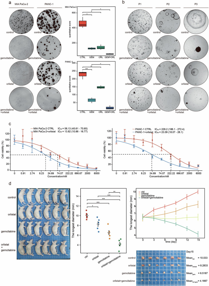
Pancreatic ductal adenocarcinoma (PDAC) comprises a group of highly malignant tumors of the pancreas. Metabolic reprogramming in tumors plays a pivotal role in promoting cancer progression. However, little is known about the metabolic alterations in tumors that drive cancer drug resistance in patients with PDAC. Here, we identified acyl-CoA thioesterase 8 (ACOT8) as a key player in driving PDAC gemcitabine (GEM) resistance. The expression of ACOT8 is significantly upregulated in GEM-resistant PDAC tissues and is closely associated with poor survival in patients with PDAC. Gain- and loss-of-function studies have shown that ACOT8 drives PDAC GEM resistance both in vitro and in vivo. Mechanistically, ACOT8 regulates cellular cholesterol ester (CE) levels, decreases the levels of phosphatidylethanolamines (PEs) that bind to polyunsaturated fatty acids and promote peroxisome activation. The knockdown of ACOT8 promotes ferroptosis and increases the chemosensitivity of tumors to GEM by inducing ferroptosis-associated pathway activation in PDAC cell lines. The combination of orlistat, an ACOT8 inhibitor, and GEM significantly inhibited tumor growth in PDAC organoid and mouse models. This study reveals the biological importance of ACOT8 and provides a potential combination therapy for treating patients with advanced GEM-resistant PDAC.


Conflict of interest statement

Competing interests: The authors declare no competing interests.

Figures

Fig. 1
Fig. 1. Acyl-CoA thioesterase 8 (ACOT8) is significantly overexpressed in patients with gemcitabine-resistant pancreatic ductal adenocarcinoma (PDAC) and is associated with high malignancy and a poor prognosis.
a, b Differences in gene expression at the transcriptional level between the gemcitabine-resistant and gemcitabine-sensitive groups. c Differential expression of ACOTs between the RESIS and SENS groups. d Data from PAAD_GSE148673 show that ACOT8 is enriched in epithelial and malignant cells in PDAC. e The expression of ACOT8 in PDAC was determined via The Cancer Genome Atlas (TCGA) database. f Scoring of samples with different staining intensities in tissue microarrays (TMAs). g ACOT8 expression was significantly higher in tumor tissues (T) than in paraneoplastic tissues (TP). h ACOT8 expression was positively correlated with malignancy. i Overall survival (OS) analysis showing that patients with high ACOT8 expression levels had a poor prognosis. For data shown in this figure, statistical analyses were conducted using methods such as the t test, one-way ANOVA, and the log-rank test. *P < 0.05, **P < 0.01, ***P < 0.001.
Fig. 2
Fig. 2. ACOT8 positively regulates the proliferative and invasive capacity of pancreatic cancer cells both in vivo and in vitro.
ac Stable ACOT8 transcripts from AsPC-1, MIA PaCa-2 and PANC-1 cells were verified via qRT‒PCR, Western blotting and immunofluorescence. d Effects of ACOT8 overexpression or knockdown on the proliferative capacity of PDAC cells. e Effects of ACOT8 overexpression or knockdown on colony formation assay results in PDAC cells. f Effects of ACOT8 overexpression or knockdown on Transwell assay results in PDAC cells. g Effects of ACOT8 overexpression or knockdown on scratch test results in PDAC cells. h Effects of ACOT8 overexpression or knockdown on subcutaneous PDAC cell tumor formation in nude mice. For data shown in this figure, statistical analyses were performed using methods such as the t test, one-way ANOVA, and two-way ANOVA. *P < 0.05, **P < 0.01, ***P < 0.001.
Fig. 3
Fig. 3. ACOT8 overexpression enhances gemcitabine resistance in pancreatic cancer cells.
a ACOT8 expression was determined after induction by the addition of gradient gemcitabine. b In three different patient-derived (patient 1/2/3) organoid models, low-dose gemcitabine (20 nM) was significantly more potent in the ACOT8-knockdown group than in the control group. c Compared with those in the control group, the morphology of MIA PaCa-2 and PANC-1 cells in the ACOT8-knockdown group was more pronounced under the effect of gemcitabine, whereas cells in the ACOT8-overexpression group maintained a more normal morphology than cells in the control group. d In the colony formation assay, low-dose (30 nM) gemcitabine significantly inhibited colony formation in the ACOT8-knockdown group but weakly inhibited colony formation in the ACOT8-overexpression group. e Determination of the IC50 of gemcitabine in AsPC-1, MIA PaCa-2 and PANC-1 cells after ACOT8 overexpression or knock down. For data shown in this figure, statistical analysis was conducted via one-way ANOVA, and log-logistic analysis was employed to establish a fitting model for the drug resistance experiment. *P < 0.05, **P < 0.01, ***P < 0.001.
Fig. 4
Fig. 4. Transcriptome sequencing clustering revealed the ferroptotic pathway, and functional assays validated the inhibitory effect of ACOT8 on ferroptosis.
a Comparisons of genes upregulated in the overexpression group, genes downregulated in the two knockout groups, and genes highly expressed in drug-resistant patients were performed, resulting in the identification of two common differentially expressed genes: ACOT8 and SLC7A11. b Pathway clustering of the differentially expressed genes revealed the ferroptotic pathway. c Differential mRNA expression of ferroptosis-related genes in different ACOT8 expression groups. d Detection of lipid peroxidation levels by flow cytometry in cells after ACOT8 knock down (with the ferroptosis probe BODIPY 581/591C11). e Detection of lipid peroxidation levels by immunofluorescence in cells after ACOT8 knock down (with the ferroptosis probe BODIPY 581/591C11). f, g Detection of ROS and MDA levels in cells after ACOT8 knock down. For data shown in this figure, statistical analysis was conducted via one-way ANOVA. **P < 0.01, ***P < 0.001, ****P < 0.0001.
Fig. 5
Fig. 5. Inducing ferroptosis in pancreatic cancer cells reduces the degree of gemcitabine resistance caused by high ACOT8 expression.
ac Erastin (ERA, 3 μM) reversed the gemcitabine resistance caused by ACOT8 overexpression. For data shown in this figure, log-logistic analysis was employed to establish a fitting model for the drug resistance experiment. ***P < 0.001.
Fig. 6
Fig. 6. Acyl-CoA thioesterase 8 (ACOT8) inhibits ferroptosis by regulating the expression of core ferroptosis-related genes, the metabolism of cholesterol and polyunsaturated phosphatidylethanolamine (PE), and the number and function of peroxisomes.
a Effects of altering ACOT8 expression on the expression of ferroptosis-related genes. b Altered expression levels of GPX4 and SLC7A11 verified by immunohistochemistry (IHC) to stain subcutaneous tumor blocks in mice. c, d Verification of the ability of 3 μM erastin to reverse ferroptosis resistance induced by ACOT8 overexpression via the ferroptosis probe BODIPY 581/591C11 and ROS detection. e Untargeted lipid transcriptome sequencing of MIA PaCa-2 cells after ACOT8 knock down. f Changes in the content and composition of cholesterol and PE in cells after ACOT8 knock down. g Typical ferroptosis in the mitochondria of cells after ACOT8 knock down was observed by transmission electron microscopy (red arrows: mitochondria; blue arrows: peroxisomes). h Altered expression of the peroxisome marker catalase in the ACOT8-knockdown group. For data shown in this figure, statistical analysis was conducted via one-way ANOVA. **P < 0.01, ***P <0.001, ****P < 0.0001.
Fig. 7
Fig. 7. The lipid-lowering drug orlistat reverses the protective effect of acyl-CoA thioesterase 8 (ACOT8) on ferroptosis in pancreatic cancer cells by inhibiting ACOT8 expression.
a Expression of ACOT8, SLC7A11, and GPX4 after 2 weeks of treatment of cells with graded concentrations of orlistat. b In the colony formation assays, 30 μM orlistat reversed the increase in AsPC-1 cell proliferation caused by ACOT8 overexpression. c, d Orlistat (30 μM in cells) reversed ferroptosis resistance caused by ACOT8 overexpression when hemin was included in the background. e, f Orlistat (30 μM in cells; 10 μM in organoids) reversed ferroptosis resistance caused by ACOT8 overexpression when hemin was included in the background. g Orlistat reversed catalase overexpression caused by ACOT8 overexpression. h Effects of orlistat on cholesterol and PE contents in the ACOT8-overexpression group. For data shown in this figure, statistical analysis was conducted via one-way ANOVA. **P < 0.01.
Fig. 8
Fig. 8. The lipid-lowering drug orlistat enhances the gemcitabine sensitivity of PDAC in vivo and in vitro.
a Effects of 30 μM orlistat and 50 nM gemcitabine alone or in combination on MIA PaCa-2 and PANC-1 cells in colony formation assays. b Effects of 20 μM orlistat and 20 nM gemcitabine alone or in combination on organoids. c Changes in the IC50 of gemcitabine in combination with orlistat. d Effects of an intraperitoneal injection of 30 mg/kg orlistat and 10 mg/kg gemcitabine alone or in combination on subcutaneous tumor formation in nude mice. *P < 0.05, **P < 0.01, ***P < 0.001.
Fig. 9
Fig. 9
ACOT8 inhibited gemcitabine-induced pancreatic cancer cell  ferroptosis by regulating ferroptosis-related gene pathways, organelle function, and lipid metabolism, thereby inducing pancreatic cancer gemcitabine resistance.

References

    1. Siegel RL, Miller KD, Jemal A. Cancer statistics, 2020. CA Cancer J Clin. 2020;70:7–30. - PubMed
    1. Rahib L, Smith BD, Aizenberg R, Rosenzweig AB, Fleshman JM, Matrisian LM. Projecting cancer incidence and deaths to 2030: the unexpected burden of thyroid, liver, and pancreas cancers in the United States. Cancer Res. 2014;74:2913–21. - PubMed
    1. Neoptolemos JP, Kleeff J, Michl P, Costello E, Greenhalf W, Palmer DH. Therapeutic developments in pancreatic cancer: current and future perspectives. Nat Rev Gastroenterol Hepatol. 2018;15:333–48. - PubMed
    1. Hunt MC, Solaas K, Kase BF, Alexson SE. Characterization of an acyl-coA thioesterase that functions as a major regulator of peroxisomal lipid metabolism. J Biol Chem. 2002;277:1128–38. - PubMed
    1. Ishizuka M, Toyama Y, Watanabe H, Fujiki Y, Takeuchi A, Yamasaki S, et al. Overexpression of human acyl-CoA thioesterase upregulates peroxisome biogenesis. Exp Cell Res. 2004;297:127–41. - PubMed

MeSH terms