. 2025 Apr;39(4):888-898.
doi: 10.1038/s41375-025-02520-x. Epub 2025 Feb 12.

Immunotherapy targeting a leader sequence cathepsin G-derived peptide

Affiliations

Immunotherapy targeting a leader sequence cathepsin G-derived peptide

Chunhua Shi et al. Leukemia. 2025 Apr.

Abstract

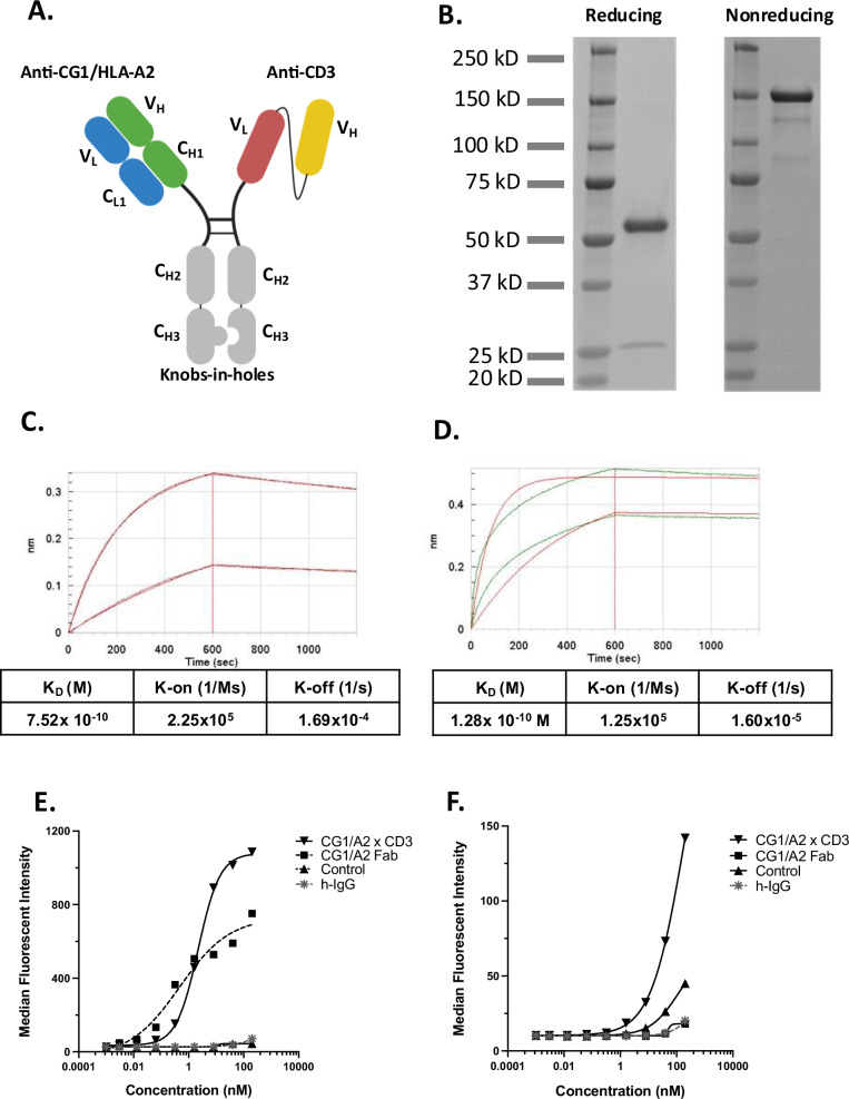
Myeloid azurophil granules provide a rich source of intracellular leukemia antigens. Cathepsin G (CG) is a serine protease that has higher expression in acute myeloid leukemia (AML) blasts in comparison to normal myeloid progenitors. Based on the unique biology of HLA-A*0201 (HLA-A2), in which presentation of leader sequence (LS)-derived peptides is favored, we focused on the LS-CG-derived peptide CG1 (FLLPTGAEA). We previously detected CG1/HLA-A2 complexes on the surface of primary HLA-A2+ AML blasts and cell lines, and immunity targeting CG1/HLA-A2 in leukemia patients. T cell receptor (TCR)-mimic (m) antibodies are immunotherapeutic antibodies that target peptide-HLA (pHLA) complexes. Here we report on the engineering, preclinical efficacy, and safety evaluation of a novel CG1/HLA-A2-targeting, T cell-engager, bispecific antibody (CG1/A2xCD3). CG1/A2xCD3 showed high binding affinity to CG1/HLA-A2 monomers, CD3-Fc fusion protein, and to AML and T cells, with potent killing of HLA-A2+ primary AML and cell lines in vitro and in vivo. This correlated with both tumor- and CG1/A2xCD3-dependent T cell activation and cytokine secretion. Lastly, CG1/A2xCD3 had no activity against normal bone marrow. Together, these results support the targeting of LS-derived peptides and the continued clinical development of CG1/A2xCD3 in the setting of AML.


Conflict of interest statement

Ethics approval and consent to participate: All methods were performed in accordance with the University of Texas MD Anderson Cancer Center guidelines and regulations for laboratory research. Research approval was obtained through the MD Anderson Cancer Center IRB-approved tissue collection protocol (Protocol# LAB99-062) and IACUC animal protocol (Protocol# 00001336-RN02). Informed consent was obtained from all study participants prior to tissue collection. This study did not contain any direct research on human subjects; only archived human tissue. No identifiable images from human research participants were included in this manuscript. Competing interests: The authors declare no competing interests.

Figures

Fig. 1
Fig. 1. CG1/A2xCD3 design and characterization.
A Structure of CG1/A2xCD3 bispecific antibody showing the CG1/HLA-A2 Fab, the anti-CD3e scFv and knobs-in-holes. B SDS-PAGE of CG1/A2xCD3 under reducing and non-reducing conditions. Reducing conditions yielded two bands, consistent with heavy chain and light chain. The lower band is primarily composed of the CG1-HLA-A2 binding moiety light chain, since the CD3e binding moiety is composed of scFv. Intact CG1/A2xCD3 is detected under nonreducing conditions. Octet® bio-layer interferometry (BLI) was used to determine the binding affinity of CG1/A2xCD3 to CD3 (C) and to CG1/HLA-A2 (D) monomers. CD3e-Fc and CG1/HLA-A2 monomers were added at 100 nM and 20 nM concentrations. The fitted (red) and actual (green) binding curves are shown. Binding of CG1/A2xCD3 to CD3e demonstrated KD = 7.52 × 10−10 M; K-on = 2.25 × 105 1/M × seconds [Ms]; K-off = 1.69 × 10−4 1/second [s]. Binding of CG1/A2xCD3 to CG1/HLA-A2 monomers demonstrated KD = 1.28 × 10−10 M; K-on = 1.25 × 105 1/Ms; K-off = 1.60 × 10−5 1/s. Cell surface binding of CG1/A2xCD3 to EM2 (E) and Jurkat T cells (F) was measured using flow cytometry. HLA-A2 + EM2 CML cells express CG1 on cell surface and therefore demonstrate high affinity binding of CG1/A2xCD3 for CG1/A2. Staining of Jurkat T cells shows high affinity binding of CG1/A2xCD3 to CD3; Jurkat cells express CD3 and lack HLA-A2, CG and CG1. As control antibodies, we used CG1/A2 Fab, human IgG (h-IgG), and Velo8/CD138-CD3e (control) bispecific antibody.
Fig. 2
Fig. 2. CG1/A2xCD3 activates T cells and lyses CG1/HLA-A2-expressing myeloid leukemia.
U937-A2 AML cells (AC), ML2 AML cells (DF) and EM2 CML cells (GI) were co-cultured with PBMC and CG1/A2xCD3 for 24 h. Addition of CG1/A2xCD3 led to T cell activation, as measured using flow cytometry staining for CD69 (A, D, G) and increased cytokine section (IFNg, IL-2, and TNF-a) (B, E, H) by T cells in comparison with control bispecific antibody. C, F, I CG1/A2xCD3 increased the killing of target myeloid leukemia calls in comparison with control bispecific antibody. ***p < 0.001.
Fig. 3
Fig. 3. CG1/A2xCD3 has potent in vivo activity against myeloid leukemia.
A Schema of experimental design. Female NSG mice were engrafted with luciferase-transduced ML2 cells via tail vein injection of 1 × 105 cells. After 1 week, 1 × 107 normal donor PBMC  were administered via tail vein injection. Four days after PBMC injection, mice were treated increasing doses of CG1/A2xCD3 (0.1, 0.05, and 0.01 mg/Kg) or PBS. B, C Bioluminescence imaging of mice shows a dose dependent decrease in bioluminescence after treatment with CG1/A2xCD3 in comparison with PBS. D Percentage of CD3+ T cells in blood after treatment over time. IHC analysis shows lower MPO staining (E, F), lower numbers of tumor cells by H&E staining (G, H), and higher bone marrow restoration of normal hematopoiesis (I, J) in the CG1/A2xCD3-treated group in contrast with the PBS-treated groups. ****p < 0.0001; ***p < 0.001; **p < 0.01.
Fig. 4
Fig. 4. CG1/A2xCD3 eliminated primary AML in vivo.
Patient primary HLA-A2+ AML sample was engrafted into NSG mice via tail vein injection. After confirming engraftment using flow cytometry staining of mouse blood for hCD45, 1 × 107 normal donor PBMC were infused intravenously to mice via tail vein. Mice were treated intraperitoneally weekly for 3 weeks with increasing doses of CG1/A2xCD3 (1.0, 0.1, and 0.01 mg/Kg) or PBS. A Leukemia burden in the blood was identified by flow cytometry staining for hCD45+/hCD3- cells. B Kaplan-Meir curves showing increased survival of CG1/A2xCD3-treated mice, in comparison with PBS.
Fig. 5
Fig. 5. CG1-bispecific antibody does not inhibit normal hematopoiesis.
Healthy donor HLA-A2+ bone marrow (BM) was cultured alone or co-cultured with peripheral blood mononuclear cells (PBMC) at increasing concentrations of CG1/A2xCD3 or control bispecific antibody. Cells were then resuspended in methylcellulose semi-solid matrix on low adhesion 6-well plates and co-cultured for 14 days. On day 14, colonies were counted and imaged using an inverted light microscope. Each group was cultured at least in duplicate and data represent four independent experiments. Cytarabine-treated BM was used as positive control.

References

    1. Holmberg-Thyden S, Dufva IH, Gang AO, Breinholt MF, Schejbel L, Andersen MK, et al. Epigenetic therapy in combination with a multi-epitope cancer vaccine targeting shared tumor antigens for high-risk myelodysplastic syndrome—a phase I clinical trial. Cancer Immunol Immunother. 2022;71:433–44. - PMC - PubMed
    1. Qazilbash MH, Wieder E, Thall PF, Wang X, Rios R, Lu S, et al. PR1 peptide vaccine induces specific immunity with clinical responses in myeloid malignancies. Leukemia. 2017;31:697–704. - PMC - PubMed
    1. Rezvani K, Yong AS, Mielke S, Savani BN, Musse L, Superata J, et al. Leukemia-associated antigen-specific T-cell responses following combined PR1 and WT1 peptide vaccination in patients with myeloid malignancies. Blood. 2008;111:236–42. - PMC - PubMed
    1. Molldrem JJ, Clave E, Jiang YZ, Mavroudis D, Raptis A, Hensel N, et al. Cytotoxic T lymphocytes specific for a nonpolymorphic proteinase 3 peptide preferentially inhibit chronic myeloid leukemia colony-forming units. Blood. 1997;90:2529–34. - PubMed
    1. Molldrem JJ, Lee PP, Wang C, Felio K, Kantarjian HM, Champlin RE, et al. Evidence that specific T lymphocytes may participate in the elimination of chronic myelogenous leukemia. Nat Med. 2000;6:1018–23. - PubMed