. 2025 Feb 12;25(1):49.
doi: 10.1186/s12906-025-04780-7.

Ribes nigrum leaf extract downregulates pro-inflammatory gene expression and regulates redox balance in microglial cells

Affiliations

Ribes nigrum leaf extract downregulates pro-inflammatory gene expression and regulates redox balance in microglial cells

Alvard Minasyan et al. BMC Complement Med Ther. .

Abstract

Background: This study focuses on the investigation of the antioxidant and anti-inflammatory activities of alcohol extracts from Ribes nigrum leaves on murine BV-2 microglial Wt and Acyl-CoA oxidase 1 deficient (Acox1-/-) cell line models, useful for the investigation of some neurodegenerative disorders.

Methods: The extract chemical composition was analyzed via LC-Q-Orbitrap HRMS. Various assays, including DPPH, MTT, and H2DCFDA, were used to assess the extract's antioxidant capacity, cell viability, and reactive oxygen species (ROS) production. Immunoblotting and RT-qPCR techniques were employed to measure protein expression and gene transcription in treated cells. Statistical analysis was conducted using GraphPad Prism, with significance determined at p < 0.05.

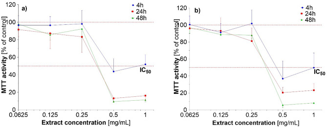
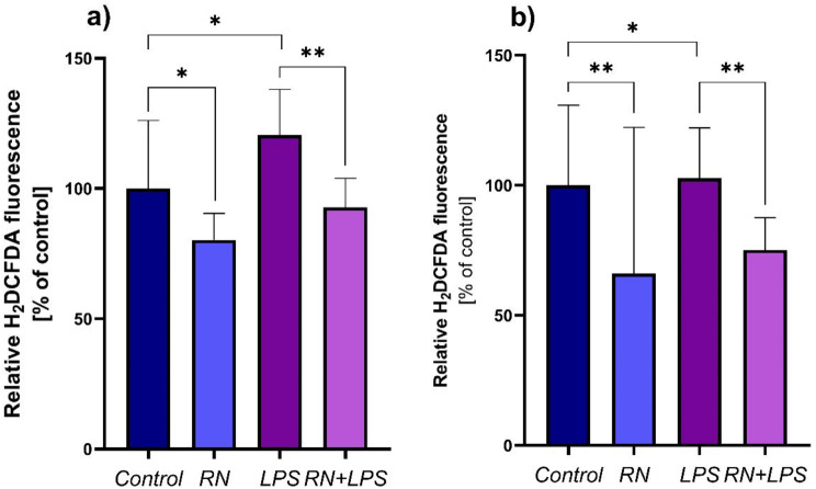
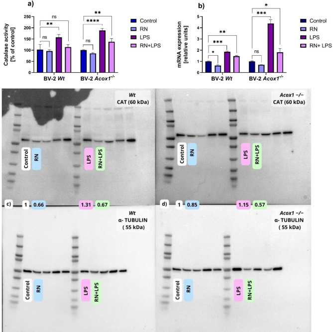
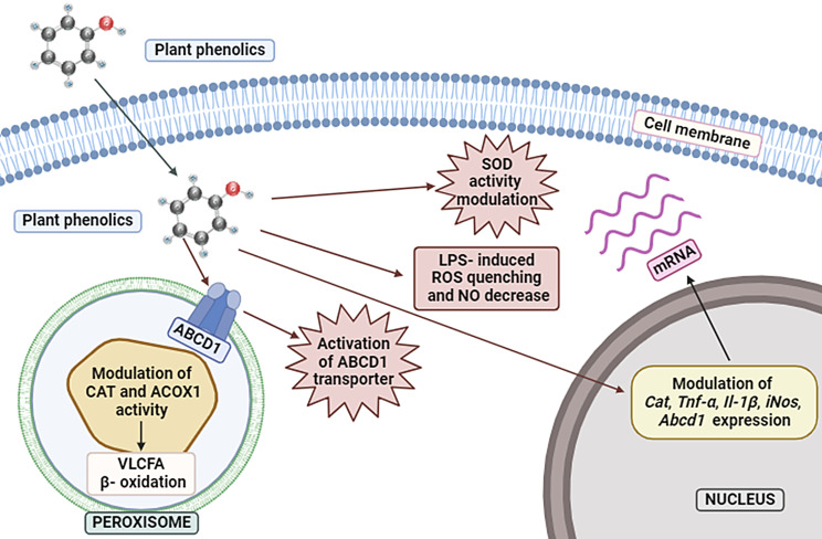
Results: Investigations showed the presence of phenolic compounds in this extract, among which flavan-3-ols, flavonols, furanocoumarins, hydroxycinnamates were major components, which are known for their biological activity in various test systems. The MTT test revealed a concentration of 0.125 mg/mL of R. nigrum extract as the highest non-toxic. The investigated extract showed high antioxidant activity in chemical-based tests. The antioxidant potential of the R. nigrum leaf extract was furtherly explored using the BV-2 microglial cell line models. Moreover, the extract was found to alter the activity of the main antioxidant enzyme, catalase and fatty acid oxidation enzyme, Acyl-CoA oxidase 1 (ACOX1) as well as the expression of appropriate genes in Wt and Acox1-/- BV-2 microglial cells such as Cat, iNos, Il-1β, Tnf-α, and Abcd1. In Wt cells, after the 24-hour treatment with R. nigrum leaf extract, ACOX1 activity was downregulated, meanwhile the catalase activity remains unchanged. Further treatment led to the downregulation of catalase and the upregulation of ACOX1 activity. However, in Acox1-/- cells, which represent a model of oxidative stress, an increase in catalase activity was observed only after 48 h of treatment. It was also observed the reduced ROS and NO formation in cells, showing the pronounced antioxidant capacity of R. nigrum extract in the investigated cell-models.

Conclusion: Our study demonstrated the protective effects of R. nigrum leaf extracts on BV-2 microglial cells by reducing oxidative and nitrosative stress, decreasing pro-inflammatory gene expression, and normalizing peroxisomal function, highlighting the potential of these extracts as therapeutic agents for managing oxidative stress and inflammation.

Keywords: Armenian flora; Neurodegenerative disorders; Oxidative stress; Polyphenols; Reactive oxygen species.


Conflict of interest statement

Declarations. Ethics approval and consent to participate: There is no need to obtain the licenses for the plant sampling as the selected plant is not wild-growing and the leaves are considered as bio-waste. Consent for publication: Not applicable. Plant ethics: The R. nigrum plant was cultivated at the Lori province (Armenia, 1600–1650 m a.s.l.) and harvested during the fruiting period (July 2019) as suggested in literature [33]. The cultivation was carried out without the treatment with any fertilizer or pesticide. The identification of the plant was carried out at the Department of Botany and Mycology, Yerevan State University (YSU), Armenia. The plant samples are available at the Department of Microbiology & Plants and Microbes Biotechnology, Biology Faculty, Yerevan State University, Yerevan, Armenia. The voucher specimen number was not provided for the cultivated plant species. There is not any permissions or licenses needed for harvesting of the cultivated plant samples. Competing interests: The authors declare no competing interests.

Figures

Fig. 1
Fig. 1
The LC chromatographic profile of R. nigrum extract monitored either by Quadrupole-Orbitrap High resolution MS (black HRMS chromatograms) or UV light (orange UV chromatograms). (The UV chromatogram of extract at 265–325 nm. The peak numbers refer to those implemented in Table 2.)
Fig. 2
Fig. 2
Effect of R. nigrum leaf extract on the cell proliferation and viability of the Wt (a) and the Acox1−/− BV-2 (b) microglial cells for 4-, 24- or 48-hours treatment period (MTT test). The untreated cells were considered as control. The results are means ± SD of two independent experiments performed in triplicate (p ≤ 0.05)
Fig. 3
Fig. 3
Effects of R. nigrum leaf extract on ROS production induced by LPS in the Wt (a) and Acox1 −/− (b) BV-2 microglial cells assessed by the H2 DCFDA (5 µM) dye test. Cells were incubated for 24 h with RN (0.125 mg/mL) in the absence or presence of LPS (1 µg/mL). All values are presented as means ± SD of two independent experiments performed in triplicate (*p < 0.1; and **p < 0.01). Statistical significance was determined using one-way ANOVA followed by Tukey’s test. The α-tocopherol did not show any statistically significant effect
Fig. 4
Fig. 4
Effect of R. nigrum extract on the NO production induced by LPS in the culture media of the Wt (a) and Acox1−/− (b) BV-2 microglial cells assessed by the Griess test. Cells were incubated for 24 h with RN (0.125 mg/mL) in the absence or the presence of LPS (1 µg/mL). All values are presented as means ± SD of two independent experiments performed in triplicate (*p < 0.1; **p < 0.01; ***p < 0.001; ****p < 0.0001 and ns, not significant). Statistical significance was determined using one-way ANOVA followed by Tukey’s test
Fig. 5
Fig. 5
Effect of R. nigrum leaf extract on the gene expression of iNos (a), Tnf-α (b), Il-1β (c) and Abcd1 (d) with or without the LPS influence. All values are presented as means ± SD of two independent experiments performed in triplicate (*p < 0.1; **p < 0.01; ***p < 0.001; ****p < 0.0001 and ns, not significant). Statistical significance was determined using two-way ANOVA followed by Tukey’s test for multiple comparisons
Fig. 6
Fig. 6
Effect of R. nigrum extract on catalase activity in the Wt and Acox1−/− BV-2 microglial cells (a), and on ACOX1 activity (b) in Wt BV-2 microglial cells. All values are presented as means ± SD of two independent experiments performed in triplicate (*p < 0.1; **p < 0.01; ***p < 0.001 and ns, not significant). Statistical significance was determined using two-way ANOVA followed by Tukey’s test for multiple comparisons (a) and one-way ANOVA followed by Tukey’s test (b). The α-tocoferol did not show any statistically significant effect
Fig. 7
Fig. 7
Effect of R. nigrum extract on the level of catalase activity (a) and on the expression of the peroxisomal Cat mRNA (b) in the Wt and in Acox1−/− BV-2 microglial cells. Panels (c) and (d) show the results of immunoblotting assay of RN effect on the expression of the peroxisomal protein CAT in Wt (c) and in Acox1−/− (d) BV-2 microglial cells. Cells were incubated for 24 h with RN (0.125 mg/mL) in the absence or presence of LPS (1 µg/mL). Cell lysates were analyzed by PAGE-SDS electrophoresis and subjected to immunoblotting. Band intensities were analyzed by densitometry and standardized to α-tubulin expression level. Table values represent standardized densitometric analysis obtained after the signal intensity quantification of different proteins. All values are presented as means ± SD of two independent experiments performed in triplicate (*p < 0.1; **p < 0.01; ***p < 0.01 and ns, not significant). Statistical significance was determined using one-way ANOVA followed by Tukey’s test for multiple comparisons
Fig. 8
Fig. 8
The schematic representation of influence of the phenolic components of RN extract on microglial BV-2 Wt cells

References

    1. Loreto Palacio P, Godoy JR, Aktas O, Hanschmann E-M. Changing perspectives from oxidative stress to Redox Signaling—Extracellular Redox Control in Translational Medicine. Antioxidants. 2022;11:1181. - PMC - PubMed
    1. Sharifi-Rad M, Anil Kumar NV, Zucca P, Varoni EM, Dini L, Panzarini E et al. Lifestyle, oxidative stress, and antioxidants: back and forth in the pathophysiology of Chronic diseases. Front Physiol. 2020;11. - PMC - PubMed
    1. Gupta N, Verma K, Nalla S, Kulshreshtha A, Lall R, Prasad S. Free radicals as a double-edged Sword: the Cancer Preventive and therapeutic roles of Curcumin. Molecules. 2020;25:5390. - PMC - PubMed
    1. Sahakyan G, Vejux A, Sahakyan N. The role of oxidative stress-mediated inflammation in the Development of T2DM-Induced Diabetic Nephropathy: possible preventive action of tannins and other Oligomeric Polyphenols. Molecules. 2022;27(24). - PMC - PubMed
    1. Santolini J, Wootton SA, Jackson AA, Feelisch M. The Redox architecture of physiological function. Curr Opin Physiol. 2019;9:34–47. - PMC - PubMed

LinkOut - more resources