. 2025 Jan 30;17(3):510.
doi: 10.3390/nu17030510.

Selected Plant Extracts Regulating the Inflammatory Immune Response and Oxidative Stress: Focus on Quercus robur

Affiliations

Selected Plant Extracts Regulating the Inflammatory Immune Response and Oxidative Stress: Focus on Quercus robur

Rawan Nehme et al. Nutrients. .

Abstract

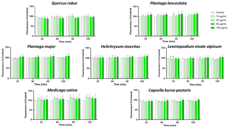
Background/Objectives: Inflammation is a vital response of the immune system, frequently linked to the development and progression of numerous chronic and autoimmune diseases. Targeting inflammation represents an attractive strategy to prevent and treat these pathologies. In this context, many pathways, including pro-inflammatory cytokines secretion, NFκB activation, reactive oxygen species (ROS) production, inflammasome activation and arachidonic acid metabolism could be highlighted and addressed. Several plant materials have traditionally been used as effective and non-harmful anti-inflammatory agents. However, well-established scientific evidence is lacking, and their mechanisms of action remain unclear. The current article compares the effects of seven plant extracts, including Quercus robur L. (Oak), Plantago lanceolata L. (narrowleaf plantain), Plantago major L. (broadleaf plantain), Helichrysum stoechas L. (immortelle or helichrysum), Leontopodium nivale alpinum Cass. (edelweiss), Medicago sativa L. (alfafa) and Capsella bursa-pastoris Moench (shepherd's purse) on different inflammatory pathways. Results: All of the plant extracts significantly affected ROS production, but their action on cytokine production was more variable. As the Quercus robur extract showed the highest efficacy in our models, it was subsequently assessed on several inflammatory signaling pathways. Quercus robur significantly decreased the secretion of IFNγ, IL-17a, IL-12, IL-2, IL-1β and IL-23 in stimulated human leucocytes, and the expression of TNFα, IL-6, IL-8, IL-1β and CXCL10 in M1-like macrophages. Additionally, a significant reduction in PGE2 secretion, COX2, NLRP3, caspase1 and STAT3 expression and NFκB p65 phosphorylation was observed. Conclusions: Our results clearly indicate that Quercus robur has a potent anti-inflammatory effect, making it a promising candidate for both the treatment and prevention of inflammation and related diseases, thereby promoting overall well-being.

Keywords: Quercus robur; antioxidant; inflammation; macrophages; plant extracts.


Conflict of interest statement

The authors declare the following financial interests/personal relationships which may be considered as potential competing interests: A. Chervet, A. Rousset and J.-Y. Berthon were employed by the company Greentech. The remaining authors declare that the research was conducted in the absence of any commercial or financial relationships that could be construed as a potential conflict of interest.

Figures

Figure 1
Figure 1
Production of ROS by blood leukocytes with different concentrations of plant extracts. Cells were stimulated with PMA (1 µM) and treated with the indicated concentrations of extracts for 30, 60, 90 and 120 min, and then ROS production was measured. Data are shown as means ± SEM (Control = 100%); * p < 0.05 compared with Control.
Figure 2
Figure 2
Effect of plant extract on leukocyte viability. Cells were treated with the indicated concentrations of extracts for 2 h, and then cell viability was measured. Data are shown as means ± SEM (Control = 100%); * p < 0.05 compared with Control.
Figure 3
Figure 3
Impact of plant extracts on PBMC cytokine secretions: Cells were incubated with or without PHA (5 µg/mL) and plant extracts (0 or 50 µg/mL) for 24 h. Cytokine levels were measured with Luminex Bio-Plex 200 System. Data are shown as mean ± SEM (Control = 100% indicated by the red line) and analyzed using one-way ANOVA followed by Dunett’s post-hoc test (n = 3). * p < 0.05, ** p < 0.01, *** p < 0.001 compared with Control.
Figure 4
Figure 4
Quercus robur extract characterization: (A) Chromatogram with compounds identified (numbers 1 to 5 in the chromatogram represent the compounds identified in (B)). (B) Compounds identified in the Quercus robur extract (comparison with analytical standard or according to literature data [20]). (C) Chemical structure of the main identified compounds.
Figure 5
Figure 5
Effect of the Quercus robur extract on macrophage polarization: THP-1 cells were supplemented with PMA to allow their activation and then treated with LPS and IFNγ for the polarization into M1-type. The effect of the Quercus robur extract (50 µg/mL) was evaluated on (A) M0-type macrophages and (B) M1-type macrophages. Gene expression was quantified by real-time PCR and normalized using β-actin as an internal control. (C) Macrophages were analyzed by flow cytometry. The representative side scatter area plots are shown for CD14 (M0) and CD80 (M1) labeling (Blue represent cells that are negative for both markers, green represent cells that are CD14+/CD80 and orange represent cells that are positive for both CD14 and CD80). (D) Percentage of M0 (CD14+) and M1 (CD14+ CD80+ and CD14 CD80+) macrophages in treated and untreated cells using the total number of labelled cells as the total number of cells. Data are expressed as mean ± SEM and * p < 0.05 and ** p < 0.01compared with Control.
Figure 6
Figure 6
Effect of Quercus robur on several inflammatory pathways and antioxidant response in blood leukocytes and PBMCs. Blood leukocytes were incubated with or without LPS (10 µg/mL) and Quercus robur (0 or 50 µg/mL). The expression level of different genes was quantified by real-time PCR and normalized using β-actin as an internal control. (A) Expression of COX-2 gene. (B) Secretion of PGE2 in supernatant compared to untreated cells. (C) Expression of inflammasome effector genes. (D) Expression of STAT-3 gene. (E) Expression of antioxidant-sensitive genes. (F) Effect of Quercus robur on p65 phosphorylation in PBMCs. Cells were incubated with or without LPS (10 µg/mL) and the extract (50 µg/mL) for 2 h. Pellets from treated cells were collected and NFκB phosphorylation was measured using the NFκB p65 (Total/Phospho) Human InstantOne™ ELISA kit. Data are expressed as mean ± SEM (Control = 100%) (n = 3). * p < 0.05, ** p < 0.01.

References

    1. Germolec D.R., Shipkowski K.A., Frawley R.P., Evans E. Markers of Inflammation. Methods Mol. Biol. Clifton NJ. 2018;1803:57–79. doi: 10.1007/978-1-4939-8549-4_5. - DOI - PubMed
    1. Askenase M.H., Sansing L.H. Stages of the Inflammatory Response in Pathology and Tissue Repair after Intracerebral Hemorrhage. Semin. Neurol. 2016;36:288–297. doi: 10.1055/s-0036-1582132. - DOI - PMC - PubMed
    1. Harvanová G., Duranková S., Bernasovská J. The Role of Cytokines and Chemokines in the Inflammatory Response. Alergol. Pol. Pol. J. Allergol. 2023;10:210–219. doi: 10.5114/pja.2023.131708. - DOI
    1. Funes S.C., Rios M., Escobar-Vera J., Kalergis A.M. Implications of Macrophage Polarization in Autoimmunity. Immunology. 2018;154:186–195. doi: 10.1111/imm.12910. - DOI - PMC - PubMed
    1. Meizlish M.L., Franklin R.A., Zhou X., Medzhitov R. Tissue Homeostasis and Inflammation. Annu. Rev. Immunol. 2021;39:557–581. doi: 10.1146/annurev-immunol-061020-053734. - DOI - PubMed

MeSH terms

LinkOut - more resources