

Identification of MCM2-Interacting Proteins Associated with Replication Initiation Using APEX2-Based Proximity Labeling Technology
- PMID: 39940790
- PMCID: PMC11816892
- DOI: 10.3390/ijms26031020
Identification of MCM2-Interacting Proteins Associated with Replication Initiation Using APEX2-Based Proximity Labeling Technology
Abstract
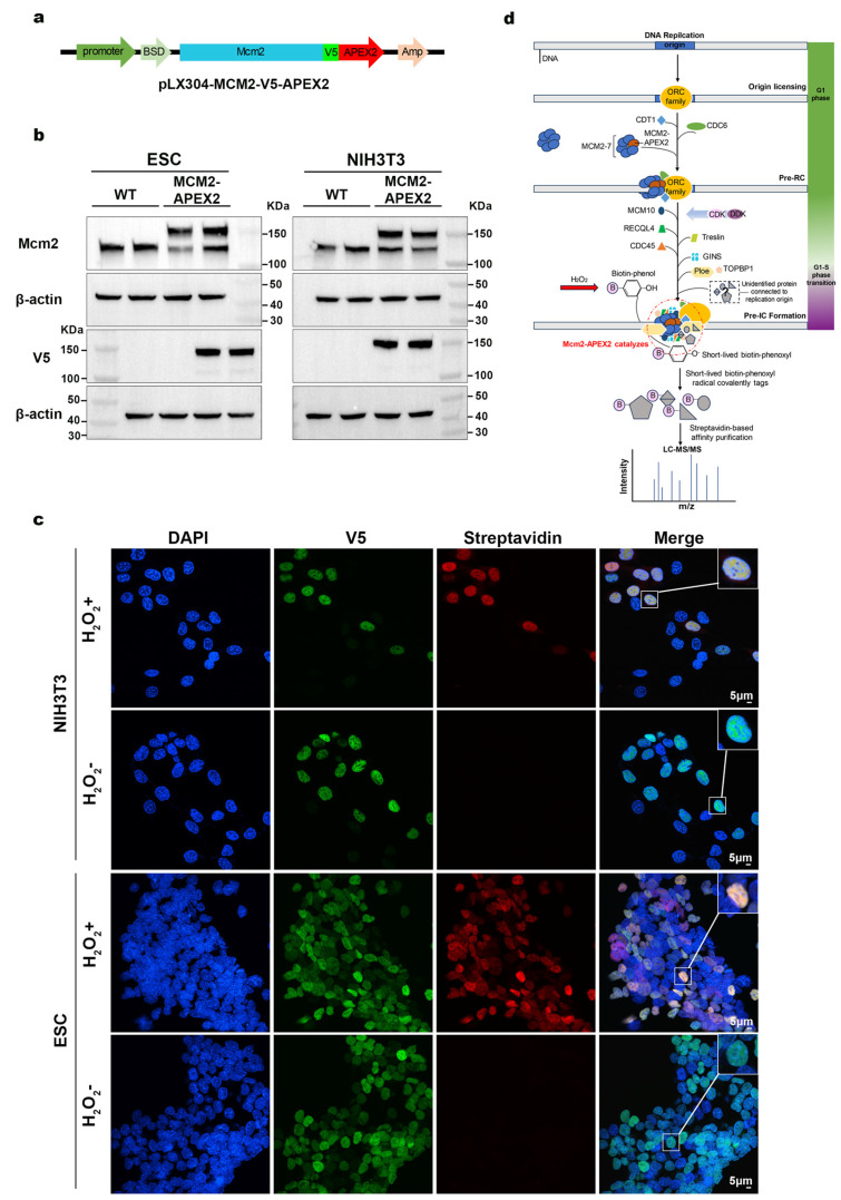
DNA replication is a crucial biological process that ensures the accurate transmission of genetic information, underpinning the growth, development, and reproduction of organisms. Abnormalities in DNA replication are a primary source of genomic instability and tumorigenesis. During DNA replication, the assembly of the pre-RC at the G1-G1/S transition is a crucial licensing step that ensures the successful initiation of replication. Although many pre-replication complex (pre-RC) proteins have been identified, technical limitations hinder the detection of transiently interacting proteins. The APEX system employs peroxidase-mediated rapid labeling with high catalytic efficiency, enabling protein labeling within one minute and detection of transient protein interactions. MCM2 is a key component of the eukaryotic replication initiation complex, which is essential for DNA replication. In this study, we fused MCM2 with enhanced APEX2 to perform in situ biotinylation. By combining this approach with mass spectrometry, we identified proteins proximal to the replication initiation complex in synchronized mouse ESCs and NIH/3T3. Through a comparison of the results from both cell types, we identified some candidate proteins. Interactions between MCM2 and the candidate proteins CD2BP2, VRK1, and GTSE1 were confirmed by bimolecular fluorescence complementation. This research establishes a basis for further study of the component proteins of the conserved DNA replication initiation complex and the transient regulatory network involving its proximal proteins.
Keywords: APEX2; DNA replication; MCM2; proximity labeling.
Conflict of interest statement
The authors declare no conflicts of interest.
Figures


References
-
- Chen L., Li N., Zhang M., Sun M., Bian J., Yang B., Li Z., Wang J., Li F., Shi X., et al. APEX2-based proximity labeling of Atox1 identifies CRIP2 as a nuclear copper-binding protein that regulates autophagy activation. Angew. Chem. Int. Ed. Engl. 2021;60:25346–25355. doi: 10.1002/anie.202108961. - DOI - PubMed
MeSH terms
Substances
Grants and funding
- 32100460/National Natural Science Foundation of China
- 2019YFA0903800/National Key R&D Program of China
- 32090031/Major Program of the National Natural Science Foundation of China
- XDB0480000/Strategic Priority Research Program of the Chinese Academy of Sciences
- 32070610/General Program of the National Natural Science Foundation of China
- 32000580/National Natural Science Foundation of China for Young Scholars
- 32101178/National Natural Science Foundation of China for Young Scholars
- 2021B1515020109/Guangdong Province Fund for Distinguished Young Scholars
- 2021A1515110377/Guangdong Basic and Applied Basic Research Foundation
- JCHZ20200005, DWKF20210001, ZTXM20190019/Shenzhen Institute of Synthetic Biology Scientific Research Program
- 2021M693301, 2021M703386/Project funded by China Postdoctoral Science Foundation
- 2023B10564003/Doublefirst-class discipline promotion project
- 2021A1515110483/Guangdong Basic and Applied Basic Research Foundation
LinkOut - more resources
Full Text Sources
Miscellaneous
