. 2025 Jan 26;26(3):1042.
doi: 10.3390/ijms26031042.

Loss of Tyrosine Phosphatase Mu Promotes Scoliosis Progression Through Osteopontin-α5β1 Integrin Signaling and PIPK1γ90 Activity

Affiliations

Loss of Tyrosine Phosphatase Mu Promotes Scoliosis Progression Through Osteopontin-α5β1 Integrin Signaling and PIPK1γ90 Activity

Mohamed Elbakry et al. Int J Mol Sci. .

Abstract

Adolescent idiopathic scoliosis (AIS) is characterized by a curvature of the spine affecting approximately 4% of the pediatric population, and the mechanisms driving its progression remain poorly understood. Whole-exome sequencing of a French-Canadian AIS cohort with severe scoliosis identified rare variants in the PTPRM gene, which encodes Protein Tyrosine Phosphatase μ (PTPµ). However, these rare variants alone did not account for the pronounced reduction in PTPµ at both mRNA and protein levels in severe AIS cases. This led us to investigate epigenetic regulators and the identification of five microRNAs (miR-103a-3p, miR-107, miR-148a-3p, miR-148b-3p, and miR-152-3p) that target PTPRM mRNA. These microRNAs were significantly elevated in plasma from severe AIS patients, and miR-148b-3p was also upregulated in AIS osteoblasts. Phenotypic analysis of bipedal Ptrprm knockout (PTPµ -/-) mice showed increased prevalence and severity of scoliosis, while quadrupedal PTPµ -/- mice did not develop scoliosis, underscoring PTPµ's role as a disease-modifying factor. Mechanistically, PTPµ deficiency was found to disrupt Gi-coupled receptor signaling in osteoblasts by enhancing the interaction between osteopontin (OPN) and α5β1 integrin, along with increased tyrosine phosphorylation of phosphatidylinositol-4-phosphate 5-kinase type I (PIPKIγ90). These findings provide novel insights into the molecular mechanisms underlying spinal deformity progression in AIS, linking PTPµ depletion to aberrant OPN-α5β1 integrin signaling and highlighting potential therapeutic targets to stop, mitigate, or prevent scoliosis.

Keywords: Gi-coupled receptor signaling; OPN-α5β1 integrin; PIPK1γ90; PTPRM variants; PTPµ; PTPµ-null mice; adolescent idiopathic scoliosis; microRNAs; osteoblast; spinal deformity.


Conflict of interest statement

This work led to a patent application (pending) owned by CHU Sainte-Justine. The authors have declared that no other potential conflicts of interest exist.

Figures

Figure 1
Figure 1
Analysis of PTPRM gene and protein expression in osteoblasts derived from scoliotic and non-scoliotic subjects. Panel (A) illustrates a qPCR analysis to assess the expression levels of PTPRM in primary osteoblasts derived from scoliotic patients (AIS) compared to non-scoliotic trauma cases considered here as healthy controls (HC) and revealed a two-fold reduction in PTPRM mRNA level in osteoblasts from scoliotic patients compared to the HC group (*** p > 0.001, t-test). Panel (B) shows a representative Western blot analysis of cell lysates demonstrating a 50% reduction in PTPµ protein levels in osteoblasts from idiopathic scoliosis patients compared to non-scoliotic controls. The experiment was repeated multiple times using different healthy controls and AIS patients. Another representative blot is shown in Supplementary Figure S1, showing 4 HC and 4 AIS patients. PTPµ protein levels were quantified by densitometry using ImageJ software and normalized to Na+/K+ ATPase. PTPµ protein levels were significantly reduced in AIS osteoblasts compared to HC. Data are representative of multiple independent experiments.
Figure 2
Figure 2
Effect of PTPµ-deficiency on plasma OPN and scoliosis in bipedal mice. Panel (A) represents a quantitative comparison of plasma osteopontin (OPN) levels in bipedal wild-type (WT) and PTPµ −/− mice. Plasma OPN levels were determined by ELISA at the indicated time point after bipedal surgery in 33 wild-type mice and 58 PTPµ −/− mice. Error bars represent standard deviation. Panel (B) summarizes the frequency of occurrence of scoliosis as induced by bipedal surgery in WT and PTPµ −/− mice. Panels (C,D) are representative spine radiographies showing a striking difference in spinal curvature between (C) bipedal WT and (D) bipedal PTPµ −/− mice.
Figure 3
Figure 3
Osteoblasts from bipedal PTPµ-deficient mice demonstrate less response to GiPCR stimulation. Osteoblasts from bipedal wild-type (WT) and PTPµ −/− mice were stimulated with increasing concentrations of Apelin-17 (A), oxymethazolin (B), or somatostatin (C). Concentration–response curves were generated with Prism software (version 6.03) using data normalized to the response achieved at maximal stimulation in osteoblasts from bipedal WT mice. (*** p > 0.001, t-test). Data are expressed as mean ± standard error of the mean of experiments performed three times in duplicate for at least 12 mice per genotype.
Figure 4
Figure 4
Osteoblasts from bipedal PTPµ-deficient mice demonstrate less response to GiPCR stimulation following rOPN treatment. Osteoblasts from bipedal wild-type (WT) and PTPµ −/− mice were treated with recombinant OPN (rOPN) before Gi-coupled receptor stimulation with Apelin-17 (A), oxymethazolin (B), or somatostatin (C). Concentration–response curves were generated with Prism software using data normalized to the response achieved at maximal stimulation in osteoblasts from bipedal WT mice. Data are expressed as mean ± standard error of the mean of experiments performed three times in duplicate for at least 12 mice per genotype.
Figure 5
Figure 5
OPN silencing rescues Gi-coupled receptor signaling impairment in PTPµ −/− osteoblast. Osteoblasts from bipedal wild-type (WT) and PTPµ −/− mice were transfected with scramble (Scrb) or osteopontin (OPN) siRNA (siOPN) and efficiency of siRNA was verified with qPCR 48 h after transfection (n = 12 per genotype) (A) and by Western blot (n = 2 per genotype) (B) using α-Tubulin as the internal control. The effect of OPN silencing on Gi-coupled receptor signaling was evaluated by cellular dielectric spectroscopy by treating mouse osteoblasts with increasing concentrations of Apelin-17 (C), oxymethazolin (D), or somatostatin (E). Concentration–response curves were generated with Prism software. Symbols: open circle = PTPµ −/− mice transfected with Scrb; closed circle = wild-type (WT) mice transfected with siOPN; open diamond = PTPµ −/− mice transfected with siOPN and closed diamond = wild-type (WT) mice transfected with Scrb. Data are expressed as mean ± standard error of the mean of experiments performed three times in duplicate for at least 12 mice per genotype. * p < 0.05, ** p < 0.01 versus Scrb-transfected WT group based on one-way ANOVA, then followed by the Dunnett’s post doc test.
Figure 6
Figure 6
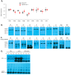
Loss of PTPµ contributes to increase the affinity of α5β1 integrin toward OPN. (A) Total RNA was extracted from osteoblasts of bipedal wild-type (WT) and PTPµ −/− mice, and mRNA expression levels of CD44 and indicated beta (β) alpha (α) integrins were examined by qPCR analysis using β-actin as the internal control. Error bars show standard error of the mean of three independent experiments performed in duplicate. (B) WT and PTPµ −/− osteoblasts (samples from 3 mice per genotype were pooled (n = 3)) were lysed and 20 µg of proteins were resolved by 10% SDS-PAGE and immunoblotted for antibodies specific for the indicated proteins. OPN served as the loading control. (C) WT and PTPµ −/− osteoblasts were lysed and subjected to immunoprecipitation (samples from 3 mice per genotype were pooled (n = 3)) with antibodies directed against each integrin subunit as indicated, including CD44 and OPN, and immunoprecipitates were resolved by 10% SDS-PAGE. Western blots were revealed with an antibody raised against OPN. OPN served as the loading control. The molecular weights for the integrins β1, β3, β5, β8, α1, α4, α5, αv, CD44, OPN are respectively 138kDa, 125kDa, 100kDa, 97kDa, 150kDa, 150kDa, 150kDa, 135kDa, 81kDa and 66kDa. (D) MC3T3-E1 cells (mouse osteoblasts) treated with PBS or rOPN were subjected to immunoprecipitation with antibodies against the Gi1, Gi2, or Gi3 alpha subunit. Precipitates were resolved by 10% SDS-PAGE and immunoblotted with an antibody directed against phospho-serine. Cells were pre-treated with antibody against β1 integrin for 30 min followed by an 18 h incubation with 0.5 µg/mL rOPN, prior to immunoprecipitation and immunoblotting. Bands shown are representative of results obtained with independent experiments. A second replicative experiment is shown in Supplementary Figure S6.
Figure 7
Figure 7
PIPKIγ90 is deregulated in osteoblasts from PTPµ −/− mice. (A) Cell lysates were prepared with osteoblasts from WT and PTPµ −/− mice and proteins subjected to immunoprecipitation with anti-PIPKIγ90 antibody. Immunoprecipitates were resolved by 10% SDS-PAGE and immunoblotted with an antibody directed against phospho-tyrosine or PIPKIγ90 as the control band (n = 1 per genotype). Data shown is a representation, of a replicated experiment. (B) Osteoblasts from WT and PTPµ −/− mice were transfected with scramble (Scrb) or OPN siRNA and efficiency of siRNA was verified with qPCR 48 h after transfection (n = 3) and by Western blot (samples from 3 mice per genotype were pooled (n = 3)) (C), with β-actin serving as the loading control. (D) Effect of PIPKIγ90 depletion on impedance signature of Gi-coupled receptor signaling was evaluated by cellular dielectric spectroscopy by challenging WT and PTPµ −/− osteoblasts with 10 µM somatostatin and analyzed with prism software. Data are expressed as mean ± SEM of experiments performed three times in duplicate for at least 12 mice per genotype. *** p < 0.001, versus (WT + Scrb) based on one-way ANOVA followed by Dunnett’s post doc test.

References

    1. Cheng J.C., Castelein R.M., Chu W.C., Danielsson A.J., Dobbs M.B., Grivas T.B., Gurnett C.A., Luk K.D., Moreau A., Newton P.O., et al. Adolescent idiopathic scoliosis. Nat. Rev. Dis. Primers. 2015;1:15030. doi: 10.1038/nrdp.2015.30. - DOI - PubMed
    1. Dickson R.A. The etiology and pathogenesis of idiopathic scoliosis. Acta Orthop. Belg. 1992;58((Suppl. 1)):21–25. - PubMed
    1. Machida M. Cause of idiopathic scoliosis. Spine. 1999;24:2576–2583. doi: 10.1097/00007632-199912150-00004. - DOI - PubMed
    1. Burwell R.G. Aetiology of idiopathic scoliosis: Current concepts. Pediatr. Rehabil. 2003;6:137–170. doi: 10.1080/13638490310001642757. - DOI - PubMed
    1. Jiang X., Liu F., Zhang M., Hu W., Zhao Y., Xia B., Xu K. Advances in genetic factors of adolescent idiopathic scoliosis: A bibliometric analysis. Front. Pediatr. 2024;11:1301137. doi: 10.3389/fped.2023.1301137. - DOI - PMC - PubMed

Substances

LinkOut - more resources