. 2025 Jan 30;26(3):1215.
doi: 10.3390/ijms26031215.

Evaluation of a Norcantharidin Nanoemulsion Efficacy for Treating B16F1-Induced Melanoma in a Syngeneic Murine Model

Affiliations

Evaluation of a Norcantharidin Nanoemulsion Efficacy for Treating B16F1-Induced Melanoma in a Syngeneic Murine Model

Gabriel Martínez-Razo et al. Int J Mol Sci. .

Abstract

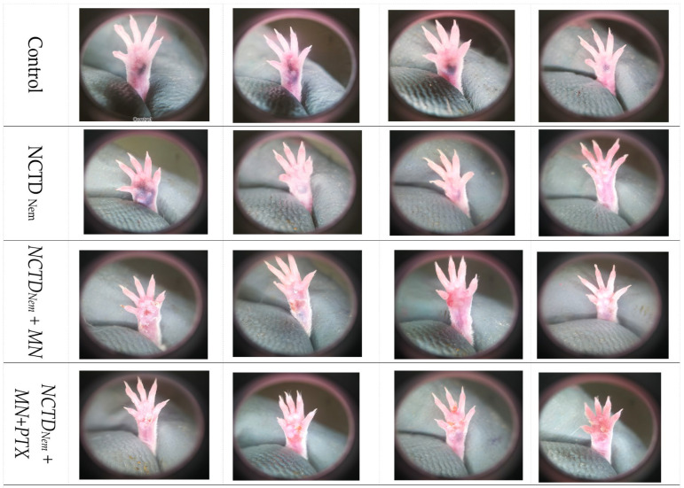
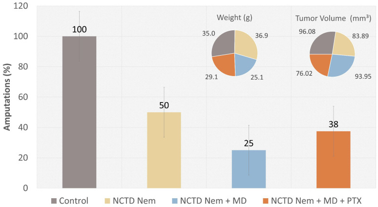
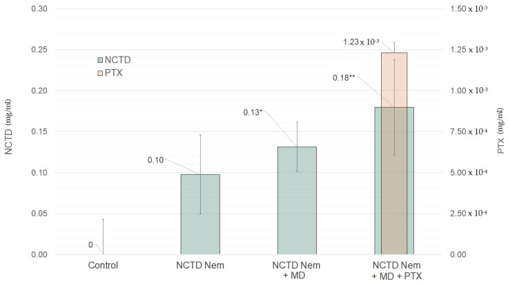
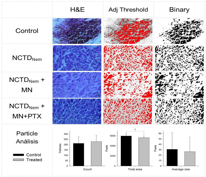
Melanoma, a lethal type of cancer originating from melanocytes, is the leading cause of death among skin cancers. While surgical excision of the lesions is the primary treatment for melanoma, not all cases are candidates for surgical procedures. New treatments and complementary options are necessary, given the increasing diagnosis rate. In the present study, a norcantharidin-containing nanoemulsion was developed and evaluated in vivo using a syngeneic graft murine model. Norcantharidin is the demethylated analog of cantharidin, known for its anticancer properties. Our model contemplates surgical excision surgery simulating the standard treatment and the role of the nanoemulsion as a potential adjuvant therapy. We observed a significant decrease in the growth rate of the melanoma lesion in the treated groups compared to the control group, both at the 20th and 30th days of treatment. Moreover, we evaluated the drug bioavailability in serum samples, and the results showed that norcantharidin was detectable in a range of 0.1 to 0.18 mg/mL in the treated groups. Furthermore, histopathological analysis was performed on the amputated tumors, where significant differences were found regarding size, mitosis rate, lymphocytic infiltration, and multispectral quantitative image analysis compared to the control group. If more clinical studies are conducted, the norcantharidin-containing nanoemulsion could be a potential alternative or adjuvant therapy. Topical nanosystems can become or complement standard therapies, which is needed as melanoma affects not only in terms of mortality but also the patient's morbidity and life quality.

Keywords: melanoma; nanoemulsion; nanosystems; nanotechnology; norcantharidin; topical administration.


Conflict of interest statement

The authors declare no conflicts of interest.

Figures

Figure 1
Figure 1
Representative Photographs of Melanoma Lesions After 20 Days of Treatment Across Different Groups. The images depict the condition of the melanoma lesions in four representative mice from each treatment group on the 20th day of treatment. Top row: Control group, which received no treatment, showing advanced tumor progression. Second row: NCTD nanoemulsion-treated group (NCTDNem), displaying varying degrees of tumor progression. Third row: NCTD nanoemulsion applied with microneedles (NCTDNem + MD), demonstrating a reduction in tumor size and progression. Bottom row: NCTD nanoemulsion with microneedle application supplemented with pentoxifylline (NCTDNem + MD + PTX), showing further reduction in tumor size and evidence of potential regression.
Figure 2
Figure 2
Representative Photographs of Melanoma Lesions After 30 Days of Treatment Across Different Groups. The images show the condition of the melanoma lesions in four representative mice from each treatment group on the 30th day of treatment. Top row: Control group, showing continued tumor progression with more prominent growth and pigmentation. Second row: NCTD nanoemulsion-treated group (NCTDNem), where varying levels of tumor progression are observed, including some cases of advanced growth. Third row: NCTD nanoemulsion applied with microneedles (NCTDNem + MD), displaying minimal tumor growth and signs of potential tumor regression. Bottom row: NCTD nanoemulsion with microneedle application supplemented with pentoxifylline (NCTDNem + MD + PTX), showing further reduction in tumor size and less pigmentation compared to other groups.
Figure 3
Figure 3
Impact of Treatments on Surgical Excision Rates, Group Mean Weight, and Macroscopic Tumor Volume. The vertical bars represent the percentage of surgical excisions within each treatment group, with standard error indicated. The pie charts illustrate the mean weight (left pie chart) and macroscopic tumor volume (right pie chart) across treated groups. The groups include Control (no treatment), NCTD nanoemulsion (NCTDNem), NCTD nanoemulsion applied with microneedle (NCTDNem + MD), and NCTD nanoemulsion applied with microneedle and supplemented with pentoxifylline (NCTDNem + MD + PTX).
Figure 4
Figure 4
Systemic bioavailability of NCTD and PTX in serum samples. The data is presented as milligrams (mg) per mL of serum across different treatment groups. The green bars represent norcantharidin (NCTD) concentration, while the orange bar represent the groups’ pentoxifylline (PTX) concentration. The groups are Control, Norcantharidin nanoemulsion (NCTDNem), Norcantharidin nanoemulsion applied with microneedle (NCTDNem + MD), and Norcantharidin nanoemulsion applied with microneedle supplemented with pentoxifylline (NCTDNem + MD + PTX). Statistical significance: *: p < 0.05; **: p < 0.01.
Figure 5
Figure 5
Representative high-resolution micrographs of H&E-stained melanoma samples. Size data is presented as the mean size of tumors in centimeters (cm) ± standard deviation (S.D.). Mitoses are counted per 1 mm of high-power field (HPF). Lymphocyte infiltration (L.I.) is evaluated qualitatively, with the scale ranging from less to more (* < **). Tumor regression (Reg) is indicated as presence (+) or absence (−). Groups are (A) Control, (B) Norcantharidin nanoemulsion (NCTDNem), (C) Norcantharidin nanoemulsion applied with microneedle (NCTDNem + MD), and (D) Norcantharidin nanoemulsion applied with microneedle and supplemented with pentoxifylline (NCTDNem + MD + PTX).
Figure 6
Figure 6
Multispectral analysis of micrographs from tumor lesion fixed in and tainted histologically. First column hematoxylin and eosin (H&E) tainted samples. Second column: adjusted threshold using the image blue channel. Third column: Binary selection of particle analysis. Groups are Control, Norcantharidin nanoemulsion NCTDNem, Norcantharidin nanoemulsion applied with microneedle (NCTDNem + MD), Norcantharidin nanoemulsion applied with microneedle and supplemented with pentoxifylline (NCTDNem + MD + PTX). Bottom: Particle analysis presented as particle count, total area (pixels), and pixel average particle size (pixels). Groups are Control v. Treated. Significance *: p < 0.05.
Figure 7
Figure 7
Nanoemulsion preparation, characterization and application to the syngeneic graft inoculation area. (1) Pre-emulsion preparation by mixing the oil phase into the water phase. (2) Nanoemulsion obtention through high-energy sonication. (3) Characterization of the nanoemulsion in droplet size and PDI. (4) Different forms of application forms, direct and using microneedle.

Similar articles

References

    1. Sladden M.J., Balch C., Barzilai D.A., Berg D., Freiman A., Handiside T., Hollis S., Lens M.B., Thompson J.F. Surgical excision margins for primary cutaneous melanoma. Cochrane Database Syst. Rev. 2009;2007:CD004835. doi: 10.1002/14651858.CD004835.pub2. - DOI - PubMed
    1. IARC Publications. [(accessed on 28 June 2024)]. Available online: https://publications.iarc.fr/
    1. Eggermont A.M.M., Spatz A., Robert C. Cutaneous melanoma. Lancet. 2014;383:816–827. doi: 10.1016/S0140-6736(13)60802-8. - DOI - PubMed
    1. Welch H.G., Woloshin S., Schwartz L.M. Skin biopsy rates and incidence of melanoma: Population-based ecological study. BMJ. 2005;331:481. doi: 10.1136/bmj.38516.649537.E0. - DOI - PMC - PubMed
    1. Tromme I., Legrand C., Devleesschauwer B., Leiter U., Suciu S., Eggermont A., Francart J., Calay F., Haagsma J.A., Baurain J.-F., et al. Melanoma burden by melanoma stage: Assessment through a disease transition model. Eur. J. Cancer. 2016;53:33–41. doi: 10.1016/j.ejca.2015.09.016. - DOI - PubMed

MeSH terms

LinkOut - more resources