. 2025 Jan 21;14(3):661.
doi: 10.3390/jcm14030661.

Five-Year Comparative Study of Zygomatic and Subperiosteal Implants: Clinical Outcomes, Complications, and Treatment Strategies for Severe Maxillary Atrophy

Affiliations

Five-Year Comparative Study of Zygomatic and Subperiosteal Implants: Clinical Outcomes, Complications, and Treatment Strategies for Severe Maxillary Atrophy

Rafal Zielinski et al. J Clin Med. .

Abstract

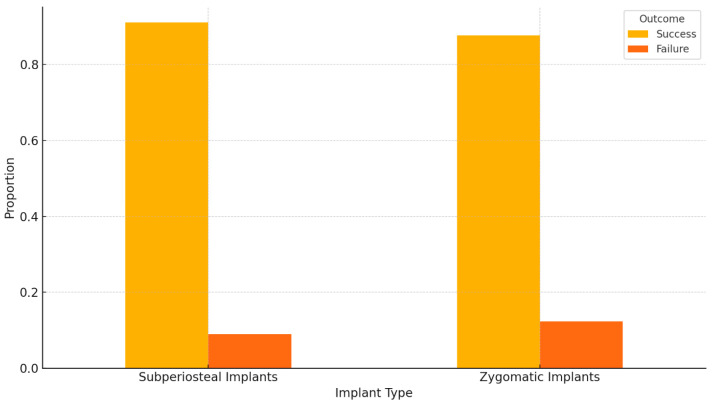
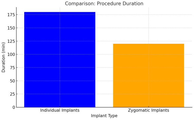
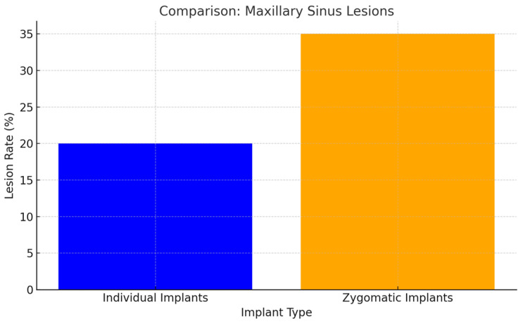
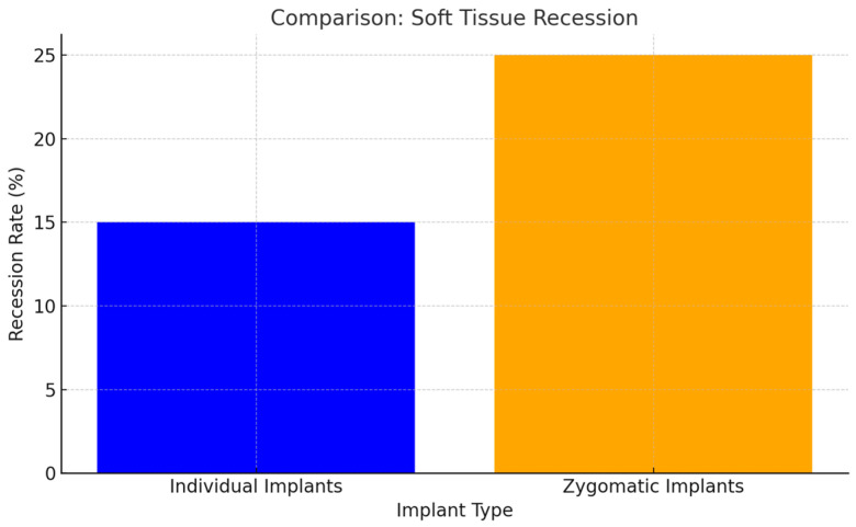
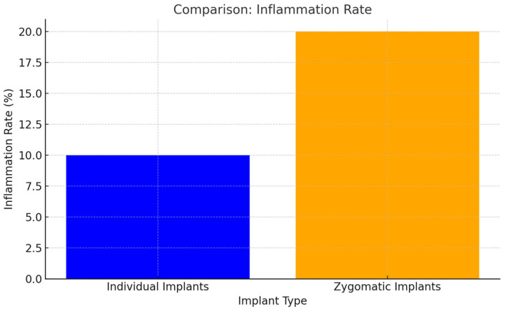
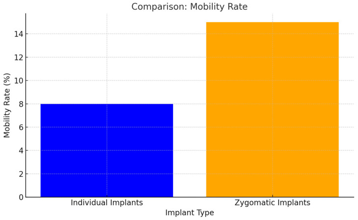
Background/Objectives: Severe maxillary atrophy presents challenges in maxillofacial rehabilitation. This study compares the clinical outcomes of zygomatic and subperiosteal implants, focusing on implant survival, soft tissue management, and postoperative complications over a five-year follow-up. Methods: A retrospective cohort study analyzed 150 patients divided into two groups based on the type of implant. Zygomatic implants were assessed for immediate functional loading, procedural efficiency, and complications such as sinus-related issues and orbital damage. Subperiosteal implants were evaluated for their customized design, keratinized mucosa integration, and adaptation to severe anatomical limitations. Statistical analyses, including Chi-square tests, were used to determine significant differences (p < 0.05). Results: This study demonstrated differences in complication rates (sinus-related complications: 12.4% for zygomatic implants; peri-implantitis: 5.6% for subperiosteal implants). Implant survival rates were comparable (zygomatic: 96.3%, subperiosteal: 97.1%, p = 0.278). Zygomatic implants demonstrated higher incidences of sinus-related complications (12.4%) and risks of orbital damage. Subperiosteal implants exhibited superior soft tissue stability with fewer cases of peri-implantitis (5.6%, p < 0.05). Procedural duration was shorter for zygomatic implants (177 min vs. 123 min); however, subperiosteal implants allowed for re-implantation after failure, providing flexibility that was unavailable with zygomatic implants. Conclusions: Zygomatic implants excel in immediate functional loading and reduced procedural time but require advanced surgical expertise to mitigate anatomical risks. Subperiosteal implants offer a safer, customizable solution, particularly in anatomically complex cases. These findings emphasize the importance of individualized treatment planning and technological advancements in implant design to optimize clinical outcomes for patients with severe maxillary atrophy.

Keywords: maxillary atrophy; subperiosteal implants; zygomatic implants.


Conflict of interest statement

The authors declare no conflicts of interest.

Figures

Figure 1
Figure 1
MaI Implant® (Integra Implants®, Lodz, Poland) placement secured with self-tapping screws. The photo was taken before the implant was covered with autologous bone chips.
Figure 2
Figure 2
Process of designing a template to reduce bone on the alveolar ridge. (a) template put on alveolar ridge before bone reduction; (b,f) MaI Implant® (Integra Implants®, Lodz, Poland) on the bone; (c) template put on alveolar ridge after bone reduction; (d,e) multiunits on reduced bone.
Figure 3
Figure 3
Clinical application of template (ad) and the MaI Implant® (Integra Implants®, Lodz, Poland) placement after bone adjustment (e,f).
Figure 4
Figure 4
Bichat fat pad cover over the MaI Implant® (Integra Implants®, Lodz, Poland). The fat pad can be sutured using resorbable sutures, in which case it is attached to the periosteum, or it can be sutured using non-resorbable sutures. However, in the latter case, the knot must be left above the gingiva so that it can be removed after a minimum of 10 days.
Figure 5
Figure 5
(a,b) bone chips were harvested using a disposable bone scraper and placed on the rami of the MaI Implant® (Integra Implants®, Lodz, Poland). The aim of the bone chips was to reduce the risk of dehiscence of MaI Implant®.
Figure 6
Figure 6
Extra-maxillary zygomatic implant placement.
Figure 7
Figure 7
This picture shows the placement of a zygomatic implant using the Stella method (a) and bone chips harvested from the anterior maxillary sinus placed on the zygomatic implant in order to separate the implant from periosteal (b).
Figure 8
Figure 8
These pictures show soft tissue dehiscence around the zygomatic implant in the region of tooth 23 (a,b). An attempt was made to cover the exposed implant with a vascularized flap from the palate (c) after smoothing the implant with a bur. However, the gingiva became exposed again, and it was decided to remove the implant due to recurrent maxillary sinus infections. After unscrewing the implant (d), the cavity left by the removed implant was covered with Bichat’s fat pad (e,f).
Figure 9
Figure 9
Bar chart comparing the success and failure rates of subperiosteal and zygomatic implants. p-value: 0.65 means there is no statistically significant difference in the success and failure rates between subperiosteal and zygomatic implants based on the data provided.
Figure 10
Figure 10
The longer procedure time for individual implants was due to the adjustments required to fit the bone to the template. Additionally, for subperiosteal implants, bone grafting from the lineaobliqua was performed, which further extended the duration of the procedure (p < 0.05).
Figure 11
Figure 11
The design and placement technique of individual implants minimized the risk of sinus membrane perforation and associated complications (p < 0.05).
Figure 12
Figure 12
Extra-maxillary zygomatic implant placement carries a higher risk of soft tissue dehiscence (p < 0.05).
Figure 13
Figure 13
Sinusitis associated with extra-maxillary implant placement is a cause of inflammation observed both clinically around zygomatic implants and in CBCT imaging (p < 0.05).
Figure 14
Figure 14
Anchorage in the zygomatic bone of zygomatic implants creates a cantilever effect. As a result, mobility at the distal end of zygomatic implants becomes apparent when pressure is applied with a dental instrument (p < 0.05).

Similar articles

Cited by

References

    1. Albrektsson T., Zarb G., Worthington P., Eriksson A.R. The long-term efficacy of currently used dental implants: A review. Int. J. Oral Maxillofac. Implant. 1986;1:11–25. - PubMed
    1. Papaspyridakos P., Chen C.J., Singh M., Weber H.P., Gallucci G.O. Success criteria in implant dentistry: A systematic review. J. Dent. Res. 2012;91:242–248. doi: 10.1177/0022034511431252. - DOI - PubMed
    1. Aparicio C., Manresa C., Francisco K., Aparicio A., Nunes J., Claros P., Potau J.M. Zygomatic implants placed with two different techniques in edentulous maxillae: A 5-year retrospective study. Int. J. Oral Maxillofac. Implant. 2014;16:627–642.
    1. Chrcanovic B.R., Abreu M.H. Zygomatic implants for the rehabilitation of atrophic posterior maxilla: A review. Oral Maxillofac. Surg. 2013
    1. Zielinski R., Okulski J., Simka W., Kozakiewicz M. The Zygomatic Anatomy-Guided Approach, Zygomatic Orbital Floor Classification, and ORIS Criteria—A 10-Year Follow-Up. J. Clin. Med. 2023;12:6681. doi: 10.3390/jcm12206681. - DOI - PMC - PubMed

LinkOut - more resources