

Brain Imaging in Patients with Non-Small Cell Lung Cancer-A Systematic Review
- PMID: 39941379
- PMCID: PMC11818832
- DOI: 10.3390/jcm14030708
Brain Imaging in Patients with Non-Small Cell Lung Cancer-A Systematic Review
Abstract
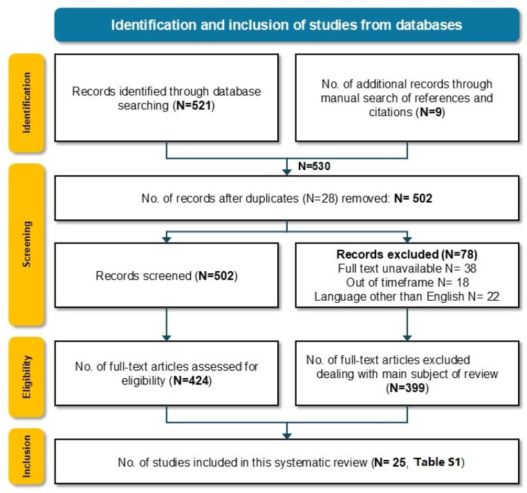
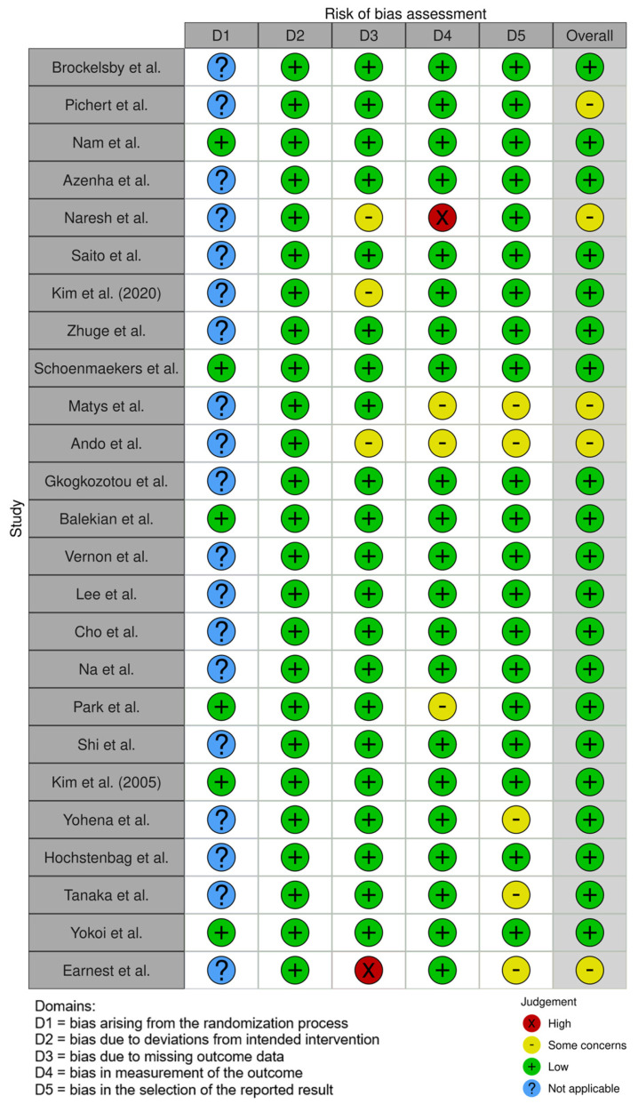
Background: Lung cancer frequently metastasizes to the brain, liver, and adrenal glands with a significant negative prognostic impact on overall survival and quality of life (QoL). To optimize treatment and prognosis, adequate staging with the detection of distant metastases is crucial. The incidence of brain metastases in potentially resectable early-stage non-small cell lung cancer (NSCLC) is as low as 3%; hence, the need for preoperative brain imaging has been a constant matter of debate, especially in stage II. In stages III and IV NSCLC, neuroimaging is an essential part of staging. Methods: A systematic literature search was performed. Publications from 1999 to 2024, focusing on preoperative brain imaging (BI) in the staging of stages I-IV NSCLC, were included. Data extraction included study population characteristics, the modality of BI, the incidence of brain metastases (BMs), and the main outcomes of the studies. The final included studies were selected according to the PRISMA criteria. In the second step, guidelines on BI in NSCLC staging of major importance were identified and compared. Results: A total of 530 articles were identified, of which 25 articles were selected. Four prospective studies and 21 retrospective investigations were included. Most of the investigations focused on BI in the early stages. The main imaging modality for BI was magnetic resonance imaging (MRI), followed by computed tomography (CT). Besides the identified 25 studies, the most important internationally applied guidelines on brain imaging in the staging of NSCLC were reviewed. While some guidelines agree on preoperative BI in NSCLC stage III (Union for International Cancer Control-UICC eighth edition) patients, other guidelines recommend earlier BI starting from clinical stage II. All mentioned guidelines homogenously recommend BI in patients with symptoms suggestive of brain pathologies. Conclusions: BI in NSCLC staging is recommended in neurologically symptomatic patients suggestive of brain metastases as well as NSCLC patients with stage III disease. Neuroimaging in stage IA patients, as well as in pure GGO (Ground-Glass Opacity) lesions, was considered unnecessary. The predominantly applied imaging modality was ce-MRI (contrast-enhanced magnetic resonance imaging). Inconsistency exists concerning BI in stage II. The identification of prognostic factors for developing BM in patients with early-stage NSCLC could help to clarify which subgroup might benefit from preoperative BI.
Keywords: MRI; brain imaging; brain metastases; non-small cell lung cancer (NSCLC); preoperative staging.
Conflict of interest statement
The authors declare no conflicts of interest.
Figures
References
-
- BWS CPW World Cancer Report 2014. [(accessed on 11 March 2024)]. Available online: https://publications.iarc.fr/Non-Series-Publications/World-Cancer-Report....
-
- Global Burden of Disease Cancer Collaboration. Fitzmaurice C., Dicker D., Pain A., Hamavid H., Moradi-Lakeh M., MacIntyre M.F., Allen C., Hansen G., Woodbrook R. The Global Burden of Cancer 2013. JAMA Oncol. 2015;1:505–527. doi: 10.1001/jamaoncol.2015.0735. Erratum in JAMA Oncol. 2015, 1, 690. https://doi.org/10.1001/jamaoncol.2015.2892 . - DOI - PMC - PubMed
Publication types
LinkOut - more resources
Full Text Sources
