

Dermoscopy of Basal Cell Carcinoma Part 1: Dermoscopic Findings and Diagnostic Accuracy-A Systematic Literature Review
- PMID: 39941860
- PMCID: PMC11816234
- DOI: 10.3390/cancers17030493
Dermoscopy of Basal Cell Carcinoma Part 1: Dermoscopic Findings and Diagnostic Accuracy-A Systematic Literature Review
Abstract
Introduction: Basal cell carcinoma (BCC) is the most common malignant skin tumor. While rarely fatal, it can cause local tissue damage. Part I of the review summarizes the dermoscopic features of BCC and the diagnostic accuracy of dermoscopy in the diagnosis of BCC.
Methods: A search of the PubMed database was performed for studies reporting on the diagnostic accuracy of dermoscopy or dermoscopic findings in BCC, either pigmented or non-pigmented, located anywhere on the body, of any histopathologic subtype, size and at any age of onset.
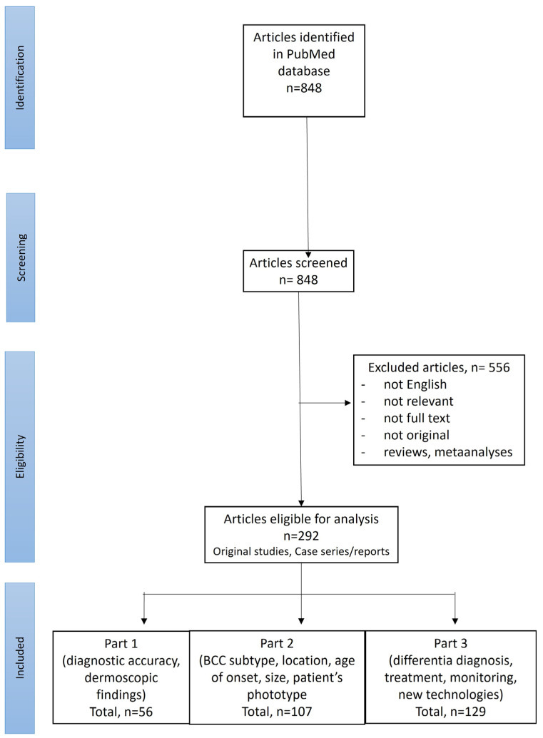
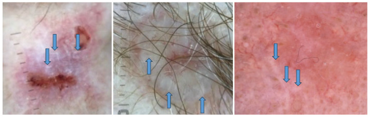
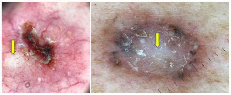
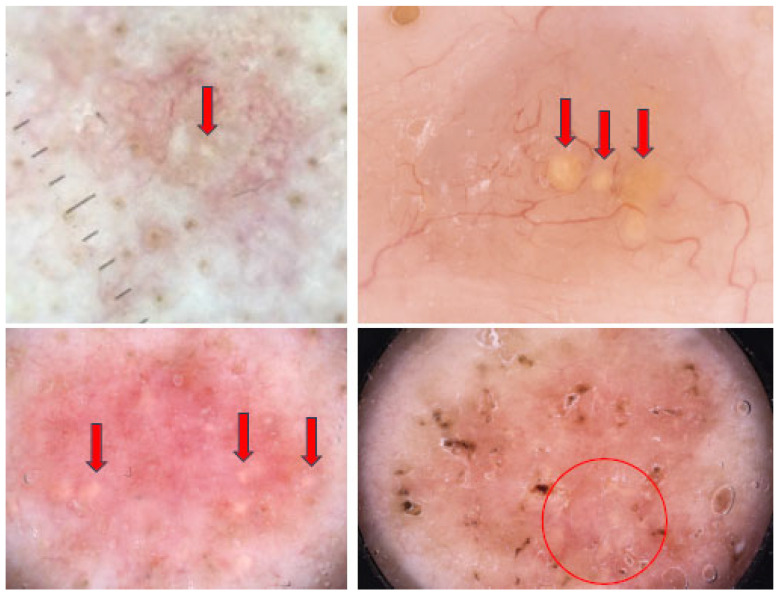
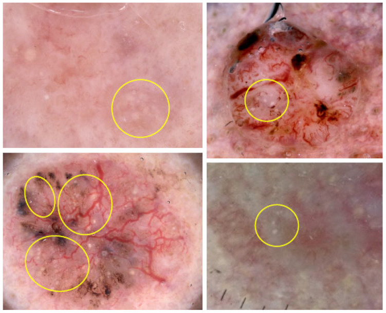
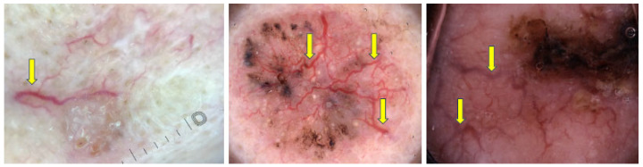
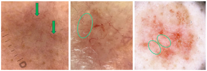
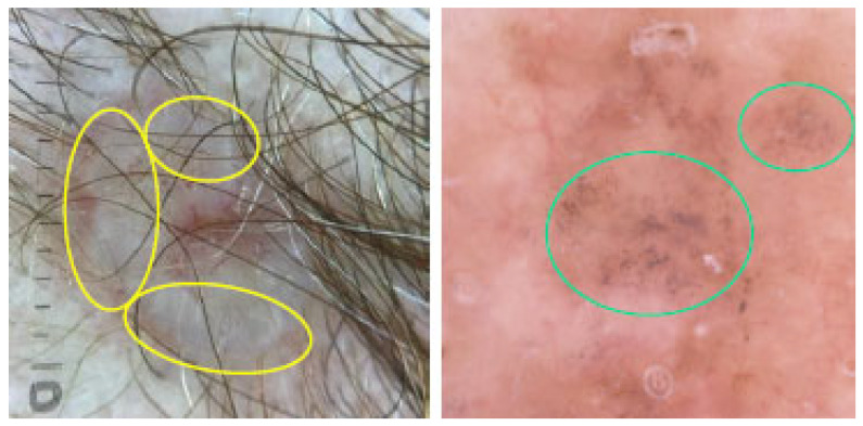
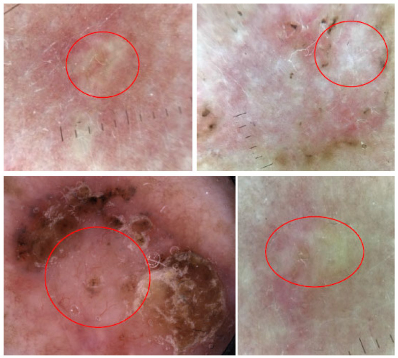
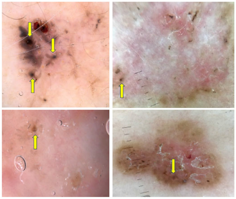
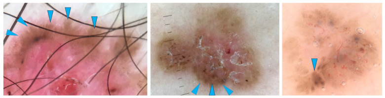
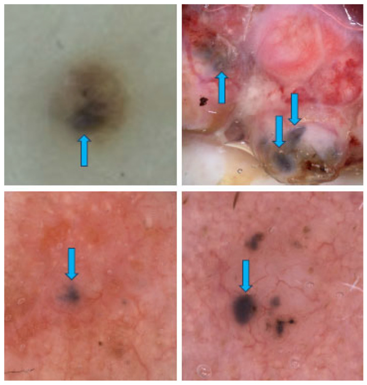
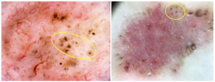
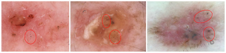
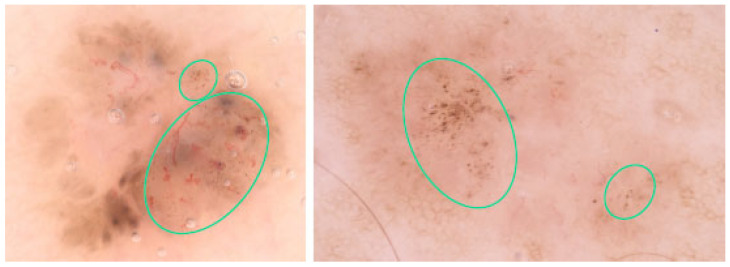
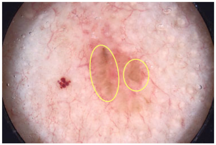
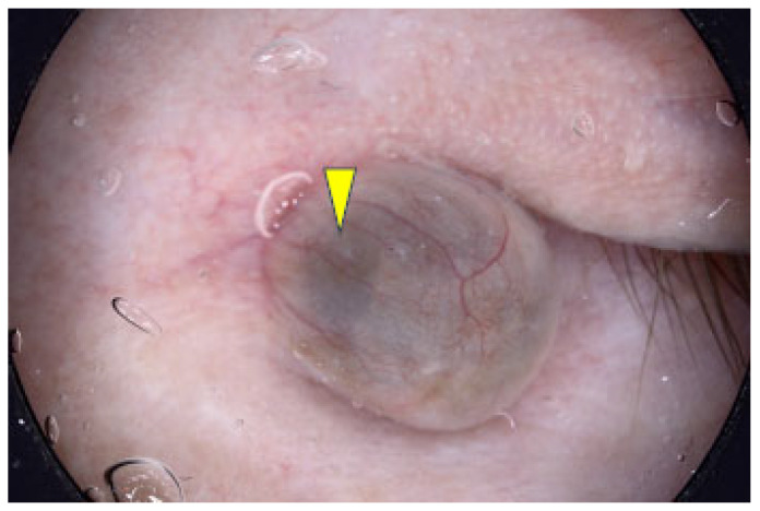
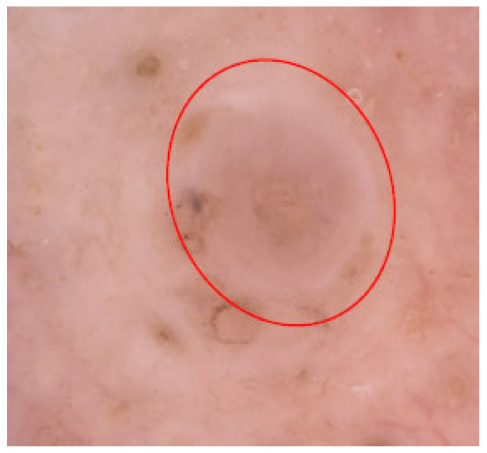
Results: BCC was found to present with a wide range of dermoscopic features, including white structures (shiny white lines, shiny white areas, rosettes), yellow structures (milia-like cysts, yellow lobular-like structures), multiple aggregated yellow-white globules (MAY globules), blue structures (blue ovoid nests), vascular structures (arborizing vessels, short fine telangiectasias), multiple small erosions/ulcerations, features of regression (pepper-like structures, white scar-like areas) and pigmented structures (spoke-wheel areas, maple leaf-like areas (MLLAs), blue/gray dots). Dermoscopy showed a sensitivity of 67.6-98.6% and a positive predictive value (PPV) of 85.9-97% in identifying BCC. The physician's experience and training improve the accuracy, however, BCCs on the trunk and extremities, particularly of superficial subtypes, may still constitute a challenge.
Conclusions: Dermoscopy, especially when performed by a trained physician, increases the accuracy of early BCC detection.
Keywords: basal cell carcinoma; dermatoscopy; dermoscopy; non-polarized; polarized.
Conflict of interest statement
The authors declare no conflicts of interest.
Figures

Similar articles
-
Dermoscopic features of basal cell carcinoma and its subtypes: A systematic review.J Am Acad Dermatol. 2021 Sep;85(3):653-664. doi: 10.1016/j.jaad.2019.11.008. Epub 2019 Nov 7. J Am Acad Dermatol. 2021. PMID: 31706938 Free PMC article.
-
Dermoscopy of Basal Cell Carcinoma Part 2: Dermoscopic Findings by Lesion Subtype, Location, Age of Onset, Size and Patient Phototype.Cancers (Basel). 2025 Jan 8;17(2):176. doi: 10.3390/cancers17020176. Cancers (Basel). 2025. PMID: 39857958 Free PMC article. Review.
-
Dermoscopic criteria and basal cell carcinoma.G Ital Dermatol Venereol. 2012 Apr;147(2):135-40. G Ital Dermatol Venereol. 2012. PMID: 22481576 Review.
-
Dermoscopic patterns of superficial basal cell carcinoma.Int J Dermatol. 2008 Oct;47(10):1015-8. doi: 10.1111/j.1365-4632.2008.03731.x. Int J Dermatol. 2008. PMID: 18986346
-
Dermoscopic features of basal cell carcinoma in skin of color: A retrospective cross-sectional study from Puducherry, South India.Indian J Dermatol Venereol Leprol. 2023 Jan-Mar;89(2):254-260. doi: 10.25259/IJDVL_420_20. Indian J Dermatol Venereol Leprol. 2023. PMID: 33969659
Cited by
-
Polarized Dermoscopy and Ultraviolet-Induced Fluorescence Dermoscopy of Basal Cell Carcinomas in the H- and Non-H-Zones of the Head and Neck.Dermatol Ther (Heidelb). 2025 Jun;15(6):1507-1522. doi: 10.1007/s13555-025-01432-z. Epub 2025 Apr 28. Dermatol Ther (Heidelb). 2025. PMID: 40293692 Free PMC article.
-
Dermoscopy of Basal Cell Carcinoma Part 3: Differential Diagnosis, Treatment Monitoring and Novel Technologies.Cancers (Basel). 2025 Mar 19;17(6):1025. doi: 10.3390/cancers17061025. Cancers (Basel). 2025. PMID: 40149358 Free PMC article. Review.
References
-
- Longo C., Guida S., Mirra M., Pampena R., Ciardo S., Bassoli S., Casari A., Rongioletti F., Spadafora M., Chester J., et al. Dermatoscopy and reflectance confocal microscopy for basal cell carcinoma diagnosis and diagnosis prediction score: A prospective and multicenter study on 1005 lesions. J. Am. Acad. Dermatol. 2024;90:994–1001. doi: 10.1016/j.jaad.2024.01.035. - DOI - PubMed
-
- Coppola R., Barone M., Zanframundo S., Devirgiliis V., Roberti V., Perrella E., Donati M., Palese E., Tenna S., Persichetti P., et al. Basal cell carcinoma thickness evaluated by high-frequency ultrasounds and correlation with dermoscopic features. Ital. J. Dermatol. Venerol. 2021;156:610–615. doi: 10.23736/S2784-8671.20.06576-1. - DOI - PubMed
Publication types
LinkOut - more resources
Full Text Sources
