

Improving Stroke Treatment Using Magnetic Nanoparticle Sensors to Monitor Brain Thrombus Extraction
- PMID: 39943310
- PMCID: PMC11820568
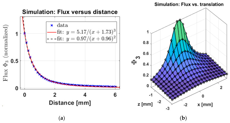
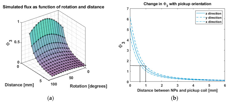
- DOI: 10.3390/s25030672
Improving Stroke Treatment Using Magnetic Nanoparticle Sensors to Monitor Brain Thrombus Extraction
Abstract
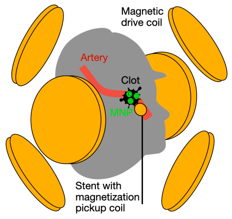
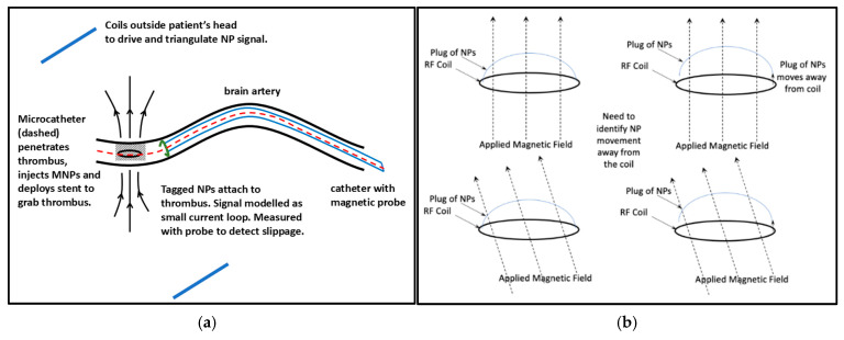
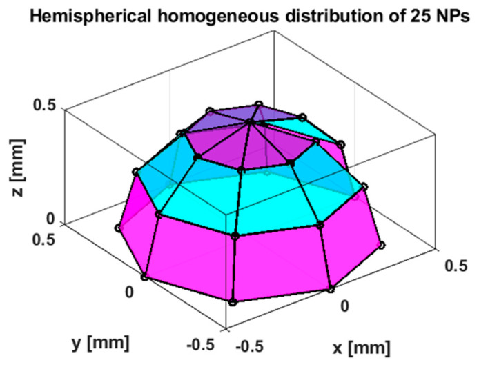
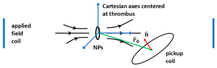
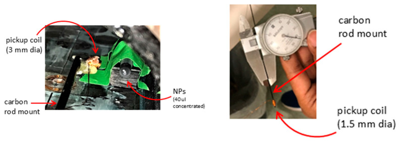
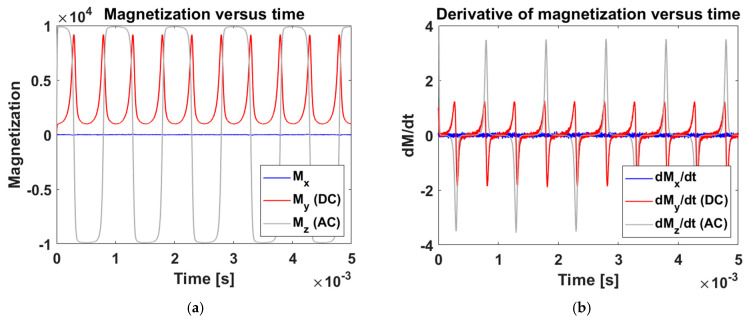
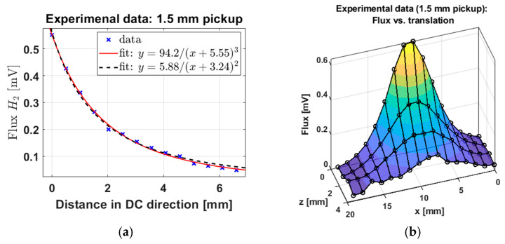
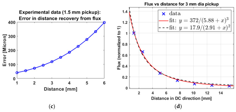
(1) Background: Mechanical thrombectomy (MT) successfully treats ischemic strokes by extracting the thrombus, or clot, using a stent retriever to pull it through the blood vessel. However, clot slippage and/or fragmentation can occur. Real-time feedback to a clinician about attachment between the stent and clot could enable more complete removal. We propose a system whereby antibody-targeted magnetic nanoparticles (NPs) are injected via a microcatheter to coat the clot, oscillating magnetic fields excite the particles, and a small coil attached to the catheter picks up a signal that determines the proximity of the clot to the stent. (2) Methods: We used existing simulation code to model the signal from NPs distributed on a hemispherical clot with three orthogonally applied magnetic fields. An in vitro apparatus was built that applied fields and read out signals from a 1.5 mm pickup coil at a variable distance and orientation angle from a sample of 100 nm iron oxide core/shell NPs. (3) Results: Our simulations suggest that the sum of the voltages induced in the pickup coil from three orthogonal applied fields could localize a clot to within 180 µm, regardless of the exact orientation of the pickup coil, with further precision added via rotation-correction formulae. Our experimental system validated simulations; we estimated an in vitro distance recovery precision of 41 µm with a pickup coil 1 mm from the clot. (4) Conclusions: Magnetic NP sensing could be a safe and real-time method to estimate whether a clot is attached to the stent retriever during MT.
Keywords: clot; magnetic dipole field; magnetic nanoparticles; mechanical thrombectomy; simulations; stroke; thrombus.
Conflict of interest statement
LCD Nanotech is held by J.W. DJ and SG employed by LCD Nanotech. The remaining authors declare that the research was conducted in the absence of any commercial or financial relationships that could be construed as a potential conflict of interest.
Figures
Similar articles
-
Combined stent-retriever and aspiration intra-arterial thrombectomy performance for fragmentable blood clots: A proof-of-concept computational study.J Mech Behav Biomed Mater. 2022 Nov;135:105462. doi: 10.1016/j.jmbbm.2022.105462. Epub 2022 Sep 14. J Mech Behav Biomed Mater. 2022. PMID: 36116343
-
Preventing vessel perforations in endovascular thrombectomy: feasibility and safety of passing the clot with a microcatheter without microwire: the wireless microcatheter technique.J Neurointerv Surg. 2019 Jul;11(7):653-658. doi: 10.1136/neurintsurg-2018-014267. Epub 2018 Dec 7. J Neurointerv Surg. 2019. PMID: 30530771
-
Adjustment of Stent Retriever Length to Clot Extent Affects First-Pass Reperfusion in Endovascular Treatment of Acute Ischemic Stroke.Cerebrovasc Dis. 2020;49(3):277-284. doi: 10.1159/000508028. Epub 2020 Jun 16. Cerebrovasc Dis. 2020. PMID: 32544906
-
Development of the Trevo ProVue Retriever for intracranial clot removal in acute ischemic stroke.Ann N Y Acad Sci. 2014 Nov;1329:107-15. doi: 10.1111/nyas.12579. Ann N Y Acad Sci. 2014. PMID: 25399522 Review.
-
In vitro models for the experimental evaluation of mechanical thrombectomy devices in acute ischemic stroke.Interv Neuroradiol. 2023 Dec;29(6):759-767. doi: 10.1177/15910199221118404. Epub 2022 Aug 15. Interv Neuroradiol. 2023. PMID: 35971288 Free PMC article. Review.
References
-
- Tsao C.W., Aday A.W., Almarzooq Z.I., Anderson C.A., Arora P., Avery C.L., Baker-Smith C.M., Beaton A.Z., Boehme A.K., Buxton A.E. Heart disease and stroke statistics—2023 update: A report from the American Heart Association. Circulation. 2023;147:e93–e621. - PubMed
-
- Goyal M., Menon B.K., van Zwam W.H., Dippel D.W., Mitchell P.J., Demchuk A.M., Dávalos A., Majoie C.B., van der Lugt A., de Miquel M.A., et al. Endovascular thrombectomy after large-vessel ischaemic stroke: A meta-analysis of individual patient data from five randomised trials. Lancet. 2016;387:1723–1731. doi: 10.1016/S0140-6736(16)00163-X. - DOI - PubMed
MeSH terms
Substances
Grants and funding
LinkOut - more resources
Full Text Sources
Medical
Miscellaneous
