Meta-Analysis
. 2025 Jan 29:38:13794.
doi: 10.3389/ti.2025.13794. eCollection 2025.

Association of Inflammatory Profile During Ex Vivo Lung Perfusion With High-Grade Primary Graft Dysfunction: A Systematic Review and Meta-Analysis

Affiliations
Meta-Analysis

Association of Inflammatory Profile During Ex Vivo Lung Perfusion With High-Grade Primary Graft Dysfunction: A Systematic Review and Meta-Analysis

Andrea Costamagna et al. Transpl Int. .

Abstract

PGD3 is the manifestation of ischemia-reperfusion injury which results from inflammation and cell death and is associated with poor outcome. This systematic-review and meta-analysis of non-randomized controlled trials on patients undergoing Ltx with reconditioned lungs via EVLP, aims to assess the association between the levels of proinflammatory biomarkers during EVLP and PGD3 development within the firsts 72 h post-Ltx. Biomarkers were categorized by timing (1-hour, T0 and 4-hours, Tend from EVLPstart) and by their biological function (adhesion molecules, chemokines, cytokines, damage-associated-molecular-patterns, growth-factors, metabolites). We employed a four-level mixed-effects model with categorical predictors for biomarker groups to identify differences between patients with PGD3 and others. The single study and individual measurements were considered random intercepts. We included 8 studies (610 measurements at T0 and 884 at Tend). The pooled effect was 0.74 (p = 0.021) at T0, and 0.90 (p = 0.0015) at Tend. The four-level model indicated a large pooled correlation between developing PGD3 at 72 h post-Ltx and inflammatory biomarkers values, r = 0.62 (p = 0.009). Chemokine group showed the strongest association with the outcome (z-value = 1.26, p = 0.042). Pooled panels of inflammation markers, particularly chemokines, measured at T0 or at Tend, are associated with the development of PGD3 within the first 72 h after Ltx.

Systematic review registration: https://osf.io/gkxzh/.

Keywords: biomarkers; ex vivo lung perfusion; inflammation; ischemia-reperfusion injury; primary graft dysfunction.


Conflict of interest statement

The authors declare that the research was conducted in the absence of any commercial or financial relationships that could be construed as a potential conflict of interest.

Figures

FIGURE 1
FIGURE 1
Risk of bias assessment using the Risk Of Bias In Non-randomised Studies - of Interventions (ROBINS-I) tool for the selected studies included in the meta-analysis.
FIGURE 2
FIGURE 2
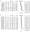
Forest plot of studies assessing inflammatory biomarkers. These are detailed in the column labeled “Marker,” accompanied by their corresponding groups denoted as “Group.” The experimental group corresponds to the PGD grade 3 at 72 h group, while the control group represents the non-PGD grade 3 group. The standardized mean difference (SMD), along with its respective 95% confidence interval (95% CI) and the individual weight for each study, is reported on the right. In the forest plot, squares placed to the right—considering 0 as the midpoint—indicate higher marker levels in the experimental group. (A) Forest plot for overall studies, excluding outliers, (B) all the studies. Abbreviations: AD, adhesion molecules; CKs, chemokines; DAMPs, damage-associated molecular patterns; Hematop GF, growth factors; sE-selectin, endothelial selectin; sICAM, intercellular adhesion molecule; vCAM, vascular cell adhesion molecule; ET-1, endothelin-1; Big ET-1, big endothelin-1; IL-8, interleukin-8; MCP, monocyte chemoattractant protein; GROα, growth-related oncogene alpha; MIP-1α, macrophage inflammatory protein-1 alpha; MIP-1β, macrophage inflammatory protein-1 beta; IL-1β, interleukin-1 beta; IL-6, interleukin-6; TNF-α, tumor necrosis factor alpha; M30, M30; HMGB, high mobility group box 1; nuDNA, nuclear DNA; mtDNA, mitochondrial DNA; M-CSF, macrophage colony-stimulating factor; G-CSF, granulocyte colony-stimulating factor.
FIGURE 3
FIGURE 3
Forest plot of studies assessing inflammatory biomarkers at Tend corresponding to 4 h from EVLP start. Each plot represents a specific group of biomarkers: adhesion molecules [AD, (A)], chemokines [Chem, (B)], cytokines [CKs, (C)], damage-associated molecular patterns [DAMPs, (D)], growth factors [HGF, (E)], and endogenous metabolites produced during inflammatory phenomena such as carbon monoxide and nitric oxide metabolite [Met, (F)]. The experimental group corresponds to the PGD grade 3 at 72 h, while the control group represents the non-PGD grade 3. The standardized mean difference (SMD), accompanied by its respective 95% confidence interval (95% CI) and the individual weight for each study, is reported on the right. In the forest plot, the placement of squares to the right of the plot—taking 0 as the midpoint—indicates higher marker levels in the experimental group. Abbreviations: sE-selectin, endothelial selectin; sICAM, intercellular adhesion molecule; vCAM, vascular cell adhesion molecule; ET-1, endothelin-1; Big ET-1, big endothelin-1; IL-8, interleukin-8; MCP, monocyte chemoattractant protein; GROα, growth-related oncogene alpha; MIP-1α, macrophage inflammatory protein-1 alpha; MIP-1β, macrophage inflammatory protein-1 beta; IL-1β, interleukin-1 beta; IL-6, interleukin-6; TNF-α, tumor necrosis factor alpha; M30, M30; HMGB, high mobility group box 1; nuDNA, nuclear DNA; mtDNA, mitochondrial DNA; M-CSF, macrophage colony-stimulating factor; G-CSF, granulocyte colony-stimulating factor; CO, carbon monoxide; NOx, nitric oxide metabolite.

References

    1. Cypel M, Yeung JC, Liu M, Anraku M, Chen F, Karolak W, et al. Normothermic Ex Vivo Lung Perfusion in Clinical Lung Transplantation. N Engl J Med (2011) 364(15):1431–40. 10.1056/NEJMoa1014597 - DOI - PubMed
    1. Chao BT, McInnis MC, Sage AT, Yeung JC, Cypel M, Liu M, et al. A Radiographic Score for Human Donor Lungs on Ex Vivo Lung Perfusion Predicts Transplant Outcomes. J Heart Lung Transpl (2024) 43:797–805. 10.1016/j.healun.2024.01.004 - DOI - PubMed
    1. Ayyat KS, Okamoto T, Niikawa H, Sakanoue I, Dugar S, Latifi SQ, et al. A CLUE for Better Assessment of Donor Lungs: Novel Technique in Clinical Ex Vivo Lung Perfusion. J Heart Lung Transpl Off Publ Int Soc Heart Transpl (2020) S1053-2498(20):31665-X. 10.1016/j.healun.2020.07.013 - DOI - PubMed
    1. Costamagna A, Steinberg I, Simonato E, Massaro C, Filippini C, Rinaldi M, et al. Clinical Performance of Lung Ultrasound in Predicting Graft Outcome During Ex Vivo Lung Perfusion. Minerva Anestesiol (2021) 16. 10.23736/s0375-9393.21.15527-0 - DOI - PubMed
    1. Mazzeo AT, Fanelli V, Boffini M, Medugno M, Filippini C, Simonato E, et al. Feasibility of Lung Microdialysis to Assess Metabolism During Clinical Ex Vivo Lung Perfusion. J Heart Lung Transpl (2018) 38:267–76. 10.1016/j.healun.2018.12.015 - DOI - PubMed

LinkOut - more resources