. 2025 Jan 29:38:12859.
doi: 10.3389/ti.2025.12859. eCollection 2025.

γδ T Cells' Role in Donor-Specific Antibody Generation: Insights From Transplant Recipients and Experimental Models

Affiliations

γδ T Cells' Role in Donor-Specific Antibody Generation: Insights From Transplant Recipients and Experimental Models

Xavier Charmetant et al. Transpl Int. .

Abstract

The generation of donor-specific antibodies (DSA) requires that alloreactive B cells receive help from follicular helper T (TFH) cells. Recent works have suggested that γδ T cells could contribute to T cell-dependent humoral responses, leading us to investigate their role in DSA generation. Analysis of a cohort of 331 kidney transplant recipients found no relation between the number of circulating γδ T cells and the risk to develop DSA. Coculture models demonstrated that activated γδ T cells were unable to promote the differentiation of B cells into plasma cells, ruling out that they can be "surrogate" TFH. In line with this, γδ T cells preferentially localized outside the B cell follicles, in the T cell area of lymph nodes, suggesting that they could instead act as "antigen-presenting cell" (APC) to prime αβ TFH. This hypothesis was proven wrong since γδ T cells failed to acquire APC functions in vitro. These findings were validated in vivo by the demonstration that following transplantation with an allogeneic Balb/c (H2d) heart, wild-type and TCRδKO C57BL/6 (H2b) mice developed similar DSA responses, whereas TCRαKO recipients did not develop DSA. We concluded that the generation of DSA is unfazed by the absence of γδ T cells.

Keywords: B cell; donor specific antibody (DSA); gamma delta T cell; humoral response; translational science.


Conflict of interest statement

The authors declare that the research was conducted in the absence of any commercial or financial relationships that could be construed as a potential conflict of interest.

Figures

FIGURE 1
FIGURE 1
Serological and cellular follow-up of kidney transplant recipients. (A) Kaplan-Meier curve of DSA incidence after kidney transplantation in the cohort. (B, C) Kidney transplant recipients T cells were phenotyped by flow cytometry the day of the transplantation (D0) and 2 years later (M24). (B) Representative flow cytometry profiles of the gating strategy used to assess the numbers of the different γδ T cells subtypes. (C) The number of total γδ T cells (left panel), Vδ2+ γδ T cells (middle panel) or Vδ2 γδ T cells (right panel) measured on the day of the transplantation (D0) and M24 were compared. The median (solid line) and the 25th and 75th percentiles (dotted lines) are represented. Mann-Whitney test, ****P < 0.0001. (D) The number of Vδ2 γδ T cells measured on the day of the transplantation (D0) and M24 were compared in patients with no positive CMV PCR before M24 (left panel) and in patients with ≥ 1 positive CMV PCR before M24 (right panel). The median (solid line) and the 25th and 75th percentiles (dotted lines) are represented. Mann-Whitney test, ****P < 0.0001. (E) Relative increase in Vδ2 γδ T cells between D0 and M24 in patients with or without CMV PCR positivity during the first 2 years. Mann-Whitney test, ****P < 0.0001.
FIGURE 2
FIGURE 2
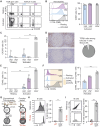
T follicular helper-like function of human γδ T cells. (A–C) PBMCs were cultured in the presence or absence of beads coated with anti-CD3 and anti-CD28 mAbs. (A) Representative flow cytometry profiles for the expression of CD40L and CXCR5 in resting (upper row) and activated (lower row) T cells. (B) Left: Representative histograms for the expression of CD69 in resting (dotted line) or activated (full line) Vδ2+ (up, purple), Vδ2 (middle, blue) or control CD4+ αβ T cells (down, grey). Right: individual values for percentages of CD69+ cells. (C) Individual values for percentages of CXCR5+ cells. (D) Left: immunohistochemical sections of a human lymph node, stained for TCRβ (upper thumbnail) and TCRδ (lower thumbnail). Right: pie-chart representing the proportion of TCRβ+ and TCRδ+ cells among follicular T cells after quantification by computer-assisted morphometry. (E) Individual values for percentages of CD40L+ cells. (F) Left: Representative histograms for the expression of CD40L in resting (dotted line) or activated (full line) Vδ2+ (up, purple), Vδ2 (middle, blue) or control CD4+ αβ T cells (down, grey). Right: individual MdFI values for CD40L+ cells. (G–I) Human B cells were cocultured with allogeneic CD4+ T or γδ T cells in the presence of IgM F(ab′)2 (signal 1), and (H) the percentage of divided cells among alive B cells was evaluated by flow cytometry, as well as (I) the trogocytosis between B and T cells. (G) Schematic representation of the experiment. (H) Left: Representative histograms. Right: Individual coculture values. (I) Left: The flow cytometry gating strategy for the assessment of trogocytosis. Right: percentage of B cells that have experienced trogocytosis in each coculture. Data are presented as median ± IQR. Data were analyzed by Mann-Whitney test when two groups were compared, Kruskal-Wallis test when more than two groups were compared, and two-way ANOVA when there was a within-group comparison between two different conditions. *P < 0.05, **P < 0.01 and ***P < 0.001.
FIGURE 3
FIGURE 3
TFH-helper function of human γδ T cells (A) Left: immunohistochemical sections of human lymph node, stained for TCRβ (upper thumbnail) and TCRδ (lower thumbnail). Right: the density of TCRβ+ and TCRδ+ cells in the follicles were quantified by computer-assisted morphometry. Pie-chart representing the distribution of TCRδ+ T cells (up) and the proportion of TCRβ+ and TCRδ+ cells among extra-follicular T cells (down). (B–E) PBMCs were cultured in the presence or absence of beads coated with anti-CD3 and anti-CD28 mAbs. (B) Up: Representative histograms for the expression of HLA-DR in Vδ2+ (up, purple), Vδ2 (middle, blue) γδ T cells or control B cells (down, grey). Down: individual MdFI values for HLA-DR+ cells. The dashed line represents the negative control. (C) Representative flow cytometry profiles for the expression of CD80 and CD86 in resting (upper row) and activated (lower row) T cells. (D, E) Individual values for percentages of (D) CD80+ and (E) CD86+ cells. (F, G) BCR-primed human B cells were cocultured with allogeneic CD4+ T in the presence or absence of syngeneic γδ T cells. (F) Schematic representation of the experiment. (G) The percentage of divided cells among alive B cells was evaluated by flow cytometry. Left: Representative histograms. Middle: individual B cell division index values. Right: individual B cell proliferation index values. Data are presented as median ± IQR. Data were analyzed by Mann-Whitney test when two groups were compared, Kruskal-Wallis test when more than two groups were compared, and two-way ANOVA when there was a within-group comparison between two different conditions. *P < 0.05 and **P < 0.01.
FIGURE 4
FIGURE 4
Assessment of the role of γδ T cells in a mouse model of heart transplantation (A) Schematic representation of the mouse strains used as recipient of an allogeneic heart transplant regarding αβ and γδ T cells compartments. (B) Presentation of the mouse model. Allogeneic Balb/c nude (H-2d) hearts were transplanted to wild-type (WT), TCRαKO, TCRδKO or CD3εKO C57BL/6 (H-2b) recipient mice. Results are from one experiment. Heart grafts were harvested from Balb/c nude and Balb/c WT donors and digested with collagenase. Heart graft cell suspensions were analyzed by flow cytometry. Representative flow cytometry profiles are shown. (C) Evolution of normalized DSA titers in the circulation of recipients is shown for wild-type (grey, n = 3), TCRαKO (red, n = 4), TCRδKO (blue, n = 4) and CD3εKO (black, n = 5) C57BL/6 mice. (D) DSA titers were compared at the peak of the response between wild-type (grey, n = 3) and TCRδKO (blue, n = 4) C57BL/6 mice. (E) The avidity of DSA produced by wild type (grey, n = 2) and TCRδKO (blue, n = 4) C57BL/6 recipients were compared at day 28 by assessing the stability of DSA binding to Balb/c splenocytes in the presence of increasing concentrations of urea used as chaotropic agent. (F) DSA isotypes were tested at the peak of the response for wild-type (grey, n = 2) and TCRδKO (blue, n = 4) C57BL/6 mice. Data are presented as mean ± SD. Data are presented as median ± IQR. Abbreviations: TCR, T-cell receptor; WT, wild-type.

References

    1. Everly MJ, Rebellato LM, Haisch CE, Ozawa M, Parker K, Briley KP, et al. Incidence and Impact of De Novo Donor-Specific Alloantibody in Primary Renal Allografts. Transplant (2013) 95(3):410–7. 10.1097/TP.0b013e31827d62e3 - DOI - PubMed
    1. Wiebe C, Gibson IW, Blydt-Hansen TD, Karpinski M, Ho J, Storsley LJ, et al. Evolution and Clinical Pathologic Correlations of De Novo Donor-Specific HLA Antibody Post Kidney Transplant. Am J Transpl (2012) 12(5):1157–67. 10.1111/j.1600-6143.2012.04013.x - DOI - PubMed
    1. Sicard A, Phares TW, Yu H, Fan R, Baldwin WM, Fairchild RL, et al. The Spleen Is the Major Source of Antidonor Antibody-Secreting Cells in Murine Heart Allograft Recipients. Am J Transpl Off J Am Soc Transpl Am Soc Transpl Surg (2012) 12(7):1708–19. 10.1111/j.1600-6143.2012.04009.x - DOI - PMC - PubMed
    1. Chen CC, Pouliquen E, Broisat A, Andreata F, Racapé M, Bruneval P, et al. Endothelial Chimerism and Vascular Sequestration Protect Pancreatic Islet Grafts From Antibody-Mediated Rejection. J Clin Invest (2018) 128(1):219–32. 10.1172/JCI93542 - DOI - PMC - PubMed
    1. Pouliquen E, Koenig A, Chen CC, Sicard A, Rabeyrin M, Morelon E, et al. Recent Advances in Renal Transplantation: Antibody-Mediated Rejection Takes Center Stage. F1000prime Rep (2015) 7:51. 10.12703/P7-51 - DOI - PMC - PubMed

Substances

LinkOut - more resources