. 2025 Jan 29:12:1524133.
doi: 10.3389/fbioe.2024.1524133. eCollection 2024.

Establishing rabbit critical-size bone defects to evaluate the bone-regeneration potential of porous calcium phosphate ceramics

Affiliations

Establishing rabbit critical-size bone defects to evaluate the bone-regeneration potential of porous calcium phosphate ceramics

Wei Lei et al. Front Bioeng Biotechnol. .

Abstract

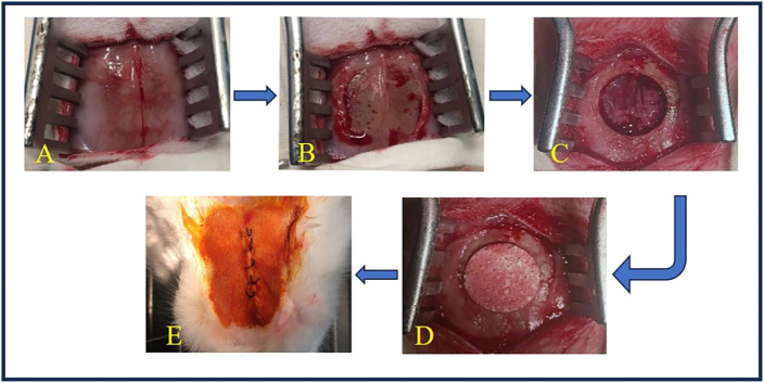
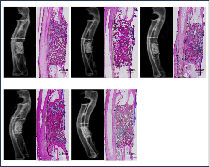
Critical-size bone defects (CSDs), which are those that do not self-repair in a given period, are essential for evaluating bone-regeneration strategies. We established CSDs models in the rabbit cranium and ulna, and the bone-regeneration capacities of porous calcium phosphate (CaP) ceramics were assessed. A 12.6-mm cranial defect was confirmed as a CSDs after 12 weeks, with submicron surface-structured biphasic calcium-phosphate (BCP) implants [consisting of 20% hydroxyapatite and 80% tricalcium phosphate (TCP)] demonstrating significantly higher bone formation (32.2% ± 10.6%) than micron surface-structured TCP (TCP-B) implants (17.8% ± 4.6%, p = 0.0121). Ulna defects (15.0 mm in length) failed to heal spontaneously within 24 weeks when the periosteum was removed from both the ulna and radius, and the radius was covered with an expanded polytetrafluoroethylene (ePTFE) membrane. No bone bridging (i.e., union) was observed in the BCP implants at 12 weeks, whereas 80% of BCP implants (four out of five) achieved union by 24 weeks. Furthermore, the bone area within the available space of BCP implants increased significantly from 19.3% ± 7.3% at 12 weeks to 37.7% ± 8.5% at 24 weeks (p = 0.0063), accompanied by significant BCP resorption (14.8% at 12 weeks and 30.2% at 24 weeks). This study offers two rabbit CSDs models for evaluating bone-regeneration strategies (including bone substitution), and the overall data obtained in the current study indicate the possibility of repairing CSDs with CaP ceramics demonstrating improved bone-forming ability given adequate implantation time.

Keywords: bone regeneration; bone substitutes; calcium phosphate ceramic; critical-size bone defect; submicron surface topography.


Conflict of interest statement

Authors JB and HY were employed by Kuros Biosciences BV. The remaining authors declare that the research was conducted in the absence of any commercial or financial relationships that could be construed as a potential conflict of interest.

Figures

FIGURE 1
FIGURE 1
Surgery of cranial implantation. (A) The skin was incised to expose the surgical field. (B) The periosteum was detached to reveal the bone surface. (C) A circular bone defect with a diameter of 12.6 mm was created using a high-speed drill. (D) TCP-B or BCP was implanted into the bone defect. (E) The wound was closed in layers with silk sutures and disinfected with iodine.
FIGURE 2
FIGURE 2
Surgery of ulna implantation. (A) A longitudinal incision was made on the forelimb of the rabbit. (B) The muscle was dissected to expose the ulna and radius, and the periosteum was elevated from the ulna and radius. (C) A 15.0-mm ulna defect was created using a high-speed burr, followed by ePTFE membrane coverage of the radius, secured with 6–0 silk sutures. (D) BCP cylinders (ø5.0 × 15.0 mm) were loaded in the defects and fixed with 6–0 silk sutures.
FIGURE 3
FIGURE 3
Physicochemical characteristics of materials used. (A) Scanning tunneling microscopy (STM) images of BCP and TCP-B; scale bars = 1 mm. (B) Surface morphology characterized by scanning electron microscopy (SEM); scale bars = 5 μm. (C) Strut pore size distribution of TCP-B and BCP. (D) X-ray diffraction.
FIGURE 4
FIGURE 4
Bone formation at week 12 in the sham defects of cranial defects (X-ray and histological overviews).
FIGURE 5
FIGURE 5
Bone formation in CaP materials at week 12. (A) Radiological and histological images of TCP-B. (B) Radiological and histological images of BCP. (C) Area percentage of bone in TCP-B and BCP implants in cranial defects at week 12. (D) Residual area percentage of TCP-B implants at different time points. (E) Residual area percentage of BCP implants at different time points.
FIGURE 6
FIGURE 6
Bone formation in ulna sham at week 24.
FIGURE 7
FIGURE 7
Bone formation in ulna defects with BCP at week 12.
FIGURE 8
FIGURE 8
Bone formation in ulna defects with BCP at week 24.
FIGURE 9
FIGURE 9
Quantification of bone. (A) Area percentage of bone in BCP implanted in cranial defects at weeks 12 and 24. (B) Area percentage of CaP ceramics in BCP implants at different time points.

References

    1. Albrektsson T., Johansson C. (2001). Osteoinduction, osteoconduction and osseointegration. Eur. Spine J. 10 (Suppl. 2), S96–S101. 10.1007/s005860100282 - DOI - PMC - PubMed
    1. Barradas A. M., Yuan H., van Blitterswijk C. A., Habibovic P. (2011). Osteoinductive biomaterials: current knowledge of properties, experimental models and biological mechanisms. Eur. Cell Mater 21, 407–429. 10.22203/ecm.v021a31 - DOI - PubMed
    1. Bauer T. W., Muschler G. F. (2000). Bone graft materials. An overview of the basic science. Clin. Orthop. Relat. Res. 371 (371), 10–27. 10.1097/00003086-200002000-00003 - DOI - PubMed
    1. Bohner M., Santoni B. L. G., Döbelin N. (2020). β-tricalcium phosphate for bone substitution: synthesis and properties. Acta Biomater. 113, 23–41. 10.1016/j.actbio.2020.06.022 - DOI - PubMed
    1. Bouler J. M., Pilet P., Gauthier O., Verron E. (2017). Biphasic calcium phosphate ceramics for bone reconstruction: a review of biological response. Acta Biomater. 53, 1–12. 10.1016/j.actbio.2017.01.076 - DOI - PubMed

LinkOut - more resources