. 2025 Jan 29:16:1522651.
doi: 10.3389/fimmu.2025.1522651. eCollection 2025.

Functional and structural characterization of mouse Factor H-related B protein unveils a novel dimerization domain shared by FHR-B and FH

Affiliations

Functional and structural characterization of mouse Factor H-related B protein unveils a novel dimerization domain shared by FHR-B and FH

Adrián Martín-Ambrosio Doménech et al. Front Immunol. .

Abstract

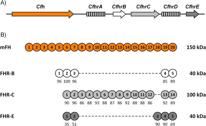
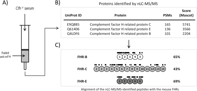
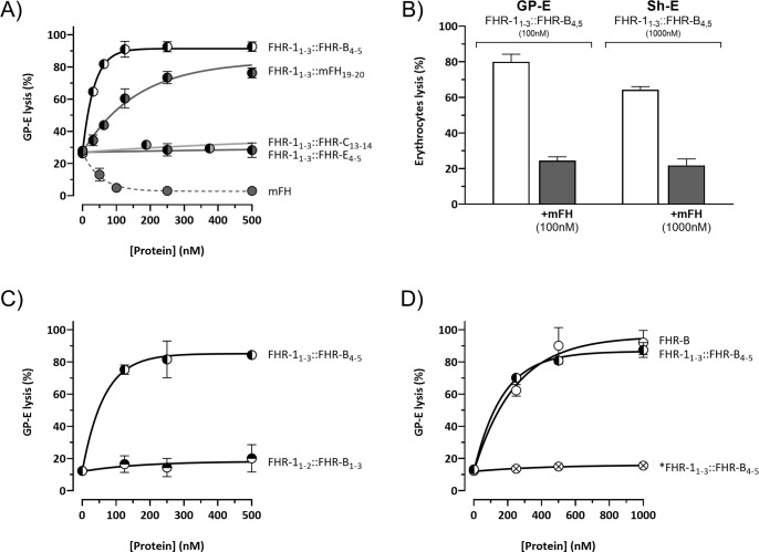
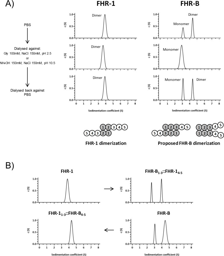
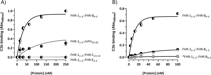
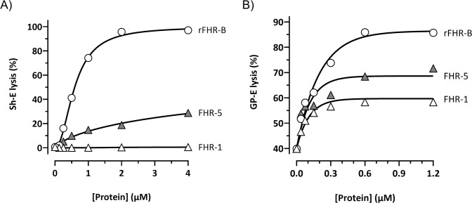
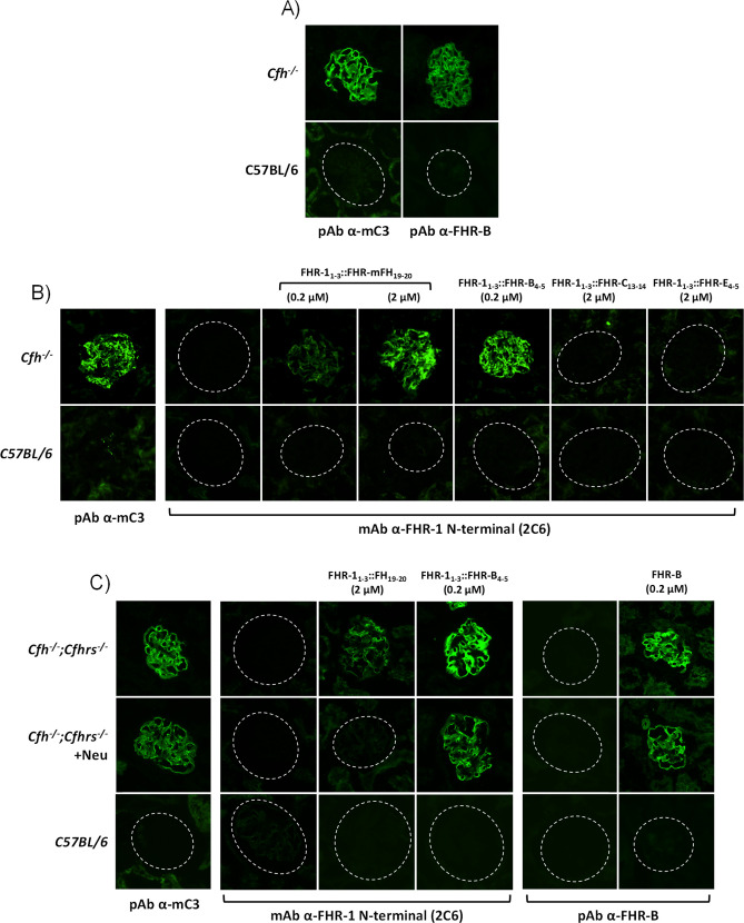
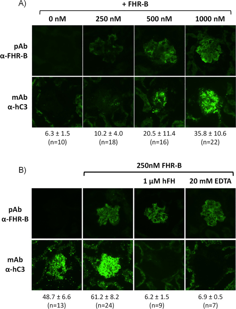
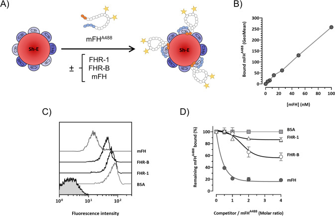
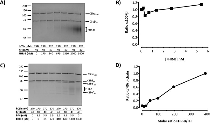
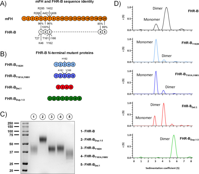
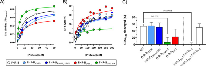
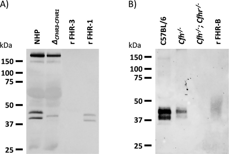
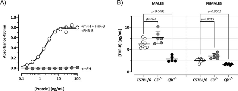
Factor H-related proteins (FHRs) are found in mice, but their equivalence to human FHRs remains uncertain. This study identifies three FHRs in mouse plasma (FHR-B, FHR-C, and FHR-E) and focuses on characterizing FHR-B. Using purified plasma proteins and recombinant mutants, FHR-B was found to form dimers and bind strongly to C3, C3b, iC3b, and C3dg. It also competes with mouse Factor H (mFH) for binding to C3b-coated surfaces and disrupts mFH regulation in hemolysis assays with sheep and guinea pig erythrocytes. These functions are localized to the C-terminal region and are dependent on FHR-B dimerization. Dimerization occurs through the N-terminal region (SCR1-3), which differs from mFH SCR5-7 by only four amino acids and also shares significant homology with human FHR-3 and human FH SCR5-7. In contrast to FHR-1, AUC experiments indicate that FHR-B dimerization is pH-sensitive, reversible and that the monomers in the dimer present the same head to tail orientation. Mutant analyses revealed that mFH SCR5-7 also forms dimers, but less efficiently than FHR-B. Notably, substituting FHR-B Tyr162 (a key residue homologous to the disease-associated Tyr402 in human FH) for His reduces dimerization. We also found that a recombinant FHR-B with a duplicated dimerization domain formed stable dimers but lacked functional activity. Overall, FHR-B shows structural and functional similarities with various human FHRs, suggesting convergent evolution between mouse and human FHRs. Furthermore, this study reveals a novel dimerization domain shared by FHR-B and mouse FH and illustrates the importance of dimerization and monomer orientation in FHRs activity. It also underlines notable differences between human and mice FHRs that should be further explored before modeling FHR-associated human diseases in mice.

Keywords: complement; complement-related diseases; dimerization; factor H; factor-H related proteins; regulation.


Conflict of interest statement

The authors declare that the research was conducted in the absence of any commercial or financial relationships that could be construed as a potential conflict of interest.

Figures

Figure 1
Figure 1
Cfhrs genes in mice. (A) Genomic organization of the mouse Cfhrs genes. Downstream of Cfh, there are five Cfhrs genes. Genes lacking evidence of transcription are represented by squared boxes. (B) Alignment of FHR-B, FHR-C, and FHR-E with mFH. SCRs are numbered inside the circles, and their amino acid sequence similarity to the corresponding mFH SCRs is indicated as percentages.
Figure 2
Figure 2
Identification of FHR-B, FHR-C and FHR-E in mice plasma using a proteomic approach. (A) Mouse FHR proteins were purified from C57BL/6 Cfh-/- plasma by affinity chromatography using protein G-purified rabbit IgG polyclonal antibody anti-mFH (see Material and Methods). (B) Eluted proteins were digested and analyzed by nLC-MS/MS, which identified several peptides unique to mouse FHR-B, FHR-C, and FHR-E proteins (See Supplementary Materials for a list of these peptides and additional proteins identified in these analysis). No peptides corresponding to FHR-A nor FHR-D were found. Importantly, no peptides unique to mouse FHRs proteins were identified in the analysis using a control Sepharose column coupled to a non-immune rabbit IgG. (C) Lines above SCRs identify the position of the unique peptides to mouse FHR-B, FHR-C, and FHR-E proteins, to illustrate that they provide an almost complete coverage of these proteins.
Figure 3
Figure 3
The C-terminal domain of FHR-B is functionally equivalent to the C-terminal domain of human FHR-1. (A) Hemolytic assay using GP-E and human serum of the hybrid proteins containing the N-terminal region of FHR-1 and the C-terminals domains of mFH (FHR-11-3::mFH19-20), FHR-B (FHR-11-3::FHR-B4-5), FHR-C (FHR-11-3::FHR-C14-15) or FHR-E (FHR-11-3::FHR-E4-5). Only hybrid proteins containing the C-terminal region of mFH and FHR-B exhibit de-regulation activity, measured as GP-E lysis, in these assays. (B) Hemolytic assay using GP-E or Sh-E and human serum to illustrate that the C-terminal region of FHR-B antagonizes FH regulation in both GP-E and Sh-E hemolysis assays. Figure also shows that addition of mFH antagonizes the de-regulation activity of FHR-B. (C) Hemolytic assay using GP-E and human serum to illustrate that the N-terminal domain of FHR-B (FHR-11-2::FHR-B1-3) lacks FH de-regulation activity, measured as GP-E lysis. (D) Hemolytic assay using GP-E and human serum shows that full-length FHR-B exhibit similar FH de-regulation activity than the hybrid protein FHR-11-3::FHR-B4-5. Since a monomeric variant of the hybrid protein FHR-11-3::FHR-B4-5 (*FHR-11- 3::FHR-B4-5) lacks FH de-regulation activity, these data suggests that FHR-B is a dimer. These experiments were performed in triplicate, and the figures present a representative result from at least three independent assays.
Figure 4
Figure 4
FHR-B dimerizes. (A) Full-length rFHR-B was analyzed by AUC, using FHR-1 for comparison. In PBS pH7, FHR-B resolves in 2 peaks, corresponding to FHR-B monomers and FHR-B dimers. FHR-B dimers disassembled at both high and low pH conditions, but reassemble when neutral pH is restored. Data also indicate that dimers of FHR-B have smaller sedimentation coefficient than dimers of FHR-1, which may suggest a more globular conformation of FHR-B. Diagrams illustrate the organization of the monomers in the FHR-1 dimers and a proposal for the organization of the FHR-B monomers in the dimers. (B) AUC experiments with FHR-1 and FHR-B hybrids in which we have switched the N-terminal domains. The data illustrate that the dimerization characteristics of FHR-1 and FHR-B are determined by their N-terminal domains.
Figure 5
Figure 5
C-terminal domain of FHR-B binds strongly to C3b. (A) ELISA experiments performed with C3b-coated plates to analyze the capacity of the hybrid proteins including the C-terminal regions of FHR-B (FHR-11-3::FHR-B4-5), FHR-C (FHR-11-3::FHR-C14-15) or FHR-E (FHR-11-3::FHR-E4-5) to bind C3b. A hybrid protein with the C-terminal region of mFH (FHR-11-3::mFH19-20) was included as a reference. (B) Similar ELISA experiments as in A to show that an hybrid protein with the N-terminal domains of both FHR-1 and FHR-B (FHR-11-2::FHR-B1-3) lacks C3b binding capacity. Similarly, these experiments show that the monomeric variant of the FHR-11-3::FHR-B4-5 hybrid, called *FHR-11-3::FHR-B4-5, lacks C3b binding capacity, which indicates that the strong FHR-B binding to C3b-coated surfaces requires FHR-B dimerization. These experiments were performed in triplicate, and the figures present a representative result from at least three independent assays.
Figure 6
Figure 6
FHR-B promotes much stronger complement de-regulation in Sh-E and GP-E hemolysis assays than FHR-1 and FHR-5. (A) Hemolytic assay using Sh-E and mouse serum of the FHR-B, FHR-5 and FHR-1 proteins shows a higher capacity of FHR-B to promote complement de-regulation than FHR human proteins. De-regulating activity is measured as Sh-E lysis. (B) Hemolytic assay using GP-E and mouse serum of the FHR-B, FHR-5 and FHR-1 proteins shows a higher capacity of FHR-B to promote complement de-regulation than FHR human proteins. De-regulating activity is measured as GP-E lysis. These experiments were performed in duplicate, and the figures present a representative result from at least three independent assays.
Figure 7
Figure 7
FHR-B binds strongly to C3-opsonized surfaces independently of the presence of sialic acids. (A) Cryostat kidney sections from C57BL/6 and Cfh-/- mice immunostained for mC3 and FHR-B reveal that FHR-B is naturally deposited in the C3-opsonized mice glomeruli. (B) Hybrid proteins with the C-terminal region of FHR-B (FHR-11-3::FHR-B4-5), FHR-C (FHR-11-3::FHR-C14-15), FHR-E (FHR-11-3::FHR-E4-5) and mFH (FHR-11-3::mFH19-20) were added to cryostat kidney sections of Cfh-/- mice and detected with a mouse monoclonal antibody directed to the N-terminal region of FHR-1 (2C6) that is present in all these hybrid proteins. Data illustrate that the hybrid FHR-11-3::FHR-B4-5 shows a much stronger binding to C3-opsonized surfaces than the hybrid including the C-terminal region of mFH (FHR-11-3::mFH19-20). No binding was observed with FHR-11-3::FHR-C14-15 and FHR-11-3::FHR-E4-5 hybrids. (C) Same binding assay on cryostat kidney sections of Cfh-/-;Cfhrs-/- mice was performed with the hybrid proteins FHR-11-3::mFH19-20, FHR-11-3::FHR-B4-5 and the full length rFHR-B. Much stronger binding was again observed for FHR-11-3::FHR-B4-5 than FHR-11-3::mFH19-20 and this binding was similar to that obtained for the rFHR-B protein. Interestingly, binding of FHR-11-3::FHR-B4-5 and rFHR-B, but not of FHR-11-3::mFH19-20 was preserved after desialylation of the cryostat kidney sections using Clostridium perfringens neuraminidase. Importantly, different antibodies were used to detect the hybrid proteins (2C6 anti-FHR-1 N-terminus) and full length rFHR-B (in-house pAb ant-FHR-B). As cryostat kidney sections of Cfh-/-;Cfhrs-/- mice were used, the in-house pAb ant-FHR-B shows no backgorund staining.
Figure 8
Figure 8
Surface bound FHR-B promotes complement activation through the alternative pathway. (A) Cryostat kidney sections from Cfh-/-;Cfhrs-/- mice were incubated with 10% NHSΔFHRs supplemented with 1.67 µM hFH (to prevent basal hC3 activation) in the absence and presence of varying amounts of rFHR-B. Increasing amounts of FHR-B led to increased FHR-B binding to the glomeruli, which in turn enhanced complement activation and hC3 deposition in the glomeruli. (B) Complement activation on cryostat kidney sections from Cfh-/-;Cfhrs-/- mice using no-hFH-supplemented NHSΔFHRs. Basal hC3 deposition is increased by surface-bound FHR-B. hC3 deposition promoted by the surface-bound FHR-B is inhibited by adding hFH or EDTA, indicating that C3 deposition requires formation of the alternative pathway C3-convertase and it is modulated by the FHR-B/FH ratio. The mean fluorescence intensity of C3 deposits is indicated below each panel in arbitrary units, together with the numbers of glomeruli analyzed.
Figure 9
Figure 9
FHR-B competes binding of mFH to human C3b-opsonized surfaces. (A) Experiment design: mFH labeled with alexa F488 (mFHAF488) binds Sh-E coated with human C3 activated fragments in a doses-dependent manner and can be detected by flow cytometry. The capacity of rFHR-B to compete mFH binding was tested by incubating mFHAF488 in the presence of FHR-B. FHR-1 and non-labeled mFH were included as a negative and a positive control, respectively. (B) Calibration curve of the mFHAF488 binding to human C3b. (C) Competition of mFHAF488 binding to C3 coated Sh-E in the presence of mFH, rFHR-B, FHR-1 and BSA at a molar ratio of 1:4 (mFHAF488/competitor protein) measured as mean fluorescence intensity. (D) Competition of mFHAF488 binding to C3 coated Sh-E in the presence of mFH, rFHR-B, FHR-1 and BSA at different molar ratios. These experiments were performed in duplicate, and the figures present a representative result from at least three independent assays.
Figure 10
Figure 10
FHR-B does not have FI-cofactor activity for the cleavage of C3b, but enhances the FI-cofactor activity of limited amounts of FH. (A) Fluid phase assay with purified proteins shows that FHR-B does not have cofactor activity of FI in the proteolytic cleavage of C3b. hC3b 270nM and hFI 40nM were incubated with different concentrations of rFHR-B for 45 minutes at 37°C and loaded in an SDS-PAGE stained with Coomassie. No cleavage of the α’-chain of C3b was observed at any concentration. (B) Densitometry of the α’-chain and β-chains of C3b are represented as a α’-chain/β-chain ratio. (C) Fluid phase assay with purified proteins shows that FHR-B enhances the FI-cofactor activity of FH for the proteolytic cleavage of C3b in a dose-dependent manner. hC3b 270nM, hFI 40nM and hFH 3.5nM were incubated with different concentrations of rFHR-B for 45 minutes at 37°C and loaded in an SDS-PAGE stained with Coomassie. The FI-cofactor activity of FH is illustrated by the disappearance of the α’-chain of C3b and the appearance of the α60 and α40 C3b fragments. (D) Densitometry of the α40 and β-chains of C3b are represented in as a α40/β-chain ratio. The figures present a representative result from at least three independent assays.
Figure 11
Figure 11
AUC analyses of FHR-B dimerization domain mutants. (A) Alignment between mFH and FHR-B, highlighting the differences in the three N-terminal region SCRs of FHR-B. (B) List of FHR-B mutants with alteration in the N-terminal region. (C) Coomassie stained SDS-PAGE under non-reducing conditions of the FHR-B N-terminal mutants produced. (D) AUC analyses (in PBS pH 7) of the FHR-B mutant proteins. FHR-BT161A,I166V, mimicking mFH SCR5-7 preserves an active dimerization domain, although the proportion of FHR-B dimers is reduced compared to the FHR-B native protein. Similar situation is observed when residue Tyr162 is mutated to His (FHR-BY162H). As expected, a duplication of SCR1-3 of FHR-B (FHR-BDup.1-3) results in a complete dimeric molecule. Notably, however, deletion of SCR1(FHR-BDel.1) has no impact in the dimerization capacity of FHR-B. As a whole, these data localize the FHR-B dimerization domain to SCR2 and SCR3.
Figure 12
Figure 12
Functional analysis of FHR-B dimerization domain mutants. (A) ELISA experiments performed with human C3b-coated plates to analyze the capacity of the FHR-B mutant proteins including FHR-BY162H, FHR-BT161A,I166V, FHR-BDel.1 and FHR-BDup.1-3 to bind C3b. FHR-B protein was included as a reference. (B) A GP-E hemolysis assay with human serum was used to determine the FH de-regulation activity of the FHR-B N-terminal mutants. FHR-BT161A,I166V and FHR-BY162H preserve this activity compared with the WT FHR-B protein. A slight increase in FH de-regulation activity was observed in the FHR-B mutant carrying a deletion of SCR1 (FHR-BDel.1), whereas these assays show that the FHR-B mutant with a duplication of the dimerization domain (FHR-BDup.1-3) lacks almost completely FH de-regulation activity. (C) We also tested the capacity of the different FHR-B mutants to potentiate the FI-cofactor activity of limited amounts of FH. Data show that a fully dimerized FHR-B like FHR-BDup.1-3 or FHR-11-3::FHR-B4-5 lack this functionality, whereas a monomeric form of FHR-B or mutants with decreased proportion of dimers (FHR-BT161A,I166V and FHR-BY162H) preserve this activity. Data also suggests that FHR-B SCR1 is relevant for this activity. Student’s t-test statistical analysis for independent samples was performed using FHR-B as reference. P<0.05 was considered significant. These experiments were performed in triplicate, and the figures present a representative result from at least three independent assays.
Figure 13
Figure 13
Western blot characterization of a rabbit polyclonal antibody raised against purified recombinant FHR-B. (A) Analysis of normal human plasma and a plasma from an individual deficient FHR-3 and FHR-1 show that the antibody cross-reacts with human FH, human FHL-1 and human FHR-1, but do not recognize human FHR-3. (B) The same antibody used in A was extensively adsorbed using a Sepharose column coupled to mFH and was tested in western blot with serum samples of C57BL/6, Cfh-/- and Cfh-/-; Cfhr-/- mice. No mouse FHR proteins other than FHR-B were detected. Notice the important decrease in FHR-B levels in the serum of Cfh-/- mice (see also Figure 14 ).
Figure 14
Figure 14
FHR-B levels in mouse plasma. (A) ELISA experiments to confirm that the rabbit polyclonal antibody generated in-house against the purified recombinant FHR-B protein and extensively adsorbed against mFH lacks mFH cross-reactivity. In these experiments Cfh-/-; Cfhr-/- mice plasma was reconstituted with different concentrations of mFH and/or FHR-B to show that while FHR-B was perfectly detected, mFH was not detected. (B) Using this ELISA we measure the plasma levels in male and female C57BL/6 mice. We also found that circulating levels of FHR-B were slightly increase in C3-/- mice and significantly decreased in Cfh-/- mice, suggesting that plasma FHR-B levels are influenced by the amount of C3 deposited in opsonized surfaces. Student’s t-test statistical analysis for independent samples was performed using C57BL6 mice serum as reference. P<0.05 was considered significant. These experiments were performed in duplicate, and the figures present a representative result from at least three independent assays.

Similar articles

References

    1. Vik DP, Muñoz-Cánoves P, Kozono H, Martin LG, Tack BF, Chaplin DD. Identification and sequence analysis of four complement factor H-related transcripts in mouse liver. J Biol Chem. (1990) 265:3193–201. doi: 10.1016/s0021-9258(19)39753-4 - DOI - PubMed
    1. Cserhalmi M, Csincsi ádám I, Mezei Z, Kopp A, Hebecker M, Uzonyi B, et al. . The murine factor H-Related protein FHR-B promotes complement activation. Front Immunol. (2017) 8:1145/BIBTEX. doi: 10.3389/FIMMU.2017.01145/BIBTEX - DOI - PMC - PubMed
    1. Hellwage J, Skerka C, Zipfel PF. Biochemical and functional characterization of the factor-H-related protein 4 (FHR-4). Immunopharmacology. (1997) 38:149–57. doi: 10.1016/S0162-3109(97)00075-1 - DOI - PubMed
    1. Hellwage J, Jokiranta TS, Koistinen V, Vaarala O, Meri S, Zipfel PF. Functional properties of complement factor H-related proteins FHR-3 and FHR-4: binding to the C3d region of C3b and differential regulation by heparin. FEBS Lett. (1999) 462:345–52. doi: 10.1016/S0014-5793(99)01554-9 - DOI - PubMed
    1. Hellwage J, Eberle F, Babuke T, Seeberger H, Richter H, Kunert A, et al. . Two factor H-related proteins from the mouse: Expression analysis and functional characterization. Immunogenetics. (2006) 58:883–93. doi: 10.1007/S00251-006-0153-Y/FIGURES/8 - DOI - PubMed

LinkOut - more resources