. 2025 Sep 1;110(9):2076-2090.
doi: 10.3324/haematol.2024.286887. Epub 2025 Feb 13.

Overall cancer risk in people with deleterious germline DDX41 variants

Affiliations

Overall cancer risk in people with deleterious germline DDX41 variants

Sophia C Korotev et al. Haematologica. .

Abstract

Germline loss-of-function (LoF) DDX41 variants predispose to late-onset hematopoietic malignancies (HM), predominantly of myeloid lineage. Among 43 families with germline DDX41LoF variants, bone marrow (BM) biopsies in those without (N=8) or with malignancies (N=21) revealed mild dysplasia in peripheral blood (57%) and BM (88%), long before the average age of DDX41-related HM onset. Therefore, we recommend baseline BM biopsies in people with germline DDX41LoF alleles to avoid over-diagnosis of myelodysplastic syndromes. A variety of solid tumors were also observed in our cohort, with 24% penetrance by age 75. Although acquired DDX41 mutations are common in HM, we failed to identify such alleles in solid tumors arising in those with germline DDX41LoF variants (N=15), suggesting an alternative mechanism driving solid tumor development. Furthermore, 33% of pedigrees in which ≥15% of first-degree relatives including the proband were diagnosed with a solid tumor had second germline deleterious variants in other cancer-predisposition genes, likely serving as primary cancer drivers. Finally, both lymphoblastoid cell lines and primary peripheral blood from individuals with germline DDX41LoF variants exhibited differential levels of inflammation-associated proteins. These data provide evidence of inflammatory dysfunction mediated by germline DDX41LoF alleles that may contribute to solid tumor growth in the context of additional germline cancer- associated variants. For those with HM and personal/family histories of solid tumors, we recommend broad germline testing. DDX41 may be an indirect modifier of solid tumor pathogenesis compared to its tumor suppressor function within hematopoietic tissues, a hypothesis that can be addressed in future work.


Figures
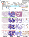
Figure 1.
Figure 1.
Family-associated germline DDX41 variants and morphologic features of baseline and malignant peripheral blood and bone marrow in individuals with deleterious germline DDX41 variants at different ages. (A) Deleterious germline DDX41 variants identified in patients and families with hematopoietic malignancies (HM) are shown above the protein schematic, and variants of uncertain significance (VUS; blue circles) are shown below. Pathogenic variants are indicated by red diamonds, and likely pathogenic variants by orange diamonds. Non-protein coding variants are listed in the bottom right. The likely pathogenic copy number variant (CNV) is indicated by an orange line. Novel variants are shown with glow and previously identified variants are shown without glow. DDX41 protein domains are indicated by color: RecA-like domain 1 (light blue), RecA-like domain 2 (lilac), and zinc finger (ZnF, light green). (B-Q) Images shown include (B, F, J, N) peripheral blood, (C, G, K, O) bone marrow (BM) aspirate, (D, H, L, P) BM trephine, and (E, I, M, Q) immunohistochemistry on trephine. (B-E) A 17-year-old (yo) female with a pathogenic DDX41 variant (p.M1?), mild dysplastic changes in erythroid lineage (red arrow) and megakaryocytic lineage (yellow arrow), but insufficient for diagnosis of myelodysplastic syndromes (MDS). (F-I) A 46yo female with a likely pathogenic DDX41 deletion of exons 12-17, significant (>10%) dysplastic changes in both erythroid and megakaryocytic lineages, but no granulocytic dysplasia. (J-M) A 73yo male (father of (F-I)) with a likely pathogenic DDX41 deletion of exons 12-17, 4.6% blasts and significant dyserythropoiesis and dysmegakaryopoiesis as well as dysgranulopoiesis manifested mainly by abnormal nuclear morphology including hyposegmentation, dense chromatin and nuclear membrane projections (orange arrow), but not cytoplasmic hypogranulation, diagnosed with MDS with multilineage dysplasia. (N-Q) A 61yo male with a likely pathogenic DDX41 variant (p.P258L), 18% blasts, and multilineage dysplasia (particularly prominent in granulocytes), diagnosed MDS with excess blasts-2 progressing toward acute myeloid leukemia.
Figure 2.
Figure 2.
Disease breakdown by DDX41 variant and timelines of solid tumors and hematopoietic malignancies in patients with multiple malignancies. (A) DDX41 protein schematic showing all malignancies identified in individuals with loss-of-function (LoF) DDX41 alleles plotted by corresponding variant. Diseases represented include hematopoietic malignancies (HM) (red), and solid tumors such as breast (pink), prostate (orange), melanoma (yellow), colon (light green), gastric (black), endometrial (light blue), lung (dark blue), tonsillar (magenta), ovarian/vulvar (purple), renal (brown), neuroendocrine carcinoma (peach), mastocytosis (dark green), head and neck (green), small bowel (teal), basal cell carcinoma (gray), fallopian tube (fuchsia), mesothelioma (light pink), and kidney (salmon) cancers. DDX41 protein domains are indicated by color: RecA-like domain 1 (light blue), RecA-like domain 2 (lilac), and zinc finger (ZnF) (light green). (B) Age at cancer diagnoses and treatments in individuals with DDX41LoF alleles who were diagnosed with more than 1 cancer. HM are shown in red. Solid tumors represented are breast (pink), prostate (orange), colon (light green), lung (dark blue), renal (brown), basal cell carcinoma (grey), gastric (black), ovarian/vulvar (purple), tonsillar (magenta), and neuroendocrine carcinoma (peach). Solid tumor cancer therapies are indicated by: radiation therapy (R); chemotherapy (C); hormonal therapy (H); surgery (S); and unknown (?). Mean latency refers to the average years between the onsets of solid tumors and HM, whereas the range refers to the minimum and maximum latencies present.
Figure 3.
Figure 3.
RNA sequencing, cytokine arrays, and Luminex assays reveal inflammatory dysregulation in DDX41var/ + patient-derived lymphoblastoid cell lines. (A) Principal component analysis (PCA) plot of RNA-sequencing data for DDX41var/+ (N=5, purple) and DDX41WT (N=3, green) lymphoblastoid cell lines (LCL). Noted clustering in gene expression is demonstrated between DDX41P258L/+ and DDX41A500Cfs*9/+, DDX41A492Gfs*17/+ and DDX41del ex12-17/+, and of DDX41WT LCL. (B) Volcano plot showing significantly (CI=95%) upregulated (red) and downregulated (blue) genes in DDX41var/+ LCL (N=5) compared to DDX41WT LCL (N=3). Genes with no statistically significant change are in grey. (C) Normalized enrichment plot of genes from 24 hallmark signaling pathways. Increases in overall gene expression in DDX41var/+ LCL from wild-type (WT) are in red, while decreases from WT are in blue. P values were determined by Pearson’s correlation. (D) Heat map of cytokine array data showing fold changes in pixel densities of increased cytokines in patient-derived DDX41var/+ (purple) LCL-conditioned media compared to DDX41WT. Fold changes range from 0.1 (blue) to >10 (red). (E) Heat map of commercial Luminex data showing fold changes in pixel densities of increased cytokines in patient-derived DDX41var/+ (purple) LCL-conditioned media compared to DDX41WT. Fold changes range from 0.1 (turquoise) to >2 (magenta). (F-I) Bar graphs showing data from a custom Luminex panel. P values were determined using two-tailed t tests with Welch’s correction and confirm higher levels of (F) ANG, (G) CXCL13, (H) CXCL8, and (I) IL-9 in DDX41var/+ (purple) LCL-conditioned media compared to DDX41WT (green).
Figure 4.
Figure 4.
Testing the proposed mechanism of inflammatory dysregulation in germline DDX41var/+ lymphoblastoid cell lines. (A) Western blots to quantify NF-kB (p65 subunit) in nuclear and cytoplasmic protein fractions from patient-derived DDX41var/+ (purple) and DDX41WT (green) lymphoblastoid cell lines (LCL). Histone H3 was used as a nuclear marker and loading control while GAPDH was used as a cytoplasmic marker and loading control. (B) Average NF-kB pixel densities in nuclear protein fractions from patient-derived DDX41var/+ (purple) and DDX41WT (green) LCL normalized to histone H3. Higher levels of NF-kB were detected in DDX41var/+ LCL compared to DDX41WT (P=0.008) according to a two-tailed t test with Welch’s correction. (C) Average NF-kB pixel densities in cytoplasmic protein fractions from patient-derived DDX41var/+ (purple) and DDX41WT (green) LCL normalized to GAPDH. No significant change in NF-kB was detected according to a two-tailed t test with Welch’s correction. (D) Visual summary of cytokine array, Luminex, and western blot data. Cytokines whose levels were higher in DDX41var/+ than in DDX-41WT LCL-conditioned media according to cytokine array and Luminex are shown in red. Increased activation and translocation of NF-kB (gold) is indicated by red upward arrows. Direct protein interactions are indicated by solid black arrows. Indirect activation of NF-kB by inflammatory cytokine signaling is indicated by a dotted red arrow. Proteins/receptors that were identified in literature but were not quantified are shown in grey. Created in https://BioRender.com.
Figure 5.
Figure 5.
Inflammatory dysregulation in UK Biobank participants with likely germline DDX41LoF variants. (A) Volcano plot showing proteins that are increased (red; 95% confidence interval [CI]) or decreased (blue) in individuals with likely germline loss-of-function (LoF) DDX41 variants compared to wild-type (WT) controls. (B) Protein-protein interaction network showing proteins found to decrease in individuals with likely germline DDX41LoF variants compared to WT controls. Only proteins with “high confidence” (0.700) or “highest confidence” (0.900) interactions (N=30) according to the STING database are shown. The level of significance with which proteins were found to decrease are indicated by color: P<0.001 (green), P<0.01 (light blue), or P<0.05 (dark blue). (C) Enrichment plot showing pathways enriched among the 114 proteins found to decrease in individuals with likely germline DDX41LoF variants compared to WT controls according to the STRING database.

References

    1. Arber DA, Orazi A, Hasserjian RP, et al. International Consensus Classification of Myeloid Neoplasms and Acute Leukemias: integrating morphologic, clinical, and genomic data. Blood. 2022;140(11):1200-1228. - PMC - PubMed
    1. Döhner H, Wei AH, Appelbaum FR, et al. Diagnosis and management of AML in adults: 2022 recommendations from an international expert panel on behalf of the ELN. Blood. 2022;140(12):1345-1377. - PubMed
    1. Khoury JD, Solary E, Abla O, et al. The 5th edition of the World Health Organization Classification of Haematolymphoid Tumours: Myeloid and Histiocytic/Dendritic Neoplasms. Leukemia. 2022;36(7):1703-1719. - PMC - PubMed
    1. Makishima H, Bowman TV, Godley LA. DDX41-associated susceptibility to myeloid neoplasms. Blood. 2023;141(13):1544-1552. - PMC - PubMed
    1. Polprasert C, Schulze I, Sekeres MA, et al. Inherited and somatic defects in DDX41 in myeloid neoplasms. Cancer Cell. 2015;27(5):658-670. - PMC - PubMed

Substances

LinkOut - more resources