. 2025 Mar 18;99(3):e0208524.
doi: 10.1128/jvi.02085-24. Epub 2025 Feb 13.

PLSCR1 suppresses SARS-CoV-2 infection by downregulating cell surface ACE2

Affiliations

PLSCR1 suppresses SARS-CoV-2 infection by downregulating cell surface ACE2

Ruiyi Ma et al. J Virol. .

Abstract

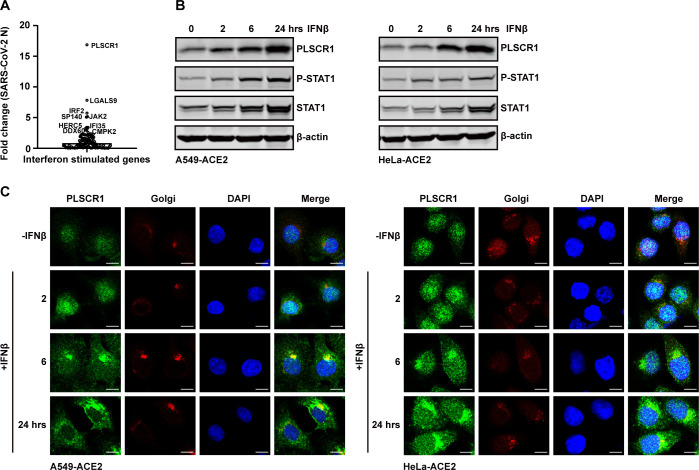
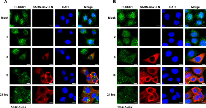
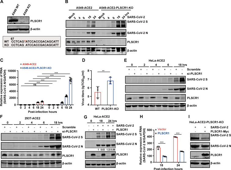
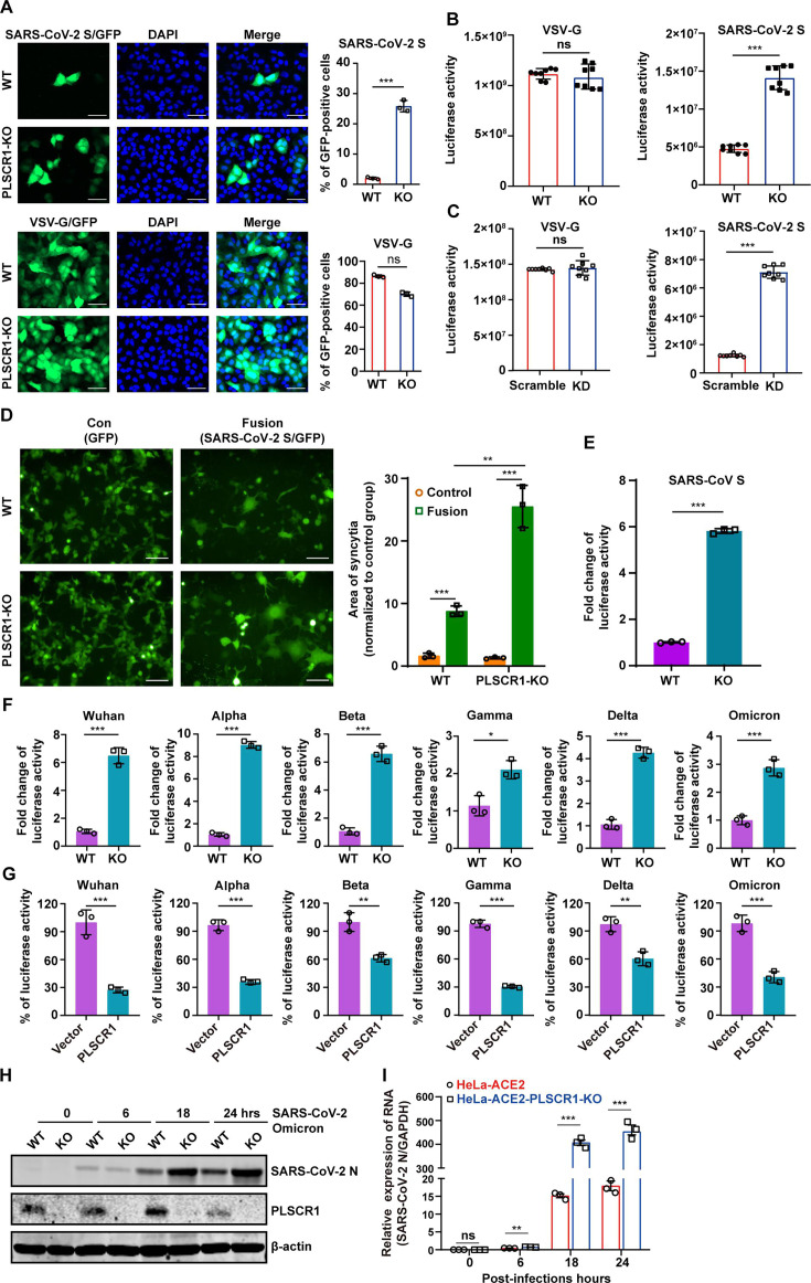
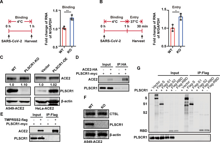
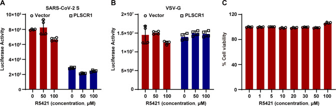
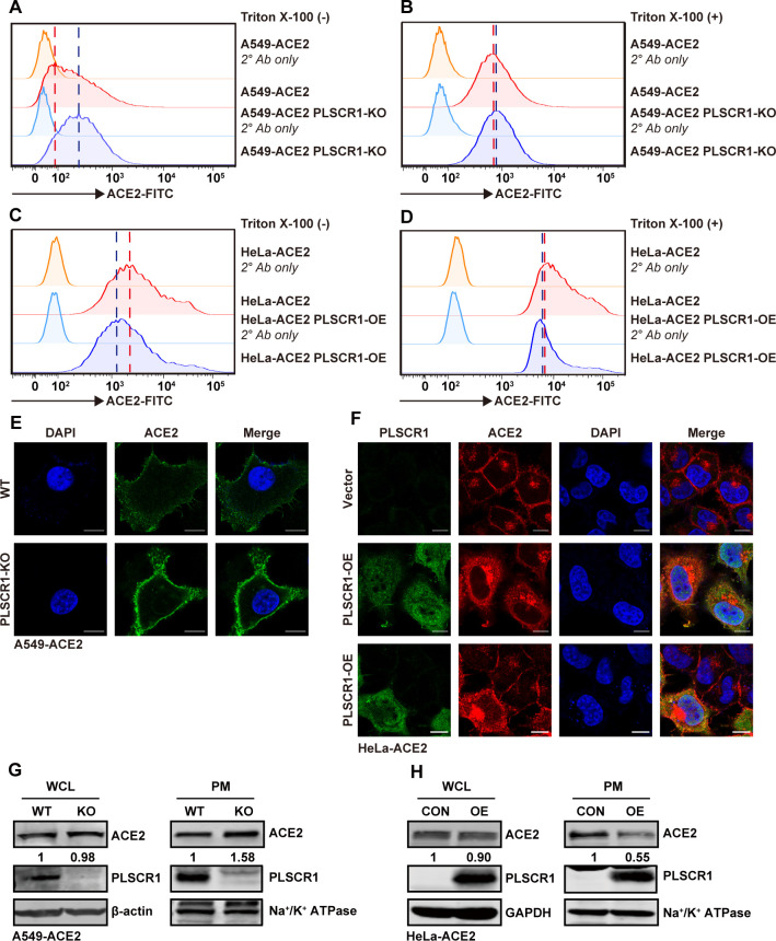
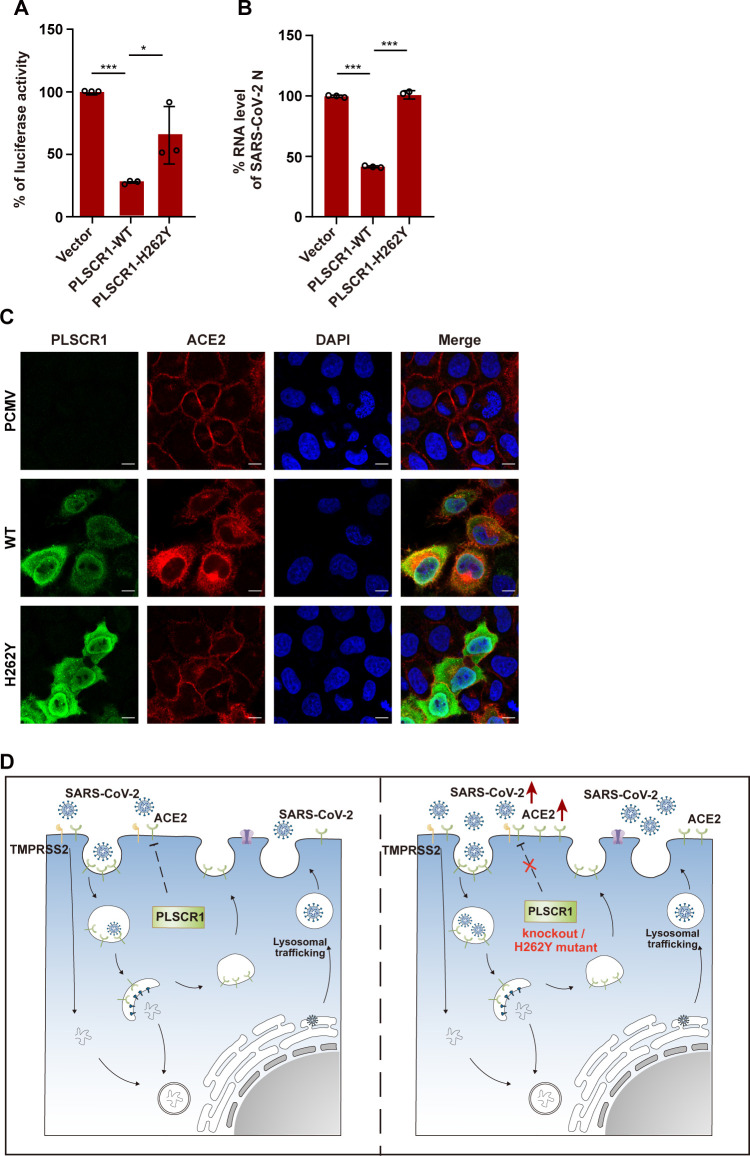
Type I interferons exert their antiviral effects against SARS-CoV-2 by inducing the expression of interferon-stimulated genes (ISGs), including but not limited to LY6E, CH25H, IFITM2/3, and IFIH1. However, the antiviral effect and underlying mechanisms of action of most ISGs in SARS-CoV-2 infection are not yet fully understood. By screening 109 ISG-knockout cell lines, we identify that phospholipid scramblase 1 (PLSCR1), an interferon-inducible protein, acts as a crucial restriction factor against SARS-CoV-2 infection. Cells lacking PLSCR1 are highly susceptible to SARS-CoV-2 infection. Conversely, overexpression of PLSCR1 inhibits SARS-CoV-2 infection. Depletion of PLSCR1 enhances cellular entry of both pseudotyped and authentic SARS-CoV-2. Mechanistically, PLSCR1 inhibits SARS-CoV-2 entry by specifically downregulating plasma membrane expression of ACE2, the virus's receptor, without affecting the overall levels of ACE2 within the cell. As such, we unraveled previously unappreciated mechanisms by which PLSCR1 exerts its restrictive effect on SARS-CoV-2. These data provide new insights into the interplay between host innate antiviral immunity and SARS-CoV-2 and shed light on novel antiviral therapeutics.

Importance: Phospholipid scramblase 1 (PLSCR1) has been identified as a critical host restriction factor against SARS-CoV-2 infection. In this study, we demonstrated that PLSCR1 inhibited SARS-CoV-2 entry by downregulating the plasma membrane expression of ACE2, the primary receptor for viral entry. Our findings elucidate a novel host-pathogen interaction that not only deepens our understanding of the innate immune response to SARS-CoV-2 but offers potential strategies for therapeutic interventions against COVID-19.

Keywords: ACE2; PLSCR1; SARS-CoV-2.


Conflict of interest statement

The authors declare no conflict of interest.

Figures

Fig 1
Fig 1
PLSCR1 was identified as a crucial restriction factor against SARS-CoV-2 infection by screening ISG-knockout cell lines, and it can be induced by type I interferon. (A) 109 ISG-knockout cell lines were infected with SARS-CoV-2 at an MOI = 0.2 for 24 h. Cells were then harvested, and total RNA was extracted. SARS-CoV-2 RNA levels were quantified using quantitative real-time PCR. (B) A549-ACE2 cells and HeLa-ACE2 cells were treated with 500 U/mL of recombinant human IFN-β for 0, 2, 6, and 24 h. PLSCR1 expression, STAT1 level, and the phosphorylation of STAT1 were detected by western blot analyses. (C) A549-ACE2 cells and HeLa-ACE2 cells were treated as described in panel (B). Cells were fixed and stained with indicated antibodies. Scale bar, 10 µm. All experiments were performed at least twice, and one representative is shown.
Fig 2
Fig 2
SARS-CoV-2 infection induces the redistribution of PLSCR1. (A) A549-ACE2 and (B) HeLa-ACE2 cells were infected with SARS-CoV-2 at an MOI = 0.5. At the indicated time points (0, 2, 6, 18, and 24 h post-infection), the cells were fixed and stained to visualize SARS-CoV-2 N (red), PLSCR1 (green), and nuclei (blue). Scale bar, 10 µm. All experiments were performed at least twice; one representative is shown.
Fig 3
Fig 3
PLSCR1 restricts SARS-CoV-2 infection. (A) PLSCR1-KO cells were generated by CRISPR-Cas9 genome editing. The upper panel shows PLSCR1 expression in WT and KO cells. The lower panel displays the sequences of the targeted region. (B) A549-ACE2 and A549-ACE2-PLSCR1-KO cells were infected with SARS-CoV-2 at an MOI = 0.5. At the indicated time points (0, 2, 4, 6, 18, and 24 h), cell lysates were collected and analyzed by western blot. (C) The cells treated identically to those in panel (B) was analyzed by qRT-PCR to measure viral RNA levels. (D) A549-ACE2 and A549-ACE2-PLSCR1-KO cells were infected with SARS-CoV-2 at an MOI = 0.5 for 24 h. Supernatants were collected, and the virus titers were determined by TCID50 assay. (E-F) HeLa-ACE2 and 293T-ACE2 cells were transfected with siRNA targeting PLSCR1. After 48 h, the cells were infected with SARS-CoV-2 at an MOI = 0.5. SARS-CoV-2 replication was assessed by western blot at the indicated time points. (G) HeLa-ACE2 and HeLa-ACE2-PLSCR1-OE (overexpressing PLSCR1) cells were infected with SARS-CoV-2 at an MOI = 0.5. Whole-cell lysates were collected at 18 and 24 h post-infection and analyzed by western blot. (H) Total RNA extracted from cells treated as in panel (G) was extracted and analyzed by qRT-PCR to measure viral RNA levels. (I) HeLa-ACE2-PLSCR1-KO cells were transfected with either a vector plasmid or a plasmid expressing PLSCR1. After 24 h of transfection, the cells were infected with SARS-CoV-2 at an MOI = 0.5 for an additional 24 h. Cell lysates were collected and analyzed by western blot. All data are mean value ± SD. These experiments were repeated at least twice. P < 0.05 (*), P < 0.01(**) and P < 0.001(***).
Fig 4
Fig 4
PLSCR1 inhibits SARS-CoV-2 spike pseudovirus infectivity. (A) The A549-ACE2 and A549-ACE2-PLSCR1-KO cells were infected with SARS-CoV-2/VSV pseudovirus carrying a GFP reporter gene. Infectivity was indicated by GFP expression, and cell nuclei were stained with DAPI (blue). The histogram on the right displays the percentage of GFP-positive cells. Scale bar, 50 µm. (B) Similar to panel (A), but the pseudovirus carries a luciferase reporter gene. Infectivity in the A549-ACE2 and A549-ACE2-PLSCR1-KO cells was quantified using a luciferase assay. (C) This panel is similar to panel (B), except PLSCR1 was knockdown (KD) in A549-ACE2 cells. (D) HEK293T cells were transfected with plasmids encoding SARS-CoV-2 S and GFP. After 24 h of transfection, the cells were co-cultured with A549-ACE2 or A549-ACE2-PLSCR1-KO cells for 24 h. Syncytia formation (cell fusion) was observed under a microscope. Scale bar, 100 µm. The histogram on the right displays the area of syncytia, which was measured using Image J. (E) A549- ACE2 and A549-ACE2-PLSCR1-KO cells were infected with pseudoviruses-bearing SARS-CoV-S protein. (F) A549-ACE2 and A549-ACE2-PLSCR1-KO cells were infected with pseudoviruses bearing S proteins from various SARS-CoV-2 variants, including Wuhan, Alpha, Beta, Gamma, Delta, and Omicron. Infectivity was measured using a luciferase assay. (G) Similar to panel (F), but HeLa-ACE2 and HeLa-ACE2-PLSCR1-OE cells were used. Infectivity of pseudoviruses was again measured by luciferase assay. (H) HeLa-ACE2 and HeLa-ACE2-PLSCR1-KO cells were infected with SARS-CoV-2 Omicron strain at an MOI = 0.5. After 0, 6, 18, and 24 h post-infection, whole cell lysates were collected and subjected to western blot analyses using antibodies specific for PLSCR1, SARS-CoV-2 N protein, and β-actin. (I) Total RNA extracted from cells treated as described in (H) were analyzed by qRT-PCR. All data are mean value ± SD. These experiments were done in triplicates and repeated at least twice. P < 0.05 (*), P < 0.01(**) and P < 0.001(***).
Fig 5
Fig 5
CRISPR/Cas9-mediated PLSCR1 knockout promotes SARS-CoV-2 entry. (A) A549-ACE2 and A549-ACE2-PLSCR1-KO cells were infected with SARS-CoV-2 at an MOI = 5. Cells were incubated on ice for 1 h to allow virus binding without internalization. Cells were then harvested and subjected to qRT-PCR to measure SARS-CoV-2 N mRNA levels. (B) Cells were treated as in panel (A) and were then incubated at 37°C for 30 min to allow viral internalization. Cells were harvested and subjected to qRT-PCR analysis. These experiments were done in triplicates and repeated at least two twice. P < 0.05 (*), P < 0.01(**), and P < 0.001(***). (C) Western blot analysis ACE2 expression levels in A549-ACE2, A549-ACE2-PLSCR1-KO, and HeLa-ACE2-Vector/PLSCR1-OE cells. (D) Co-IP assay was performed in 293T cells co-expressing HA-tagged ACE2 and myc-tagged PLSCR1. (E) Co-IP assay was performed in 293T cells co-expression flag-tagged TMPRSS2 and myc-tagged PLSCR1. (F) Western blot analysis of CTSL expression in A549-ACE2 and A549-ACE2-PLSCR1-KO cells. (G) Co-IP assay performed in 293T cells expressing various forms of SARS-CoV-2 S protein (full-length S, S1, S2 subunits, or RBD) along with myc-tagged PLSCR1. These experiments were repeated at least twice.
Fig 6
Fig 6
PLSCR1 inhibits SARS-CoV-2 entry independently of its scramblase activity and interferon signaling. (A) HeLa-ACE2-PLSCR1-KO cells were transfected with either an empty plasmid or with a plasmid expressing PLSCR1. After 4 h, cells were treated with different concentrations of R5421. After 24 h of transfection, cells were infected SARS-CoV-2 spike pseudovirus for 48 h. Viral infectivity was measured via a luciferase reporter assay. (B) This panel is similar to panel (A), except cells were infected with VSV-G pseudovirus. (C) HeLa-ACE2-PLSCR1-KO cells were treated with increasing concentrations of R5421 for 48 h. Cell viability was assessed by a CCK8 assay. These experiments were done in triplicates and repeated at least two times.
Fig 7
Fig 7
PLSCR1 inhibits the surface distribution of ACE2 on the plasma membrane. (A-B) Flow cytometry (FACS) was used to analyze ACE2 expression in A549-ACE2 and A549-ACE2-PLSCR1-KO cells. Panel (A) shows the surface expression of ACE2, whereas panel (B) shows the total cellular expression of ACE2. (C-D) HeLa-ACE2-PLSCR1-KO cells were transfected with either an empty vector or a plasmid expressing PLSCR1 for 24 h. FACS plots display surface ACE2 expression (C) and total ACE2 expression (D). (E) A549-ACE2 and A549-ACE2-PLSCR1-KO cells were fixed and stained to visualize ACE2 (green) and nuclei (blue). Scale bar, 10 µm. (F) HeLa-ACE2-PLSCR1-KO cells were transfected with either an empty vector or a plasmid expressing PLSCR1 for 24 h, then fixed and stained to visualize ACE2 (red), PLSCR1 (green), and nuclei (blue). Scale bar, 10 µm. (G) A549-ACE2 and A549-ACE2-PLSCR1-KO cells were subjected to plasma membrane (PM) fractionation. Whole cell lysates and PM fractions were analyzed by western blot. Na+/K+-ATPase was used as a plasma-specific marker. (H) HeLa-ACE2-PLSCR1-KO cells were transfected with an empty vector or a plasmid expressing PLSCR1 for 24 h. PMs were isolated and analyzed similarly to panel (G). These experiments were repeated at least three times.
Fig 8
Fig 8
Effect of the absence of PLSCR1 functional site on SARS-CoV-2. (A-B) The HeLa-ACE2 cells were transfected with an empty vector, plasmids expressing PLSCR1 of WT or H262Y mutant for 24 h. Cells were then infected with pseudovirus (A) for 48 h or SARS-CoV-2 (B) for 24 h. Viral infectivity was measured using a luciferase assay (A). Total RNA was extracted, and viral RNA levels were measured by qRT-PCR (B). (C) Empty vector, PLSCR1 wild-type and H262Y mutant were transfected into HeLa-ACE2-PLSCR1-KO cell line for 24 h. Cells were fixed and stained to visualize ACE2 (red), PLSCR1 (green), and nuclei (blue). Scale bar, 10 µm. These experiments were repeated at least three times. (D) Schematic diagram illustrating the proposed mechanism by which PLSCR1 antagonizes SARS-CoV-2 infection.

Similar articles

Cited by

References

    1. Bastard P, Rosen LB, Zhang Q, Michailidis E, Hoffmann H-H, Zhang Y, Dorgham K, Philippot Q, Rosain J, Béziat V, et al. . 2020. Autoantibodies against type I IFNs in patients with life-threatening COVID-19. Science 370:eabd4585. doi:10.1126/science.abd4585 - DOI - PMC - PubMed
    1. Zhang Q, Bastard P, Liu Z, Le Pen J, Moncada-Velez M, Chen J, Ogishi M, Sabli IKD, Hodeib S, Korol C, et al. . 2020. Inborn errors of type I IFN immunity in patients with life-threatening COVID-19. Science 370:eabd4570. doi:10.1126/science.abd4570 - DOI - PMC - PubMed
    1. Unali G, Crivicich G, Pagani I, Abou-Alezz M, Folchini F, Valeri E, Matafora V, Reisz JA, Giordano AMS, Cuccovillo I, Butta GM, Donnici L, D’Alessandro A, De Francesco R, Manganaro L, Cittaro D, Merelli I, Petrillo C, Bachi A, Vicenzi E, Kajaste-Rudnitski A. 2023. Interferon-inducible phospholipids govern IFITM3-dependent endosomal antiviral immunity. EMBO J 42:e112234. doi:10.15252/embj.2022112234 - DOI - PMC - PubMed
    1. Xu F, Wang G, Zhao F, Huang Y, Fan Z, Mei S, Xie Y, Wei L, Hu Y, Wang C, Cen S, Liang C, Ren L, Guo F, Wang J. 2022. IFITM3 inhibits SARS-CoV-2 infection and is associated with COVID-19 susceptibility. Viruses 14:2553. doi:10.3390/v14112553 - DOI - PMC - PubMed
    1. Pfaender S, Mar KB, Michailidis E, Kratzel A, Boys IN, V’kovski P, Fan W, Kelly JN, Hirt D, Ebert N, et al. . 2020. LY6E impairs coronavirus fusion and confers immune control of viral disease. Nat Microbiol 5:1330–1339. doi:10.1038/s41564-020-0769-y - DOI - PMC - PubMed

Substances