. 2025 Feb 13;10(6):e185834.
doi: 10.1172/jci.insight.185834.

Effect of aging on pulmonary cellular responses during mechanical ventilation

Affiliations

Effect of aging on pulmonary cellular responses during mechanical ventilation

Aminmohamed Manji et al. JCI Insight. .

Abstract

Acute respiratory distress syndrome (ARDS) results in substantial morbidity and mortality, especially in elderly people. Mechanical ventilation, a common supportive treatment for ARDS, is necessary for maintaining gas exchange but can also propagate injury. We hypothesized that aging leads to alterations in surfactant function, inflammatory signaling, and microvascular permeability within the lung during mechanical ventilation. Young and aged male mice were mechanically ventilated, and surfactant function, inflammation, and vascular permeability were assessed. Additionally, single-cell RNA-Seq was used to delineate cell-specific transcriptional changes. The results showed that, in aged mice, surfactant dysfunction and vascular permeability were significantly augmented, while inflammation was less pronounced. Differential gene expression and pathway analyses revealed that alveolar macrophages in aged mice showed a blunted inflammatory response, while aged endothelial cells exhibited altered cell-cell junction formation. In vitro functional analysis revealed that aged endothelial cells had an impaired ability to form a barrier. These results highlight the complex interplay between aging and mechanical ventilation, including an age-related predisposition to endothelial barrier dysfunction, due to altered cell-cell junction formation, and decreased inflammation, potentially due to immune exhaustion. It is concluded that age-related vascular changes may underlie the increased susceptibility to injury during mechanical ventilation in elderly patients.

Keywords: Aging; Endothelial cells; Macrophages; Pulmonary surfactants; Pulmonology; Vascular biology.


Figures

Figure 1
Figure 1. Effect of age on the physiological respiratory function during mechanical ventilation (20 mL/kg for 3 hours).
(A) Throughout the time course of mechanical ventilation, no significant differences were observed in the PaO2/FiO2 ratio between the young and aged animals. (B) No significant differences were observed in peak inspiratory pressure. n = 8 (young) and n = 6 (aged). Unpaired t test and repeated-measures 1-way ANOVA were used.
Figure 2
Figure 2. Lung histology, lung injury score, and alveolar septal thickness analysis of young and aged nonventilated and ventilated mice.
(A) Representative images of lung histology show that, relative to young nonventilated lungs, ventilated and aged lungs, independently and in combination, appear to have thickened alveolar walls. (B) Although not significant, a trend toward increased injury was observed with ventilation and with aging. (C) A significant increase in the proportion of alveolar regions with thickened septa was observed with aging. n = 4–5; *P < 0.05; 2-way ANOVA with Tukey’s test. Scale bar: 100 μm.
Figure 3
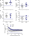
Figure 3. Effect of aging during mechanical ventilation on surfactant pool size and surfactant function.
Values for nonventilated animals are included for reference from a previous study (52) and are indicated as dashed lines (black for young; blue for aged). (AD) No significant differences were observed in total surfactant abundance (A), large (B) or small (C) aggregates, or percentage of large aggregates relative to total surfactant (D). There appeared to be an increase in minimum surface tension of surfactant in both young and aged mice when compared with historical controls (52). (E) The minimum surface tension of surfactant was significantly increased in aged versus young mice following ventilation. n = 5–8; *P < 0.05; unpaired t test and repeated-measures 1-way ANOVA.
Figure 4
Figure 4. Effect of age and mechanical ventilation on SFTP expression.
Following mechanical ventilation (20 mL/kg for 3 hours), the expression of Sftpa1 (A), Sftpb (B), Sftpc (C), and Sftpd (D) was assessed by qPCR in whole lung tissue of young and aged mice. A significant reduction in Sftpa1 was observed in aged ventilated compared with aged nonventilated mice (A). A significant decrease in Sftpb was also observed in the aged ventilated mice compared with young ventilated mice (B). No differences were observed in Sftpc (C) or Sftpd (D). n = 3–7; *P < 0.05; 2-way ANOVA with Tukey’s test.
Figure 5
Figure 5. Effect of age on the transcriptomic response to mechanical ventilation in alveolar type I + II cells.
(A and B) The volcano plot reveals only a few significantly differentially expressed genes in the young animals (A) and no significantly differentially expressed genes in the aged animals (B) following mechanical ventilation. (C) The heatmap highlights a few enriched pathways associated with cell activation that are found exclusively in the young animals following mechanical ventilation. Datasets were derived from n = 3 animals pooled per group. YNV, young nonventilated; YV, young ventilated; ANV, aged nonventilated; AV, aged ventilated.
Figure 6
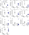
Figure 6. Effect of age and mechanical ventilation (20 mL/kg for 3 hours) on expression of inflammatory cytokines.
(AJ) The expression levels of Il6 (A), Ccl2 (B), Csf2 (C), Il1b (D), Cxcl2 (E), Cxcl1 (F), Ifng (G), Il10 (H), Il12p70 (I), and Tnfa (J) were assessed by qPCR in whole lung tissue from young and aged mice. n = 3–7. Significance is shown as *P < 0.05; **P < 0.01; ***P < 0.001; ****P < 0.0001; 2-way ANOVA with Tukey’s test.
Figure 7
Figure 7. Effect of age during mechanical ventilation on inflammatory cytokine protein abundance within the serum.
Following mechanical ventilation (20 mL/kg for 3 hours), multiplex analysis was performed to assess the abundance of IL6 (A), CCL2 (B), CSF2 (C), ILIβ (D), CXCL2 (E), CXCL1 (F), IFNγ (G), IL-10 (H), IL-12P70 (I), and TNFα (J) within the serum. n = 8 (young) and n = 6 (aged). Significance is shown as *P < 0.05; **P < 0.01; ***P < 0.001; ****P < 0.0001; unpaired t test.
Figure 8
Figure 8. Effect of age during mechanical ventilation on inflammatory cytokine protein abundance within the serum.
Following mechanical ventilation (20 mL/kg for 3 hours), multiplex analysis was performed to assess the abundance of IL6 (A), CCL2 (B), CSF2 (C), IL-1β (D), CXCL2 (E), CXCL1 (F), IFNγ (G), IL-10 (H), IL-12P70 (I), and TNFα (J) within the serum. n = 7 (young) and n = 5 (aged). Significance is shown as *P < 0.05; **P < 0.01; ***P < 0.001; ****P < 0.0001; unpaired t test.
Figure 9
Figure 9. Effect of age on the transcriptomic response to mechanical ventilation in alveolar macrophages.
(A and B) The volcano plots reveal many significantly differentially expressed genes in macrophages from the young animals following mechanical ventilation (A), with far fewer genes observed in the macrophages from the aged animals (B). (C) The heatmap highlighting enriched pathway terms regulated by differentially expressed genes in the alveolar macrophages demonstrates greater enrichment of inflammatory and activation pathways in the young mice exposed to mechanical ventilation, which is reduced in the aged mice. (D) The Circos plot displaying the degree of overlap of functional pathways between the young and aged animals in response to mechanical ventilation reveals high overlap of genes (blue lines) and pathways (purple lines) in the aged animals, as well as an abundance of genes and pathways exclusive to young animals. YNV, young nonventilated; YV, young ventilated; ANV, aged nonventilated; AV, aged ventilated.
Figure 10
Figure 10. The effect of ventilation and aging on inferred secreted signaling originating from alveolar macrophages.
(A) Inferred cell-cell communication analysis of secreted signaling pathways reveals an increase in communication from the alveolar macrophages to all other cell types in the young group following exposure to mechanical ventilation, as indicated by the red lines. (B) In contrast, the alveolar macrophages from aged animals exhibited a generally decreased response following mechanical ventilation, as indicated by the blue lines. (C) Comparison of the ventilated animals directly reveals an overall blunted response in the aged animals compared with young animals. Datasets were derived from n = 3 animals pooled per group. For circle diagrams (AC), red lines indicate more signaling in second group relative to first group; blue lines indicate less signaling in second group relative to first group. YNV, young nonventilated; YV, young ventilated; ANV, aged nonventilated; AV, aged ventilated.
Figure 11
Figure 11. Effect of aging during mechanical ventilation on protein leak within the lung.
Values for nonventilated animals are shown as reference from a previous study for the protein leak assessment (52) and are indicated as dashed lines (black for young; blue for aged). (A and B) Compared with young ventilated animals, aged ventilated animals exhibited a significant increase in protein (A) and IgM (B) in the bronchoalveolar lavage fluid. n = 8 (young) and n = 6–7 (aged). **P < 0.01; unpaired t test.
Figure 12
Figure 12. Effect of age on the transcriptomic response to mechanical ventilation in capillary type I and capillary type II cells.
(A and B) The volcano plots demonstrate several significantly differentially expressed genes in the capillary type I cells following mechanical ventilation in both young (A) and aged animals (B). (C and D) The same trend was observed for capillary type II cells. (E and F) The heatmaps highlight several enriched pathways associated with vessel development and cell-cell adhesion in both the young and aged mice in both capillary populations. Datasets were derived from n = 3 animals. YNV, young nonventilated; YV, young ventilated; ANV, aged nonventilated; AV, aged ventilated.
Figure 13
Figure 13. The effect of aging on inferred cell-cell contact autocrine signaling from the capillary cells during mechanical ventilation.
(A and B) The CellChat violin plots of inferred cell-cell contact signaling pathways reveals increased expression of several ligands and receptors associated with cell-cell adhesion in the aged ventilated compared with young ventilated mice in both capillary type I (A) and type II (B) cells (ligands and receptors shown are the most differentially enriched). (C and D) Gene ontology analysis using all differentially enriched ligands and receptors revealed that the aged ventilated animals exhibited a robust enrichment in pathways associated with cell-cell adhesion, cell junction organization, angiogenesis, and blood vessel development in both capillary type I (C) and type II (D) cells. Datasets were derived from n = 3 animals. YNV, young nonventilated; YV, young ventilated; ANV, aged nonventilated; AV, aged ventilated.
Figure 14
Figure 14. Assessment of endothelial barrier integrity in isolated PMVEC from young and aged animals, grown to confluent monolayers.
(A) Compared with young PMVEC, aged PMVEC had a significant 484% increase in monolayer permeability, as assessed by albumin flux in the transwell system. (B) Staining of adherens junctional protein, VE-cadherin, and tight junctional protein claudin-5 revealed disrupted localization in the PMVEC monolayers from aged mice compared with young mice; above are representative images from n = 5 experiments. For albumin flux experiments, n = 7. Scale bar: 50 μm. ****P < 0.0001; unpaired t test.

Similar articles

References

    1. Huppert LA, et al. Pathogenesis of acute respiratory distress syndrome. Semin Respir Crit Care Med. 2019;40(1):31–39. doi: 10.1055/s-0039-1683996. - DOI - PMC - PubMed
    1. Brown R, et al. The impact of aging in acute respiratory distress syndrome: a clinical and mechanistic overview. Front Med (Lausanne) 2020;7:589553. doi: 10.3389/fmed.2020.589553. - DOI - PMC - PubMed
    1. Acute Respiratory Distress Syndrome Network. et al. Ventilation with lower tidal volumes as compared with traditional tidal volumes for acute lung injury and the acute respiratory distress syndrome. N Engl J Med. 2000;342(18):1301–1308. doi: 10.1056/NEJM200005043421801. - DOI - PubMed
    1. Smolin B, et al. Mechanical ventilation for older medical patients in a large tertiary medical care center. Eur Geriatr Med. 2022;13(1):253–265. doi: 10.1007/s41999-021-00557-6. - DOI - PMC - PubMed
    1. Ely EW, et al. Recovery rate and prognosis in older persons who develop acute lung injury and the acute respiratory distress syndrome. Ann Intern Med. 2002;136(1):25–36. doi: 10.7326/0003-4819-136-1-200201010-00007. - DOI - PubMed

Substances