. 2025 Feb 13;10(7):e188014.
doi: 10.1172/jci.insight.188014.

Antisense oligonucleotides modulate aberrant inclusion of poison exons in SCN1A-related Dravet syndrome

Affiliations

Antisense oligonucleotides modulate aberrant inclusion of poison exons in SCN1A-related Dravet syndrome

Sheng Tang et al. JCI Insight. .

Abstract

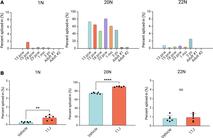
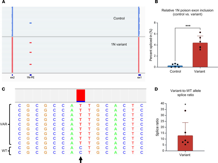
Dravet syndrome is a developmental and epileptic encephalopathy associated with pathogenic variants in SCN1A. Most disease-causing variants are located within coding regions, but recent work has shed light on the role of noncoding variants associated with a poison exon in intron 20 of SCN1A. Discovery of the SCN1A poison exon known as 20N has led to the first potential disease-modifying therapy for Dravet syndrome in the form of an antisense oligonucleotide. Here, we demonstrate the existence of 2 additional poison exons in introns 1 and 22 of SCN1A through targeted, deep-coverage long-read sequencing of SCN1A transcripts. We show that inclusion of these poison exons is developmentally regulated in the human brain, and that deep intronic variants associated with these poison exons lead to their aberrant inclusion in vitro in a minigene assay or in iPSC-derived neurons. Additionally, we show that splice-modulating antisense oligonucleotides can ameliorate aberrant inclusion of poison exons. Our findings highlight the role of deep intronic pathogenic variants in disease and provide additional therapeutic targets for precision medicine in Dravet syndrome and other SCN1A-related disorders.

Keywords: Epilepsy; Gene therapy; Genetics; Neuroscience.


Conflict of interest statement

Conflict of interest: The authors have declared that no conflict of interest exists.

Figures

Figure 1
Figure 1. Features and conservation of SCN1A poison exons.
(A) Targeted RT-PCR and long-read sequencing of SCN1A transcripts in control iNeurons shows rare inclusion of alternatively spliced exons in intron 1 (left) and intron 22 (right). Shown are representative views of reads displayed in Integrative Genomics Viewer. Note that SCN1A is located on the negative (–) strand of chromosome 2, and therefore its sequence is displayed from right to left. (B) Sashimi plots showing alternative splicing of 1N (left) and 22N (right) in iNeurons from healthy controls. PE, poison exon. (C) In-frame in silico translation starting from the nearest 5′ canonical exon shows that 1N (left) and 22N (right) each introduce a PTC (red asterisk) when spliced into the reading frame, suggesting they could serve as poison exons. The portion of the translation identical to the canonical SCN1A protein sequence is highlighted, with the unhighlighted portion representing the contribution of the poison exon. (D) Poison exons, associated pathogenic variants, and conservation. The poison exons, their related variants, the GERP score, and UCSC conservation track (Multiz alignments for vertebrate species) are shown. The GERP score is an estimate of evolutionary constraint (scale: 0 to 7; positive scores suggest evolutionary constraint) (40). From left to right: 1N is poorly conserved and overlaps with a SINE (gray bar), suggesting that it may have occurred due to a recent retrotransposition event during primate evolution. 20N, previously described, is well conserved in most mammalian species. 22N only shows limited conservation in primates.
Figure 2
Figure 2. Developmental regulation of SCN1A poison exons and NMD sensitivity in iNeurons.
(A) Developmental regulation of SCN1A poison exons. Targeted RT-PCR followed by long-read sequencing was used to assess relative inclusion of 1N, 20N, and 22N across fetal and postnatal development in the human cerebral cortex. 20N inclusion was high throughout the prenatal period and was downregulated after the first several postnatal years. In contrast, 1N and 22N inclusion was relatively low (<5%) prenatally, but still showed a trend of downregulation postnatally. Note that for display purposes, 1N and 22N are shown on a different y-axis scale. pcw, postconception weeks. n = 1 human brain sample per developmental time point, and at least 500 reads per sample were used to calculate PSI in each sample. (B) Relative inclusion of 1N, 20N, and 22N in iNeurons from healthy controls with or without NMD inhibition with 11J. 1N and 20N, but not 22N, show significantly increased relative inclusion after 4 hours of treatment with 1 μM 11J. Note that for display purposes, 1N and 22N are shown on a different y-axis scale. n = 6 biological replicate wells of iNeurons per condition, and at least 500 reads per sample were used to calculate PSI in each sample. **P < 0.01, ****P < 0.0001 by unpaired t test.
Figure 3
Figure 3. An in vitro minigene assay demonstrates aberrantly increased inclusion of 22N due to a Dravet syndrome–related variant.
(A) Generation of a splice reporter assay containing intron 22 flanked by canonical exons 22 and 23, containing the putative 22N poison exon. The minigene was inserted into a splice reporter plasmid and transfected into HEK293T cells. Note the presence of rat insulin exons, which serve as constitutively spliced control exons. The position of the Dravet syndrome–related variant is indicated on the variant construct. Created with BioRender. (B) Increased inclusion of 22N due to the presence of a Dravet-related variant in 22N (n = 5 biological replicate transfections for each of control- or variant-transfected cells). The 22N-related variant leads to decreased ΔCt (22N – canonical splice product), which represents a higher relative abundance of the 22N-containing splice product. (C) Design of splice-switching ASOs for 22N. ASO1 targets the acceptor splice site with the 3′ end of the upstream intron, whereas ASO2 targets the donor splice site with the 5′ end of the downstream intron. Created with BioRender. (D) Splice-switching ASOs ameliorate aberrantly increased inclusion of 22N. qRT-PCR data with 22N-containing splice product normalized to canonical splice product. In contrast with untreated cells or cells treated with a scrambled ASO, ASO1 and to a lesser extent, ASO2, both reduced aberrant inclusion of 22N. ASO1-treated cells demonstrate a higher ΔCt (22N – canonical splice product) compared with untreated cells, which represents a lower relative abundance of the 22N-containing splice product (n = 5 biological replicate transfections for each condition). Significance was assessed by Mann-Whitney test (B) or Kruskal-Wallis test with Dunn’s multiple-comparison test (D). *P < 0.05, **P < 0.01 (B: P = 0.0079, no ASO vs. ASO1; D: P = 0.038, no ASO vs. ASO2). NS, not significant for no ASO vs. ASO2 and no ASO vs. ASO (scr).
Figure 4
Figure 4. Aberrantly increased inclusion of 1N in iNeurons carrying a Dravet syndrome–related variant.
(A) Sample reads from targeted RT-PCR and long-read sequencing of SCN1A transcripts in iNeurons derived from a healthy control or a patient with Dravet syndrome carrying a variant within 1N. Shown are representative views of reads displayed in Integrative Genomics Viewer. PE, poison exon. (B) Targeted SCN1A RT-PCR and long-read sequencing shows increased inclusion of 1N in patient compared with control iNeurons (n = 5 biological replicate wells of iNeurons per genotype). Each sample had at least 1000 reads from which PSI (%) was calculated. (C) A sample set of reads containing 1N obtained from heterozygous patient iNeurons. The position of the variant is indicated by the arrow, with the color map above the position showing a skewed ratio of reads containing T (pathogenic variant, in red) versus C (WT variant, in blue). For this sample, there were a total 37 variant alleles to 5 WT alleles containing 1N, resulting in a splice ratio of 7.4, showing that increased 1N inclusion is strongly skewed toward the allele carrying the pathogenic variant. (D) Increased variant-to-WT allele splice ratio in reads containing 1N in patient iNeurons (n = 6 biological replicate wells of iNeurons). *P < 0.05; ***P < 0.001 by unpaired t test (B) or 1-sample t test (D).
Figure 5
Figure 5. 1N inclusion in patient iNeurons is sensitive to inhibition of nonsense-mediated decay (NMD).
(A) Sample reads from targeted RT-PCR and long-read sequencing of SCN1A transcripts in 1N variant–patient iNeurons treated with vehicle or the NMD inhibitor 11J. PE, poison exon. (B) Increased relative inclusion of 1N in 11J-treated compared to vehicle-treated patient iNeurons (n = 6 biological replicate wells of iNeurons per condition). iNeurons were treated for 24 hours with 1 μM 11J. (C) Similar variant-to-WT allele splice ratio in vehicle- or 11J-treated patient iNeurons (n = 6 biological replicate wells of iNeurons per condition). ****P < 0.0001; NS, not significant by unpaired t test (B and C)
Figure 6
Figure 6. An exon-skipping ASO ameliorates aberrant exon inclusion in patient iNeurons and shows a substantial degree of allele specificity.
(A) ASO design for targeting ASOs. The ASO_EK (exon-skipping) targeting site lies entirely within the 1N poison exon and overlaps with the 1N patient variant, and therefore has a 1-nucleotide mismatch with the WT allele. ASO_SS (splice-switching) targets the splice acceptor site with the 3′ end of the upstream adjacent intron. A scrambled version of ASO_SS was used as an additional control. PE, poison exon. Created with BioRender. (B) eSkip-Finder predictions showing increased likelihood of exon-skipping for targets closer to the acceptor splice site at the 5′ end of 1N. ASO_EK overlaps with positions 25–49 of 1N. (C) ddPCR shows that in WT iNeurons, 1N-containing transcripts account for less than 10% of total SCN1A transcripts. In contrast, patient iNeurons show similar proportions of canonical or 1N-containing transcripts. ASO_EK significantly shifted the relative abundance of canonical and 1N-containing transcripts in favor of the canonical transcript. In contrast, untreated iNeurons and ASO_SS had similar ratios of canonical and 1N-containing transcripts. The scrambled ASO shifted the relative abundance of canonical and 1N-containing transcripts slightly toward 1N (n = 6–7 biological replicate wells of iNeurons per condition). In the patient (no ASO) condition, 1 sample was excluded due a significant result on Grubb’s outlier test (P < 0.05). (D) Targeted RT-PCR and long-read sequencing was used to assess relative inclusion of 1N in untreated patient iNeurons or those treated with ASOs; ASO_EK, but not ASO_scr, improved the aberrant inclusion of 1N (n = 5–6 biological replicate wells of patient iNeurons per condition). (E) Representative set of reads containing 1N in untreated iNeurons or iNeurons treated with ASO_EK. Position of the pathogenic variant is indicated by an arrow, with the color map above showing the relative proportion of reads containing T (patient variant, in red) versus C (WT variant, in blue). Reads from untreated iNeurons are strongly skewed toward the variant allele. In contrast, reads from ASO_EK-treated iNeurons show reduced skewing toward the variant allele. (F) ASO_EK significantly decreases the variant-to-WT allele splice ratio in patient iNeurons, suggesting that it has a substantial degree of allele-specific action (n = 5–6 biological replicate wells of patient iNeurons per condition). *P < 0.05; ***P < 0.001; ****P < 0.0001 by 1-way ANOVA with Dunnett’s multiple-comparison test. NS, not significant.

References

    1. Happ HC, Carvill GL. A 2020 view on the genetics of developmental and epileptic encephalopathies. Epilepsy Curr. 2020;20(2):90–96. doi: 10.1177/1535759720906118. - DOI - PMC - PubMed
    1. Scheffer IE, Nabbout R. SCN1A-related phenotypes: epilepsy and beyond. Epilepsia. 2019;60 Suppl 3(s3):S17–S24. - PubMed
    1. Dravet C. The core Dravet syndrome phenotype. Epilepsia. 2011;52 Suppl 2(s2):3–9. doi: 10.1111/j.1528-1167.2011.02994.x. - DOI - PubMed
    1. Carvill GL, et al. Aberrant inclusion of a poison exon causes dravet syndrome and related SCN1A-associated genetic epilepsies. Am J Hum Genet. 2018;103(6):1022–1029. doi: 10.1016/j.ajhg.2018.10.023. - DOI - PMC - PubMed
    1. Carvill GL, Mefford HC. Poison exons in neurodevelopment and disease. Curr Opin Genet Dev. 2020;65:98–102. doi: 10.1016/j.gde.2020.05.030. - DOI - PMC - PubMed

MeSH terms

Substances

LinkOut - more resources