. 2025 Feb 27;68(4):4961-4987.
doi: 10.1021/acs.jmedchem.5c00026. Epub 2025 Feb 13.

Rational Design and Optimization of a Potent IDO1 Proteolysis Targeting Chimera (PROTAC)

Affiliations

Rational Design and Optimization of a Potent IDO1 Proteolysis Targeting Chimera (PROTAC)

Paige J Monsen et al. J Med Chem. .

Abstract

Indoleamine 2,3-dioxygenase 1 (IDO1) is an immunosuppressive protein that inhibits antitumor immunity through both tryptophan metabolism and nonenzymatic functions. Drugs targeting IDO1 enzyme activity have failed to improve the overall survival of patients with cancer. Developing new therapeutics that neutralize both enzyme- and nonenzyme-derived immunosuppressive IDO1 effects is therefore of high interest. We previously described a novel proteolysis targeting chimera (PROTAC), NU223612, that degrades IDO1 in cultured human glioblastoma (GBM) cells, as well as in well-established brain tumors, in vivo. In this study, we rationally optimized the structure of our lead series to create NU227326, which degrades IDO1 with a DC50 of 5 nM in human GBM cells. Mechanistic studies showed that IDO1 degradation occurred through the ubiquitin-proteasome system and was sustained for at least 2 days, supporting NU227326 as a highly potent IDO1 PROTAC suitable for further studies in GBM and other human cancers.


Conflict of interest statement

The authors declare the following competing financial interest(s): The IDO-PROTACs described herein are the subject of a U.S. patent application filed by Northwestern University and lists G.E.S., D.A.W., and P.J.M. as inventors. Dr. Lukas has received honoraria for serving on a Scientific Advisory Board for Merck, and honoraria for serving on the Speakers Bureau for Novocure. He has received research support for drug only use from BMS.

Figures

Figure 1
Figure 1
Representative IDO1 inhibitors.
Figure 2
Figure 2
(A) General PROTAC design. (B) Structures of reported IDO1 PROTACs to-date.
Figure 3
Figure 3
(A) Structures of truncated 4-substituted and 4-chloro-3-substituted type IDO1 PROTACs used in docking studies. (B, C) Surface representations of the cocrystal structure of IDO1 (blue, PDB 6AZW) with the ligand BMS-116 (green) overlaid with either the 4-substituted type analog (purple) or the 4-chloro-3-substituted type analog (gold) demonstrating the orientation of the exit vector and linker. (D, E) Binding pose of BMS-116 (green) modeled with either the 4-substituted type analog (purple) or the 4-chloro-3-substituted type analog (gold) in the IDO1 active site with representative interactions displayed to demonstrate the influence in altering the substitution position of the linker.
Figure 4
Figure 4
Design of novel IDO1 PROTACs based on modular structural optimizations of lead IDO1 PROTAC 6 (NU223612).
Figure 5
Figure 5
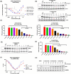
Characterization of IDO1 degradation by PROTACs 20 and 21. (A) Degradation of IDO1 by 20, 21, and NU223612 in the HiBiT assay. DC50 was calculated from the lowest concentration down to where maximum degradation was observed and excluded data points within the Hook effect range. R2 = 0.94 for 20; R2 = 0.93 for 21. (B) Dose-dependent degradation by compounds 20 and 21 on IDO1 in U87 cells treated with an extended dose range of 20 or 21 for 24 h and protein samples were analyzed by Western blotting. (C) Effects of 20 and 21 on kynurenine production in U87 human GBM cells. Cells were treated with 20 or 21 in the presence of 50 ng/mL IFNγ beginning at 24 h after plating the cells and for a total of 24 h, followed by the collection of cell culture supernatants to measure IFNγ-induced kynurenine levels using Ehrlich’s reagent. Cells cultured in the absence of IFNγ served as the control group. (D) GBM43 cells were treated with an extended dose range of 21 for 24 h, and protein samples were analyzed by Western blotting. (E) Effect of 21 on kynurenine production in GBM43 cells. Cells were treated with 21 in the presence of 10 ng/mL IFNγ for 24 h, and cell culture supernatants were collected to measure IFNγ-induced kynurenine levels using Ehrlich’s reagent. Cells cultured in the absence of IFNγ served as the control group. (F) Representative curves of percent IFNγ-induced IDO1 levels that were normalized to untreated samples as calculated in U87 and GBM43 cells to determine DC50 that produces 50% of IDO1 degradation (n = 3 independent experiments). Data are presented as mean ± SEM. Statistical significance was determined using Tukey’s multiple comparison test for comparisons between more than two groups. Significance levels are indicated as follows: P < 0.05 (*), P < 0.01 (**), P < 0.001 (***), and P < 0.0001 (****). (G) Western blotting analysis of IDO1 protein in U87 cells. IDO1 was induced in U87 cells with 50 ng/mL human IFNγ for 24 h followed by a 25, 50, or 100 nM treatment with IDO1 PROTAC (21, NU227326), inactive PROTAC (24, NU227428) or IDO1 enzyme inhibitor (4, NU223618) for 24 h before protein samples were prepared for Western blotting analysis. Percent of normalized IDO1 protein levels were derived from densitometric analysis of band intensities. Western blotting results are representative of outcomes from 3 separate experimental replicates.
Figure 6
Figure 6
Characterization of 21 (NU227326) as a potent IDO1 PROTAC. (A) Western blot analysis of IDO1 and GAPDH to determine the kinetics of 21 (NU227326)-induced IDO1 protein degradation in U87 cells. (B) Western blot analysis of IDO1 to determine the effect of a single continuous treatment of 21 (NU227326) on IDO1 protein levels at multiple time points in U87 cells. (C) Similar to (B), U87 cells were treated with 21 (NU227326) for 24 h, and cells were cultured without 21 (NU227326) for up to 48 h. Protein samples were tested in Western blot analysis to determine IDO1 levels upon withdrawal of the IDO1 PROTAC. (D) Western blotting analysis of IDO1 and GAPDH to determine the effect of parental competitive IDO1-inhibitor (4, NU223618) and noncompetitive IDO1-inhibitor (BGB-7204); (E) E3 ligase ligand (pomalidomide) and E1 ligase neddylation inhibitor (MLN4924); and (F) proteasome inhibitor (MG132) on NU227326-induced IDO1 degradation. Western blotting analysis to determine the effect of NU227326 on IFNγ-induced IDO1 levels in human adult PDX cells; PDX-6 (G) and PDX-38 (H), U87 cells exogenously overexpressing GFP-fused IDO1 (I), prostate cancer PC-3 cells (J), human pediatric KNS42 GBM cells (K), triple negative breast cancer MDA-MB-231 cells (L), pancreatic cancer SW-1990 cells (M), and ovarian SKOV-3 cells (N). Cells treated with either 21 (NU227326), 24 (NU227428), or 4 (NU223618) at indicated concentrations for 24 h and protein samples were analyzed for IDO1 and GAPDH using Western blotting analysis. Western blotting results are representative of outcomes from 3 separate experimental replicates.
Figure 7
Figure 7
Scatter plot of protein expression changes based on global quantitative proteomics. Kelly cells were treated with 1 μM compound for 24 h and analyzed as described in the Experimental Section. (A) Treatment with NU227326. (B) Treatment with NU227327.
Figure 8
Figure 8
(A) Left: BLI sensorgrams showing association and dissociation of compounds 20 (NU227327) (upper) and 21 (NU227326) (lower) with CRBN (immobilized on AR2G sensors). Steady-state data fitted as described above yielded Kd values of 520 ± 106 nM and 380 ± 63 nM for the binary complexes formed by CRBN with 20 and 21, respectively. (B) Left: BLI sensograms showing association and dissociation of compounds 20 (NU227327) (upper) and 21 (NU227326) (lower) to IDO1 protein (loaded on NI NTA sensors). Right: For each complex, the equilibrium dissociation constant (Kd) was obtained by fitting the steady state data (Req as a function of compound concentration) with a 1:1 binding model. The measured affinities for the binary complexes formed by IDO1 with compounds 20 and 21 were determined to be 370 ± 62 nM and 240 ± 49 nM, respectively. (C) BLI sensograms monitoring the association and dissociation kinetics of IDO1-20-CRBN (left) and IDO1-21-CRBN (right) ternary complexes. IDO1 protein was loaded on NiNTA biosensors. Double-referenced data sets fitted globally with a 1:1 kinetic model yielded Kd values of 321 ± 54 nM for IDO1-21-CRBN and 997 ± 143 nM for IDO1-20-CRBN. The koff values obtained from the kinetic fits translate into the following lifetimes (t1/2): IDO1-20 binary = 53 s, IDO1-20-CRBN ternary = 256 s; IDO1-21 binary = 38.6 s, IDO1-21-CRBN ternary = 1039 s.
Figure 9
Figure 9
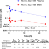
Pharmacokinetics of NU227326 (21) in mice. Compound was dosed at 50 mg/kg by IP in mice.
Scheme 1
Scheme 1. Synthesis of 4-Chloro-3-substituted and 3-Substitutied Piperidine IDO1 Binder Building Blocks 31 and 32
Reagents and conditions: (a) tert-butyl 4-((methylsulfonyl)oxy)piperidine-1-carboxylate, K2CO3, DMF, 75 °C, 12 h; (b) Pd/C, H2 (g), EtOH, 23 °C, 24–36 h; (c) (R)-2-((1S,4S)-4-(6-fluoroquinolin-4-yl)cyclohexyl)propanoic acid, EDCI, pyridine, 0 to 23 °C, 12 h; (d) 4 N HCl in dioxane, 23 °C, 12 h.
Scheme 2
Scheme 2. Synthesis of IDO1 Degraders 79
Reagents and conditions: (a) tert-butyl 3-(2-(2-aminoethoxy)ethoxy)propanoate, DIPEA, DMF, 75 °C, 12–48 h; (b) CF3COOH, CH2Cl2, 0 to 23 °C, 1 h; (c) (R)-2-((1S,4S)-4-(6-fluoroquinolin-4-yl)cyclohexyl)-N-(4-(piperidin-4-yloxy)phenyl)propanamide or 31, HATU, DIPEA, DMF, 0 to 23 °C, 6–12 h.
Scheme 3
Scheme 3. Synthesis of IDO1 Degraders 1021
Reagents and conditions: (a) tert-butyl piperazine-1-carboxylate, DIPEA, DMSO, 100 °C, 12 h; (b) CF3COOH, CH2Cl2, 0 to 23 °C, 1 h; (c) tert-butyl 3-(2-iodoethoxy)propanoate, DIPEA, DMF, 75 °C, 24 h; (d) CF3COOH, CH2Cl2, 0 to 23 °C, 1 h; (e) (c) (R)-2-((1S,4S)-4-(6-fluoroquinolin-4-yl)cyclohexyl)-N-(4-(piperidin-4-yloxy)phenyl)propanamide, 31 or 32, HATU, DIPEA, DMF, 0 to 23 °C, 1.5–24 h; (f) tert-butyl 4-(bromomethyl)piperidine-1-carboxylate, DIPEA, DMF, 75 °C, 12 h; (g) CF3COOH, CH2Cl2, 0 to 23 °C, 1–2.5 h; (h) tert-butyl 4-oxopiperidine-1-carboxylate, STAB, DMF, 23 °C, 2 h; (i) CF3COOH, CH2Cl2, 0 to 23 °C, 1 h; (j) tert-butyl 2-bromoacetate, DIPEA, DMF, 23 °C, 2–3 h; (k) CF3COOH, CH2Cl2, 0 to 23 °C, 2–4 h; (l) (c) (R)-2-((1S,4S)-4-(6-fluoroquinolin-4-yl)cyclohexyl)-N-(4-(piperidin-4-yloxy)phenyl)propanamide, 31 or 32, HATU or T3P, DIPEA, DMF, 0 to 23 °C, 1–48 h.
Scheme 4
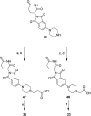
Scheme 4. Synthesis of IDO1 Degraders 22 and 23
Reagents and conditions: (a) tert-butyl 3-bromopropionate, DIPEA, DMF, 23 °C, 12 h; (b) CF3COOH, CH2Cl2, 0 to 23 °C, 24 h; (c) tert-butyl 2- bromoacetate, DIPEA, DMF, 23 °C, 2 h; (d) CF3COOH, CH2Cl2, 0 to 23 °C, 24 h; (e) 37, HATU, DIPEA, DMF, 0 to 23 °C, 30 min.
Scheme 5
Scheme 5. Synthesis of Inactive IDO1 PROTAC 24
Reagents and conditions: (a) MeI, K2CO3, DMF, 0 to 23 °C, 12 h, 82%; (b) tert-butyl piperazine-1-carboxylate, DIPEA, DMSO, 100 °C, 2.5 h, 90%; (c) CF3COOH, CH2Cl2, 0 to 23 °C, 2 h, 90%; (d) tert-butyl 4-oxopiperidine-1-carboxylate, STAB, DMF, 23 °C, 1.5 h; (e) CF3COOH, CH2Cl2, 0 to 23 °C, 12 h, 77% over 2 steps; (f) tert-butyl 2-bromoacetate, DIPEA, DMF, 23 °C, 12 h; (g) CF3COOH, CH2Cl2, 0 to 23 °C, 4 h, 72% over 2 steps; (h) 32, HATU, DIPEA, DMF, 0 to 23 °C, 30 min, 48%.

Update of

References

    1. Adamson C.; Kanu O. O.; Mehta A. I.; Di C.; Lin N.; Mattox A. K.; Bigner D. D. Glioblastoma multiforme: a review of where we have been and where we are going. Expert Opin. Investig. Drugs 2009, 18 (8), 1061–1083. 10.1517/13543780903052764. - DOI - PubMed
    1. Lukas R. V.; Wainwright D. A.; Ladomersky E.; Sachdev S.; Sonabend A. M.; Stupp R. Newly Diagnosed Glioblastoma: A Review on Clinical Management. Oncology (Williston Park) 2019, 33 (3), 91–100. - PMC - PubMed
    1. Ostrom Q. T.; Price M.; Neff C.; Cioffi G.; Waite K. A.; Kruchko C.; Barnholtz-Sloan J. S. CBTRUS Statistical Report: Primary Brain and Other Central Nervous System Tumors Diagnosed in the United States in 2016-2020. Neuro-Oncology 2023, 25 (Suppl. 4), iv1–iv99. 10.1093/neuonc/noad149. - DOI - PMC - PubMed
    1. Cao T. Q.; Wainwright D. A.; Lee-Chang C.; Miska J.; Sonabend A. M.; Heimberger A. B.; Lukas R. V. Next Steps for Immunotherapy in Glioblastoma. Cancers (Basel) 2022, 14 (16), 4023.10.3390/cancers14164023. - DOI - PMC - PubMed
    1. Eggermont A. M.; Chiarion-Sileni V.; Grob J.-J.; Dummer R.; Wolchok J. D.; Schmidt H.; Hamid O.; Robert C.; Ascierto P. A.; Richards J. M.; et al. Prolonged survival in stage III melanoma with ipilimumab adjuvant therapy. N. Engl. J. Med. 2016, 375 (19), 1845–1855. 10.1056/NEJMoa1611299. - DOI - PMC - PubMed

MeSH terms

Substances

LinkOut - more resources