Case Reports
. 2025 Feb 13;15(1):e24.00338.
doi: 10.2106/JBJS.CC.24.00338. eCollection 2025 Jan 1.

"Trapdoor" Medial Scapula Osteotomy for Resection of a Benign Subscapular Neoplasm: A Case Report

Affiliations
Case Reports

"Trapdoor" Medial Scapula Osteotomy for Resection of a Benign Subscapular Neoplasm: A Case Report

Amir M Boubekri et al. JBJS Case Connect. .

Abstract

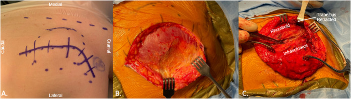
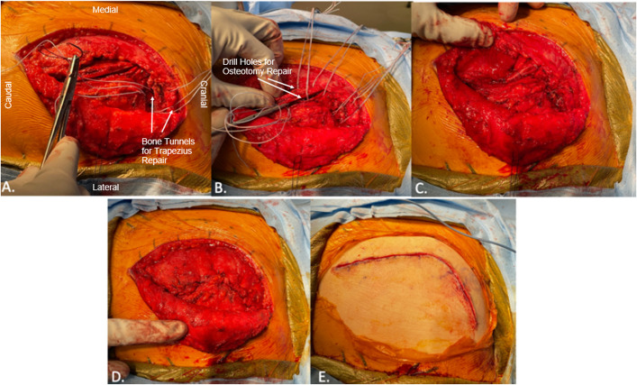
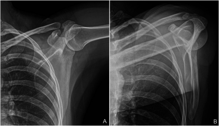
Case: Neurofibromatosis type 2 (NF2) is an autosomal dominant condition characterized by the development of neoplasms, which infrequently arise in the subscapular fossa. Surgical removal of large subscapular tumors carries the risk of shoulder dysfunction due to muscle or nerve injury. We describe the case of a patient with NF2 who presented with a hybrid subscapular neurofibroma and schwannoma tumor that was completely resected through a muscle-sparing medial scapular osteotomy approach.

Conclusion: We describe a unique muscle-sparing scapula splitting surgical approach to the subscapular region that provides excellent exposure for tumor removal and clinical outcomes with minimal postoperative scapular dyskinesis.

Level of evidence: IV (case report).


Conflict of interest statement

Disclosure: The Disclosure of Potential Conflicts of Interest forms are provided with the online version of the article (http://links.lww.com/JBJSCC/C549).

Figures

Fig. 1
Fig. 1
MRI imaging with sagittal T2 sequences demonstrated a hyperintense heterogeneously enhancing mass within the right posterior chest wall and abutting the scapula (as highlighted by the arrow) (Fig. 1-A). Interval imaging demonstrated minimal change in size (as highlighted by the arrow) (Fig. 1-B). Postoperative MRI 6 months after surgery demonstrated a complete resection (Fig. 1-C). MRI = magnetic resonance imaging.
Fig. 2
Fig. 2
The midline (dashed line), curved skin incision, and underlying tumor (dotted circle) were marked out (Fig. 2-A). Large full-thickness flaps were elevated (Fig. 2-B). The lower trapezius was elevated subperiosteally and infraspinatus fascia recessed ∼1 cm to expose osteotomy site. (Fig. 2-C).
Fig. 3
Fig. 3
The osteotomy was performed with a sagittal saw along the medial border of the scapula, just distal to the scapular spine (Fig. 3-A). The medial bone was then retracted with traction sutures, opening the “trapdoor” (Fig. 3-B). This allowed for excellent exposure to the underlying tumor bed (Fig. 3-C).
Fig. 4
Fig. 4
Both illustrations show a posterior view of an isolated right scapula. The rhomboid major and rhomboid minor insertions along the posterior medial boarder of the scapula are depicted. The illustration on the left shows the projected osteotomy trajectory represented by the dashed red line. The illustration on the right shows the scapula after the medial osteotomy. All rhomboid muscle bellies and insertions remain inviolate and undisturbed. Traction sutures are in place allowing for “trapdoor” visualization.
Fig. 5
Fig. 5
The lower trapezius was prepared for repair to its anatomic insertion along the scapular spine using #2 FiberWire sutures tied across bone tunnels (Fig. 5-A). The repair technique used #2 FiberWire sutures tied across several bone tunnels drilled into the scapular body and medial osteotomized bone, reducing it back to its anatomic position (Fig. 5-B). The osteotomy FiberWire sutures were tied sequentially first, closing the “trapdoor”, before tying down the lower trapezius repair sutures. The final repair after fascial closure was performed (Figs. 5-C and 5-D), and the skin was closed in layers (Fig. 5-E).
Fig. 6
Fig. 6
Fig. 6-A 10× CD34 image showing positive staining in the neurofibroma-like areas (green arrow) and negative staining in the schwannoma-like areas (blue arrow). Fig. 6-B 10× S100 stain showing strong, diffuse staining in the schwannoma areas (blue arrow) and more scattered staining in the neurofibroma areas (green arrow). Fig. 6-C 10× H&E stain showing a benign nerve sheath tumor with biphasic morphology, where nodules of Schwann cells with fascicular architecture and occasional Verocay bodies (blue arrow) lie within loosely textured, myxoid tissue with elongated wavy cell nuclei and collagen bundles, characteristic of a neurofibroma (green arrow).
Fig. 6
Fig. 6
Fig. 6-A 10× CD34 image showing positive staining in the neurofibroma-like areas (green arrow) and negative staining in the schwannoma-like areas (blue arrow). Fig. 6-B 10× S100 stain showing strong, diffuse staining in the schwannoma areas (blue arrow) and more scattered staining in the neurofibroma areas (green arrow). Fig. 6-C 10× H&E stain showing a benign nerve sheath tumor with biphasic morphology, where nodules of Schwann cells with fascicular architecture and occasional Verocay bodies (blue arrow) lie within loosely textured, myxoid tissue with elongated wavy cell nuclei and collagen bundles, characteristic of a neurofibroma (green arrow).
Fig. 7
Fig. 7
Fig. 7-A Anteroposterior and (Fig. 7-B) lateral radiograph views of the scapula 3 years after the osteotomy procedure. The bone has healed with no clinical dysfunction.

Similar articles

References

    1. Evans DG, Huson SM, Donnai D, Neary W, Blair V, Newton V, Harris R. A clinical study of type 2 neurofibromatosis. Q J Med. 1992;84(304):603-18. - PubMed
    1. Goel DP, Drosdowech DS, Turner RG, Lawendy AR, Garvin G. Periarticular neurofibroma of the shoulder: a case report. J Bone Joint Surg Am. 2010;92(3):715-20. - PubMed
    1. Lee DH, Hills JM, Jordanov MI, Jaffe KA. Common tumors and tumor-like lesions of the shoulder. J Am Acad Orthop Surg. 2019;27(7):236-45. - PubMed
    1. Tamura R. Current understanding of neurofibromatosis type 1, 2, and schwannomatosis. Int J Mol Sci. 2021;22(11):5850. - PMC - PubMed
    1. Ferner RE, O'Doherty MJ. Neurofibroma and schwannoma. Curr Opin Neurol. 2002;15(6):679-84. - PubMed

Publication types